Most patients at a dentist's appointment have long become accustomed to complex dental terms. Bridge, veneer, filling, premolar – these words are practically easy to understand when a doctor diagnoses a problem and prescribes treatment.
Most often, patients are confused by incomprehensible numbering . For example, when talking about wisdom teeth, the dentist can explain to the patient that the lower “eights” need treatment. Moreover, each tooth has its own numerical designation, which can reflect its type and exact location in the oral cavity.
Over the entire existence of dentistry as a science, several classifications of teeth have been invented, which are called dental formulas. Depending on the chosen numbering method, each tooth is assigned a specific number. In some systems, teeth are designated by a combination of letters, numbers and even symbols, which allows the dentition to be depicted in the form of an accurate diagram.
What is a dental formula
A dental formula or diagram is a brief description of the dental structure of the human dentition using numbers and Latin letters.
The letters are abbreviations from the Latin names of teeth accepted in medicine:
- I – these are dentes incisivi or incisors.
- C is for dentes canini or fangs.
- P stands for dentes premolars or premolars.
- M stands for dentes molars or molars.
The alphanumeric dental formula is common in many countries, including America, which is why it is sometimes called American. After the letters in the formula there is always a digital fraction, where the numerator indicates the number of teeth on the upper jaw, and the denominator on the lower jaw.
The dental formula of a healthy adult with a normal bite and 32 teeth looks like this:
This formula indicates that on each human jaw there is:
- 2 pairs of incisors;
- 1 pair of fangs;
- 2 pairs of premolars;
- 3 pairs of molars.
Dental formula in the absence of some teeth
It is not difficult to correctly number dental units with a healthy dentofacial apparatus; it is much more difficult to indicate the location of the teeth if a person does not have any molars, incisors, canines or premolars. In this case, the dentist indicates the number 0 opposite a certain group of teeth, rather than shifting the number series.
If the patient has any anomalies in the development of the dentofacial apparatus, for example, teething in the wrong place (hyperdontia, polydontia), the dentist prescribes an individual formula. In addition to numerical designations, the doctor generates a detailed report on the structure of a person’s dentition.
Symptoms of pathology
The main symptom is the presence of teeth beyond the natural set. They can have different positions, shapes and numbers. The process of teething of abnormal teeth is often accompanied by pain, fever, and inflammation of areas of the oral cavity. If measures are not taken in a timely manner, complications may develop:
- redness and swelling at the site of the impacted tooth;
- problems with chewing food and digestion;
- injuries to the mucous tissues of the oral cavity;
- violation of the position of the main teeth (dystopia) and the formation of malocclusion;
- various speech defects - mainly problems with the pronunciation of hissing sounds;
- loosening of adjacent normal teeth;
- deformation of the jaw bones.
In young children and adults, the pathology has its own nuances. They are associated with age-related characteristics of the body and affect the subsequent development of maxillofacial structures. Statistics on the spread of pathology in children and adults:
- 60% of cases are children and adolescents during the period of replacement of milk teeth with permanent ones;
- 35% of cases are adults;
- 5% of cases are small children during the period of growth of baby teeth.
Reference! Supernumerary teeth, as a rule, have a standard structure, sometimes with a smaller crown size. Less common are deformed formations of a teardrop-shaped, lumpy, chisel-shaped form.
Description, structure, functions, name and location of human teeth
It is not without reason that human teeth have different shapes, structures and sizes. Each tooth performs certain functions, and its purpose is, one way or another, reflected in the name:
- The incisors are the front teeth that fall into the smile zone. They are quite sharp as their function is to tear or cut food, hence the name. Normally, every person has lateral and central (larger) incisors.
- Canines are cone-shaped teeth that can be used to tear and hold food. They are located immediately behind the incisors.
- Premolars are the small back molars located behind the canine teeth.
- Molars are the back teeth necessary for mechanical processing of food. Wisdom teeth refer specifically to molars.
The listed types of teeth are also found in animals. But in the process of their evolution, some dental units were slightly modified:
- elephants' upper incisors became tusks;
- Poisonous snakes have poison in their fangs.
Many animals have front teeth similar to human ones, but since they are called differently, they cannot always be identified with the usual incisors and canines.
The structure of human teeth
Human teeth consist of a crown and a root part. The first is located above the level of the gum, the second is inside it. The top of the crown is covered with enamel, the strongest tissue in the body. It protects the inner layer of the tooth – dentin – from external influences. Under the dentin there is a hollow chamber with nerves and blood vessels - the pulp.
Despite its strength, enamel is vulnerable to bacteria. If left untreated, caries can affect the dentin and pulp. When the pulp chamber, which contains nerve fibers, is damaged, pulpitis develops, accompanied by acute throbbing pain. If the infection spreads to the root of the tooth, it is highly likely that it will have to be removed.
The basis of the lower part of the tooth is the root canals, which also contain arteries, veins and nerve fibers. Through the apical foramen, all of these structures are connected to the main neurovascular bundle.
The lower part of the tooth is covered with dentin and cement, which is attached to the periodontium using collagen fibers. The roots of teeth are hidden in the alveoli - depressions in the jaw bone.
Premolars
These are small molars. The crown of a small molar on the side of the closure surface is round or oval. The height of the crown is significantly less than that of the canines. The root is single, conical; in half of the cases, the root of the first upper molar can be bifurcated at the apex. We have a total of eight premolars in our mouth, four on each jaw. We use these teeth for secondary processing of food.
On what basis are teeth numbered according to different classification systems?
In addition to the Latin names of teeth, digital designations are used in dentistry. The numbering of teeth is based on the order in which they erupt. It starts from the front incisors (from the middle of the jaw) and runs to the left and right of them.
There are several generally accepted systems for numbering human teeth.
Universal system
Most often, dentists name teeth not by the Latin alphabet, but according to their location in the oral cavity (ordinal number). Moreover, using not Roman, but familiar Arabic numerals.
Names of teeth according to the universal classification system:
- two central incisors are located at number 1 and are called ones;
- the second incisors are numbered 2;
- the fangs are called triplets;
- chewing teeth or premolars are called fours and fives;
- molars are called sixes, sevens and eights.
According to the universal system of classification of dental units, the jaw is divided into 4 segments:
- top left;
- top right;
- bottom right:
- bottom left.
The further name indicates not only the serial number, but also the location of the dental unit in the human oral cavity.
The universal dental numbering system is the most popular and frequently used. It is used by dental therapists and surgeons in various countries.
The picture shows a diagram of the designation of teeth in the oral cavity of an adult according to the universal numbering system:
European system
The European Viola system is one of the newest and most advanced methods for naming human teeth. It is characterized by the division of the jaw into segments (two at the bottom and two at the top). Each segment is numbered (from 1 to 4).
Based on these numbers, each tooth receives a two-digit number. The first digit indicates the segment, and the second is the actual sequence number.
The Viola system is recognized internationally and is therefore popular all over the world. It is used in radiography, making panoramic images and allows dentists from different countries to exchange information about patients, overcoming the language barrier.
Haderup system
To designate dental units according to the Haderup system, Arabic numerals are used and segmentation into the lower and upper jaws is used:
- the “+” sign indicates that it belongs to the upper jaw;
- the “–” sign indicates the lower jaw.
The only disadvantage of such numbering is that it is necessary to additionally indicate whether the dental unit belongs to the left or right side of the jaw.
Zsigmond-Palmer system
The Zsigmond-Palmer system is considered the most imperfect, since it only indicates the numbers of teeth without their location. Standard Arabic numerals are used for numbering.
This tooth numbering system is practically not used in therapeutic and diagnostic procedures. It is used only by orthodontists and maxillofacial surgeons.
As for the placement of extra teeth, dentists distinguish the following types of disease:
- Typical hyperdontia. Applies to those patients in whom extra teeth appear only in the dentition and do not extend beyond it. Many scientists are confident that this is simply heredity, because our ancestors had a more developed dental system than modern people.
- Atypical hyperdontia. It occurs much less frequently and is characterized by the appearance of teeth outside the dentition.
In case of anomalies with baby teeth, the latter pose almost no threat. On the contrary, such a tooth can last a lifetime. But the permanent molars, over which the supernumeraries grow, should be removed, if only because it is not aesthetically pleasing.
Often, the patient grows extra fangs or incisors, or even several front teeth at once. In addition to a ruined smile, the disease can cause serious complications if the necessary measures are not taken in time.
Alternative names for human teeth
In addition to the official names, there are also alternative names for human teeth. They are not written in dental records, but have long been used in informal communication, since these teeth in people are located or grow in certain places or at certain times.
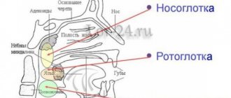
Eye teeth
The upper canines are called eye teeth because of their close location to the branches of the facial nerve. When they become inflamed, the pain radiates to the eye area and upper face. How close the incisive canals and the dental nerve are located to each other is shown in the figure:
Wisdom tooth
The wisdom tooth is the posterior third molar. He was called “wise” because he grows up in adulthood - when a person has already gained wisdom (by about 20 years).
Diagnostics
An erupted supernumerary tooth is detected during examination of the oral cavity; it is often dystopic, that is, abnormally located.
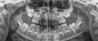
Diagnosis of unerupted (impacted) supernumerary teeth is carried out using x-ray examination. Often, X-ray diagnostics of supernumerary teeth is difficult due to the fact that in the image their contours overlap the contours of the complete teeth. For more accurate diagnosis, 3D computed tomography is used. The results of these studies make it possible to obtain comprehensive information about the relative position of erupted and impacted complete and supernumerary teeth.
Correct names of baby teeth
In Latin, baby teeth in a child are called the same as permanent teeth in an adult. But children do not have all the dental units characteristic of older people. Milk teeth are divided into:
- central incisors;
- lateral incisors;
- fangs;
- first and second molars.
There are different names for teeth, but it is better to focus on Latin terms and serial numbers. In this way, you can identify teeth even without serious knowledge of dentistry.
What does polyodontia look like?
Quite often, extra teeth are almost indistinguishable from normal ones. It is not uncommon for them to grow in the form of a drop or a thorn. These dental elements can appear either individually or fused with permanent ones. They can form tooth-like formations and entire arrays of teeth.
Also in medical practice, there are cases where polyodontia was hidden and was detected only by radiography. There are many different cases of abnormal development of the number of teeth, and if you notice symptoms, you should definitely contact the dentist.
Normal deletion
If the dentist decides that in a particular case, polyodontia can only be treated by removing an extra tooth, the patient should count on the following procedures:
- First of all, the patient should be sent for radiography. This is necessary in order to determine the size and number of roots, as well as the ratio of supernumerary and normal teeth.
- After collecting research, the doctor gives the patient anesthesia and removes excess teeth.
- In some cases, soft tissue sutures may be necessary after surgery.