Pulpitis is one of the most common dental diseases, which in most cases can be effectively treated.
The main condition for the effectiveness of the measures taken is a timely visit to a dentist-therapist. If you feel painful symptoms, you should not postpone your visit to a specialist - the sooner you go to the clinic, the greater the chance of saving the tooth. Pulpitis is characterized by an inflammatory process in the inner part of the tooth, called the “nerve.” This determines the severity of the symptoms - the pain with pulpitis is very noticeable. In most cases, the disease develops as a result of caries if the patient does not consult a doctor in time. In rare cases, pulpitis is associated with improper treatment of caries, violations of the tooth preparation process or other manipulations, and it can also be the result of injury.
Types and symptoms of pulpitis
Symptoms, as well as treatment tactics for pulpitis, depend on the type of disease. The following classification of the disease is currently used:
- Spicy. Often this type of pulpitis is the result of untreated caries. If the patient does not pay attention to tooth decay, after some time the infectious process reaches the internal structure - first dentin, and then pulp, provoking its inflammation. Symptoms of this form of the disease include acute throbbing pain, which intensifies significantly when exposed to chewing, contact of the tooth with food or high-temperature drinks. The pain often gets worse at night. Acute pulpitis, in turn, is divided into two types:
- focal - in this case the pain is “point-like” in nature, observed in one area of the tooth;
- diffuse - pain sensations spread to one or another area of the jaw as a whole.
- Chronic. It results from ignoring the symptoms of an acute form of the disease. As a rule, about 2 weeks pass from the onset of inflammation until the disease becomes chronic. Painful sensations become less pronounced, severe discomfort disappears. However, the pain continues to persist and intensifies when exposed to the causative tooth. The tooth is destroyed, and the acute pain returns from time to time.
Arsenic devitalizing paste consists of the following components:
- Arsenic anhydride.
- Elements that have an antiseptic and disinfecting effect on the pulp.
- Pain relieving components.
- Components that ensure long-lasting action of the paste.
- Additional components.
Arsenic in any form is one of the most potent poisons. A dose of 5 mg is toxic for humans, and therefore for devitalization the permissible maximum dose is 3 mg.
If the dose is exceeded, symptoms of intoxication such as vomiting and loose stools may occur.
When arsenic paste is applied to the pulp, gradual necrosis of nerve endings and blood vessels occurs. Since the blood supply is cut off, the pulp dies and the transmission of nerve impulses stops.
Many dentists prefer not to use arsenic paste for devitalization, especially when treating children, pregnant and nursing mothers. Moreover, there are safer and less toxic analogues.
What happens if pulpitis is not treated?
It can be difficult for a modern person to find time to visit a dentist in his busy schedule. The most common mistake is to try to ignore painful symptoms by “masking” them with a large number of painkillers. You should know that even if the pain was calmed, treatment of pulpitis still cannot be avoided - pathogenic organisms in the pulp chamber continue to multiply, destroying the tooth and surrounding tissues. Changes occur inside the tooth that lead to the death of the pulp, its decomposition with the formation of pus.
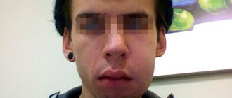
Subsequently, the pus extends beyond the root and a flux, known to many, occurs - inflammation of the periosteum. This is accompanied by severe symptoms - high body temperature, severe swelling and facial asymmetry, and also entails not only unpleasant, but also life-threatening consequences. In advanced cases, we are no longer talking about preserving the tooth, but about preserving health - there is a possibility of developing phlegmon, damage to the bone tissue of the jaw, and blood poisoning.
Chronic pulpitis can occur with the preservation of the tooth for up to several years, especially at a young age, but this does not mean that the symptoms can be ignored - it is better to visit a dentist in a timely manner in order to save the tooth and prevent serious complications.
How to deal with the disease
At the initial stage, it is not difficult to cope with pulpitis. The nerve in the tooth is preserved, and the doctor relieves the inflammation using therapeutic procedures.
The biological treatment method eliminates inflammation through antibacterial treatment of the damaged tooth, without removing the nerve. After removing carious tissue and disinfecting the pulp chamber, the dentist applies a compress with calcium hydroxide and fixes a temporary filling.
Then, after 3-7 days, during a follow-up visit, the doctor takes an x-ray. If there is no inflammatory process, then a permanent filling is installed. However, this technique requires a highly qualified doctor, so it is rarely used. For example, with traumatic inflammation of the dental nerve.
Preserving the pulp is important because... If it is present, the tooth is constantly strengthened due to the production of dentin. Conservative therapy has age restrictions - it is carried out up to 30 years.
Diagnosis of pulpitis
The diagnosis is made by a doctor based on a survey and examination of the oral cavity. As a rule, pulpitis precedes developing caries, so there is a carious cavity in the causative tooth. The doctor performs probing - places an instrument into the cavity to assess its depth and reaction. Tapping is a method that also helps the doctor find out how the tooth reacts to mechanical stress. The main task of the specialist is to determine the difference between pulpitis and deep caries - treatment tactics depend on this.
An X-ray examination is necessary in order to assess the nature of the disease, as well as determine the structure of the root system - the doctor will need this information for further endodontic treatment of tooth pulpitis (treatment and filling of canals).
Survey
To determine an accurate diagnosis, the dentist conducts an initial examination. Because If pulpitis develops against the background of other diseases, then diagnosis includes 4-5 stages. The procedure begins with communication with the patient. It is important to understand at what stage the inflammation is, which is especially important in chronic pulpitis. Therefore, the doctor asks you to describe in detail the nature of the pain. Then a “manual” examination is carried out using medical instruments (dental mirror, etc.). Additionally, the doctor checks the sensitivity of the affected tooth for temperature changes and exposure to a weak electric current charge.
The examination is completed by an X-ray examination to assess the condition of the dental nerve and canals. After collecting information and analyzing the image, the doctor plans the pulpitis treatment process and coordinates it with the patient.
Treatment of pulpitis
To avoid complications of pulpitis, it is important to start treatment in a timely manner. In rare cases, this allows you to save the pulp and avoid its extraction from the canal, but partial or complete removal of the neurovascular bundle is still more often practiced, and here’s why.
Removing the pulp allows you to completely eliminate the source of inflammation, which means preventing the spread of pathogenic microorganisms into the tissue surrounding the tooth. This is the basic principle of preserving the tooth and tissues from possible complications.
Side effects that may appear after using arsenic paste:
- If the dosage is exceeded, signs of intoxication of the body may appear - nausea, vomiting, diarrhea.
- If the paste is applied poorly, it may be washed out or leaked, accompanied by a burn of the oral mucosa with subsequent complications.
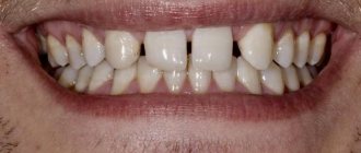
- If the paste is kept in the tooth cavity longer than intended, the dentin may change its color - turn black.
- Due to the high toxicity of the paste, periodontitis may begin.
- Swelling of the pulp due to inflammation.
- The effect of the poison can spread to bone and periosteal tissue, causing their destruction.
Treatment methods for pulpitis are divided into two broad categories:
- treatment that involves preserving living pulp;
- treatment of pulpitis of permanent teeth with pulp removal - complete or partial.
The decision to choose a method falls on the shoulders of the doctor - he assesses the condition of the pulp. The biological method (implying preservation) is used only when inflammation is observed at a very early stage, and it is also more often applicable to young patients who do not have other oral diseases.
Extirpation, or complete removal of the pulp, is the most common practice and is an effective and reliable method. It can be vital and devital - the difference is that in the first case the doctor removes the pulp under local anesthesia, and in the second, he first kills it using special means. Devital extirpation can be carried out without anesthesia, since the neurovascular bundle will no longer react to irritation, which means there will be no painful sensations. However, in practice, even in this case, an anesthetic is often given to avoid discomfort.
Preliminary devitalization consists of the fact that during the first visit, the doctor removes the destroyed tooth tissue, treats the cavity, and puts a special paste into the canals. Previously, in domestic dentistry, the method of devital amputation was used, which consisted of removing the neurovascular bundle inside the crown, while preserving the root pulp. For it, strong solutions were simply used to dry it out, but the voids in the root canals with pulp residues acted as fertile ground for the further development of the inflammatory disease. That is why today most often the pulp is completely removed - this is a reliable and effective treatment method.
A combination therapy method is the use of two or more methods. For example, removal of the entire root pulp from normal canals and partial removal (amputation) from those that have complex anatomy and are strongly curved. The prognosis in this case is more favorable than in the case of devital amputation, however, this is only true if certain conditions are met: most of the canals must be reliably sealed along the entire length, and high-quality materials must be used for this.
Modern dentistry offers a lot of opportunities for improving the method of treating pulpitis through vital extirpation. Pulp removal is carried out without first killing it, and for this purpose a rich arsenal of tools, equipment and means is used. They are designed to facilitate the process of pulp removal and canal treatment, as well as to prevent possible errors and ensure the convenience of the specialist’s work.
Modern anesthetics and devices for their controlled administration make it possible to avoid the need for devitalization - they provide effective anesthesia even for complex lower molars.
Structure
We have already examined the structure of the tooth itself, now let’s study in more detail the structure of the pulp.
Since the main substance is a loose fibrous substance, the density of the pulp is relatively low. However, this liquid amorphous colloidal system contains a huge number of pulp cells and fibers, which are quite difficult to separate from the connective tissue. Pulp fibers are nothing more than elastin and collagen.
On the surface of the fibrous structure there are odontoblasts, cylindrical cells with long processes located in the dentinal canals. It is these processes that make dentin sensitive to all external stimuli. Next are the stellate cells. The central layer contains the largest number of nerve and collagen fibers, as well as blood vessels.
The main composition of the pulp includes the most numerous cells: fibroblasts, which produce collagen fibers and create connections between cells, macrophages, histiocytes, and mast cells. In case of damage or inflammatory processes, the structure of the dental pulp also contains plasma cells, leukocytes and lymphocytes.
Stages of pulpitis treatment
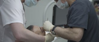
- The doctor performs anesthesia and drills out tooth tissue softened by caries, providing access to the internal structures.
- Removing the pulp - first, the doctor amputates the pulp using a bur or excavator, then performs extirpation - removes the neurovascular bundle from the root canals using thin instruments.
- Channel processing. Using small “needles” - files, reamers - the specialist passes the canals along their entire length, expanding and at the same time treating their walls with antiseptics. In this case, everything “unnecessary” is consistently washed out of the canals: bacteria, canal particles, pus, blood impurities, pulp residues, etc. Drug treatment ensures sterility - the likelihood of complications in the future depends on how carefully it is carried out. After treatment, the doctor dries the canals, places cotton pads impregnated with antiseptics there and installs a temporary filling.
- Filling. During the second visit, the dentist removes the temporary filling and medications from the canals. The treated root canals are filled to the apex - a natural narrowing or the narrowest point. For this purpose, pastes and gutta-percha pins are used that are mixed with the patient.
- Control. X-ray diagnostics are performed to assess whether the canals are filled correctly and along their entire length. Then the doctor places a filling or inlay on the crown of the tooth.
Control makes it possible to prevent some serious errors in endodontic treatment. Removal of the filling material beyond the root apex or insufficient filling are causes of possible complications in the form of the spread of the inflammatory process to the peri-root tissue, so it is important to monitor compliance with the treatment technology.
Pulp removal with preliminary devitalization is carried out according to the same principle, but in this case an additional stage is added and two visits to the doctor are required. On the first visit, the doctor applies a special paste - arsenic or arsenic-free - to the exposed pulp horn. In the first case, you need to come for an appointment within 24 hours, and if devitalization of the pulp of a multi-rooted tooth is carried out, after 48 hours. This type of therapy is more difficult to control by the doctor, because the patient may come for an appointment later than scheduled, which increases the risk of toxic effects of arsenic on the tooth root. That is why modern dentistry suggests using alternative solutions - using a paste without arsenic in the composition.
Indications during pregnancy
There is a myth that it is impossible to treat teeth while expecting a baby. This misconception can have serious consequences. Infection, for example in the form of caries, through blood vessels can spread throughout the body.
It is also a mistaken belief that x-rays harm the development of the fetus. It has been proven that modern equipment has a minimal radiation dose that does not affect the body. It is much more dangerous to bring the inflammatory process to a chronic state. Therefore, you should not delay treatment of pulpitis or caries when symptoms appear.
The process of bearing a child is associated with a decrease in immunity, and as a result, with increased sensitivity to bacteria and infections. Tooth enamel in pregnant women is not as strong, because... All nutrients are aimed at the growth of the baby. Therefore, the appearance of pulpitis and deterioration in the condition of the dentition is a normal phenomenon.
The second trimester is considered the optimal period for receiving dental care. During this period, the child is protected by the placenta from harmful substances. However, if an inflammatory process occurs in the dental nerve, it is not recommended to delay treatment so as not to expose the baby to unnecessary risk.
During pregnancy, the doctor chooses a gentle treatment method, when the filling is fixed only in the dental canals. If possible, without the use of anesthesia. After childbirth, the patient is given a permanent filling. X-ray examination is carried out only in emergency cases.
Additional methods of treating pulpitis
Today, it is possible to ensure impeccable disinfection of root canals during treatment using modern equipment - ultrasonic, laser.
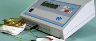
In addition, copper-calcium hydroxide depophoresis is an effective method of therapy. It has a bactericidal and fungicidal effect - it destroys pathogenic bacteria and fungi.
Physiotherapy for pulpitis is more often used as a means of treating pain after filling - it can occur due to adaptation to the material. In general, physiotherapy is used relatively rarely.
Cost of pulpitis treatment
The cost of treating pulpitis is determined by several factors, including the geography of the clinic. In a good clinic, the price already includes anesthesia, canal treatment, fillings and other materials.
The total amount is added up taking into account the number of root canals, their anatomical features, the degree of destruction of the crown and the method of its restoration, if we are talking about caries. You can find out the exact cost during a consultation with a dentist-therapist at the Dentist-K clinic.
It is important to remember that you can avoid expensive treatment by consulting a doctor in a timely manner. It is necessary to maintain hygiene and undergo professional teeth cleaning on time - this will avoid the need for pulpitis treatment.
Inflammation of the nerve in the wisdom tooth
Eights have their own specifics, and the doctor often chooses to remove the tooth. The complexity of therapy is due to the anatomical features of the third molars. Curved roots, lack of access to the tooth, partial overlap of the gums equates the treatment of pulpitis in figure eights to jewelry work. And it requires experience and high qualifications of a doctor.
An inflamed nerve in a wisdom tooth can be treated by filling the canals. Provided that there is access to the root and the dental canals have good patency. It makes sense to preserve a tooth in the presence of an antagonist tooth, if the tooth is involved in the chewing process and is used when installing a prosthesis.