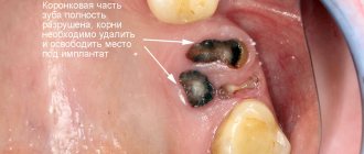
The result of dental canal treatment is the preservation and restoration of a tooth that is infected or has other pathology. The main task here remains cleaning the root canal. Infection and dying tissue must be removed from it. The resulting cavity is then securely filled. If treatment is not started in time, the inflammatory process will spread further and the tooth will have to be removed.
What is a root canal?
Each tooth, except for enamel and dentin, has an internal space - a pulp chamber, communicating with thin cavities running along the entire length of the roots of the teeth - root canals. Different dental groups have different numbers of roots, and, accordingly, canals. The front group has 1 channel, the side ones - from 2 to 4.
Narrow, winding, thin tubes with many branches - this is how to characterize the root canals of teeth. The entrance to them is located at the apex of the tooth root, it is called the apical foramen. Through it, through each root canal, passes neurovascular tissue, which is responsible for the blood supply and innervation of the tooth.
Endodontic treatment is a procedure for removing infected, inflamed or dead neurovascular tissue, followed by cleaning and disinfection of the resulting space, and completely sealing the dental canals.
Complications
Cleaning does not always go smoothly. There are unforeseen situations that disrupt its natural course. This means:
- Formation of a hole between the root wall and surrounding tissues. To eliminate the perforation, it is necessary to urgently fill it.
- Severe swelling of the cheek after administration of the anesthetic. This happens if the mucous membrane and periodontal tissues are saturated with an anesthetic drug. After all, by their nature they are quite loose and easily saturated with liquid.
- Breakage of a dental instrument. The probes that are inserted into the canal are very thin. It happens that they break off and remain inside the root. Under no circumstances should you leave them there, as severe inflammation will develop. To remove the fragment, the doctor will use special equipment.
- Allergy to materials used, medications. Anesthetics and drugs used by dentists today very rarely cause allergies. But in order to avoid a dangerous complication, before starting therapy, the doctor collects an allergy history and finds out whether the patient has drug intolerance.
If the patient strictly follows all medical instructions and sits quietly during tooth root treatment, the risk of complications is minimal. Therefore, it is important not to argue with the doctor, not to grab his hands, and not to ignore the prescriptions.
Diseases that require endodontic therapy
Treatment of tooth canals is performed only if there are strict indications, since as a result of the intervention, the tooth is deprived of the vessels and nerve endings that feed it, which makes the dental tissues less durable and more susceptible to destruction. Indications for intervention include inflammation and infections accompanying caries or traumatic tooth damage:
- Pulpitis is inflammation of the pulp (neurovascular tissue) - soft, fibrous tissue, including blood and lymphatic vessels, nerve endings and connective tissue.
- Periodontitis is inflammation of the tooth root and surrounding tissues (periodontal tissue).
These diseases are usually accompanied by the following symptoms:
- Acute, throbbing or aching pain in the tooth, increasing at night or intensifying with load on the tooth.
- Swelling, discoloration of the gums, formation of ulcers and fistulas on it.
- Putrid odor from the mouth.
In some cases, there are no pronounced symptoms and the development of pulp necrosis is detected only during a doctor’s examination. Only a specialist can accurately determine whether endodontic treatment will solve the problem and select the most effective therapeutic regimen.
Content:
- Features of canal treatment
- Processing sequence
- If pain occurs after therapy
- Complications
For pulpitis, periodontitis, and dental cysts, the canals are cleaned. This is a complex dental procedure that uses specialized equipment. The patient cannot refuse it, since there is no other way to prevent the spread of the inflammatory process.
Methods of treatment of dental canals
Not so long ago, arsenic paste was used for endodontic treatment. The dentist opened the dental chamber, put the paste into it and, after the pulp died, removed it from the tooth. Next, the tooth was filled and considered cured. The resorcinol-formalin method was also widely used - a resorcinol-formalin mixture placed in the cavity slowly hardened, stopping the disintegration of the partially removed pulp.
But such technologies have significant disadvantages:
- Arsenic and formalin saturated with resorcinol are very dangerous, toxic substances that can accumulate in various organs.
- These substances cannot always kill the nerve completely, which contributes to severe pain when trying to extract the pulp.
- Infected neurovascular tissues that are not completely removed become a source of repeated inflammation.
These methods were used because there were no sufficiently strong anesthetics that would allow removal of the affected pulp painlessly. Today they are completely irrelevant - modern dentistry treats tooth canals using a completely different technology.
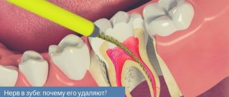
Why are patients afraid to remove nerves in a tooth?
Previously, in the process of removing a nerve in a tooth, dentists used arsenic by applying it to the affected pulp. The patient was then given a temporary filling, which was removed two days later along with the arsenic, after which the nerve was removed. This procedure took quite a long time, causing discomfort. Currently, thanks to modern technologies, the process of removing a nerve in a tooth lasts about half an hour and is carried out under local anesthesia. If the X-ray results are good, the canals and the carious cavity, cleared of bacteria, are filled.
To avoid such radical actions as removing a nerve in a tooth, you must:
- Carry out hygiene procedures as efficiently as possible
- Don't forget the importance of professional teeth cleaning
- Regularly visit the dentist, who will notice any carious lesion during a visual examination much earlier than it reaches the pulp and requires removal of the nerve.
Modern methods of dental canal treatment
There are two main methods of treating tooth root canals:
- Therapeutic (biological) in which the entire pulp or part of it is preserved. Manipulations can be carried out directly - the dentist puts a medicinal product into the dental cavity, separates the pulp chamber with an insulating gasket and installs a temporary filling. Or drugs can be delivered through the dentin, using antibiotic dressings, etc. Such drug therapy is effective only at the initial stage of inflammation.
- Surgical (pulp extirpation) - removal of all neurovascular tissue, cleaning, disinfection and sealing of dental cavities. Root canals are treated in one of the following ways: classic filling of tooth canals - filling the voids with filling material.
- Vertical condensation by injection of thermoplasticized gutta-percha.
- filling using thermophiles - hot gutta-percha on a carrier.
In some cases, endodontic treatment requires resection of the root apex - in the presence of granuloma, fibroma, cyst, perforation and other problems near the root apex.
Preparatory procedures
In order for the dentist to choose the right treatment tactics, he should obtain maximum information about the condition of the tooth. Additionally, it is necessary to find out if the patient has an allergic reaction to medications, since they are required to be used when cleaning canals.
Collection of information:
- interviewing the patient (nature of pain, sensations);
- conducting an initial examination;
- sending a patient to get an image.
If the patient is a pregnant woman, then it is more rational to choose a study performed using a visiograph. The most informative is a 3D image created by a tomograph. Based on visual data, the dentist determines the length of the canal, allowing you to accurately select the instrument. There is a more accurate way to determine the distance using an apex locator, which displays a picture on the screen that allows you to determine the distance of the inserted instrument from the apex of the root. This method prevents breakage of endodontic devices during cleaning.
X-ray of a tooth
Fact! The most difficult cleaning is carried out when the canals are obstructed and are strongly curved.
Stages of treatment
- Diagnostics
If damage to the dental canal is suspected and in order to detect a pathological focus, an X-ray examination (orthopantomogram, CT) or a study of the patient’s existing images is performed. Based on the examination results, the endodontist selects the appropriate treatment method.
- Pulpectomy
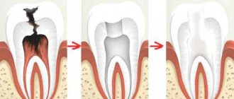
After anesthesia, the dentist opens the tooth, removes all damaged and necrotic tissue, gives the canal the required shape and rinses it with an antiseptic.
- Canal filling
Open channels are hermetically filled with filling material. For this purpose, plastic (gutta-percha, paste), hardening (cement group) and hard materials (pin structures) are used. The most progressive method is filling the canals with hot gutta-percha.
- Restoration of the crown part of the tooth
The correct anatomical shape of the tooth is restored using direct (filling) or indirect (crown, inlay) restoration.
Preparing for channel cleaning
To assess the condition and structure of the root system, the patient is prescribed a preliminary diagnosis:
- CT scan;
- radiography;
- radiography;
After this, the dentist chooses a cleaning tactic and a special tool for the job.
Before opening the coronal part, local anesthesia is administered, since in a state of inflammation, internal structures can provoke acute pain. In this case, the dentist administers the anesthetic using the following methods:
- point or infiltration, that is, when injections are made into the projection of the apexes. An injection into the cancellous bone using the same infiltration method will also numb the area well;
- The conduction technique involves anesthesia of the nerve branch that surrounds the neighboring units. Anesthesia also affects some of the soft tissues, so after the injection and treatment, the patient may continue to experience unpleasant sensations in the immobilized part of the mouth for a long time.
Features of root canal treatment under a microscope
Modern dentistry cannot be imagined without the use of innovations that take root canal treatment to a new level. One of the most advanced methods of root canal treatment is the use of a dental microscope. Powerful optics allow you to increase your view by 32 times, given that the diameter of the channel is usually no more than 1 mm, it is simply impossible to process it efficiently without the help of optics.
Advantages of dental treatment under a microscope:
- The endodontist sees in detail the mouths of the canals, the features of their structure, each branch, depth and direction. This is especially important if the tooth canal has a complex, convoluted shape.
- The microscope makes it possible to remove only diseased and dead tissues without damaging healthy ones.
- Thanks to the targeted effect on the tissue, the best result of the intervention is ensured, eliminating the likelihood of perforations, as well as other problems and complications.
The use of a dental microscope allows the dentist to successfully solve even the most complex clinical problem.
Removal process
Depulping or removing a nerve always begins with preparation. To do this, the doctor must conduct a thorough examination of the oral cavity, collect information about the person’s health, and take the necessary tests. Only after this can he begin the nerve removal procedure, which is a simple operation lasting about 1 hour.
The first stage involves the administration of an anesthetic instead of surgery. Its effect begins after about 10 minutes and lasts for 1-2 hours, depending on the dose. General anesthesia, usually in the form of a sleeping gas, may also be used. The action begins literally in 1-3 minutes.
After this, the doctor begins preparing the working area, isolating the tooth from saliva, opening it and other manipulations. Only after this does the nerve extraction procedure begin. It is carried out using a pulp extractor - a dental needle in the form of a spiral. Next, the pulp chamber is filled with an antiseptic, the canals are carefully sealed, a control photograph of the patient’s jaw is taken, and a permanent filling is fixed.
previous post
Is it possible to build up a front tooth?
next entry
Recommendations after treatment
Endodontic treatment demonstrates high success rates. After the intervention, you must adhere to the following recommendations:
- Avoid too hot, spicy, or cold foods for several days (while teeth sensitivity remains).
- Maintain good hygiene – brush your teeth twice a day, use dental floss and an irrigator.
- Do not chew anything hard (do not chew nuts, candies, etc.), as this may result in damage to the root canals and tooth destruction.
- Undergo medical examinations with a dentist and hygienist (at least 2 times a year).
Immediately after the procedure, tooth sensitivity increases greatly. This is explained by the reaction of the tissues to the filling material. The discomfort will disappear in a few days.
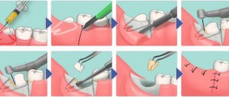
Processing sequence
Endodontic therapy includes several successive stages:
- Extraction of pulp tissues. All inflamed areas are removed from the cavity of the dental crown.
- Treatment of the tooth canal with sanitizing compounds , cleaning it from bacteria, pathogens, and necrotic accumulations. As a result of this stage, the inflammatory process is stopped.
- Changing the natural shape of the root , giving it an optimal width for further processing. The doctor needs to not only create conditions for optimal patency, but also make sure that the apex of the root reaches the apical part.
- Sealing. This is the final stage of therapy. The cleaned and expanded cavity is filled with filling material. Then a filling is placed and it is polished.
After treatment, the tooth stops hurting and acquires an aesthetic appearance. But we must take into account that now it does not receive the necessary nutrition, that is, it is essentially dead.
This means that its service life will be shorter than that of healthy neighbors. After a few years, small cracks may appear on the surface of the crown or chips may form. Therefore, it is important to take special care of the treated unit and periodically carry out a remineralizing course using special dental gels.
Expert opinion
Lyubov Ivanovna Kopylova
dentist-therapist
Experience: more than 10 years
If you have the opportunity to go to a dental clinic equipped with an electron microscope, take advantage of it! Even the most experienced dentist works with canals without a microscope by touch. This means that there is a risk of underfilling or, on the contrary, excessive deepening into the canal, leading to perforation. The use of a microscope, when the working field is clearly visualized and the doctor efficiently cleans and seals even the thinnest and most curved canals, will allow this situation to develop.
If pain occurs after therapy
Uncomfortable sensations can occur for various reasons. The most unpleasant and undesirable thing is poor quality treatment. To be sure to avoid it, you need to give preference to a proven dental clinic, which employs first-class specialists.
Common causes of pain:
- excessive processing with removal of the instrument beyond the apical foramen;
- placing an excess amount of filling compound into the root;
- root perforation;
- leaving an unclosed air cavity in the channel.
In any case, if the pain persists for more than a week, you should visit the doctor again. He will conduct a simple diagnosis and name the exact cause of the unpleasant condition.
Cost of root canal treatment in Moscow
The price of endodontic treatment depends on the number of root canals of the diseased tooth, the method used, complexity (including re-treatment) and volume of intervention, type of filling, etc. Processing 1 channel requires less work than 2 or 3, which affects the cost.
| Group of teeth | Cost of pulpitis treatment | Cost of periodontitis treatment |
| 1 canal tooth | 6500 rub. | 8500 rub. |
| 2 channel tooth | 7500 rub. | 9500 rub. |
| 3-channel tooth or more | from 8500 rub. | from 10,500 to 12,000 rub. |
The price includes the entire list of procedures performed for the treatment of dental canals. When using a dental microscope and laser equipment in complex therapy, the cost of treatment increases.
Cost of services
Treatment of dental canals Price
Treatment of dental canals from 3000 rub.
1-canal tooth (mechanical and medicinal treatment of the canal) 900 rub.
1-canal tooth (canal filling with gutta-percha pins) 750 rub.
2-canal tooth (mechanical and medicinal treatment of canals) RUB 1,500.
2-canal tooth (canal filling with gutta-percha pins) 980 rub.
3-canal tooth (mechanical and medicinal treatment of canals) RUB 2,100.
3-channel tooth (canal filling with gutta-percha pins) RUB 1,300.
Expert of the article you are reading:
Akhmedkhanov Said Rashidovich
Dental surgeon, general dentist, implantologist, orthopedic dentist, dental therapist.
You may also be interested in:
Dental treatment during pregnancy Dental consultation Treatment of dental cysts Increased sensitivity of teeth: causes and methods of treatment Filling of dental canals Treatment of pulpitis Treatment of fistula on the gums Treatment of flux (periostitis)
Show more