Chronology of the use of core inlays in dentistry: stages
Stage 1
At first, dentists used composite core inlays for crowns, reinforced with polymer fibers. Compared to metal pins, they provoked root fractures less frequently, but had a significant aesthetic drawback - the black color of the reinforcing fiber.
The technology was also not simple, since an adhesive or primer was required, etching was performed, and adhesives were applied five times to the canal walls. An opaque layer was used to mask the black color of the pin. Despite all the disadvantages, fiber structures also have tangible advantages. In the future, they can be easily removed from the canal by drilling with a regular bur.
Stage 2
The next step is white reinforcing fiber. The material began to be used for the manufacture of bridges and reinforced composite crowns. This technology has taken root in dental laboratories and moved away to orthopedic dentistry clinics. However, today it has nevertheless found application in periodontics and orthodontics - for splinting teeth.
Stage 3
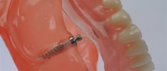
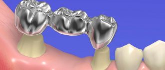
With the advent of fiberglass pins manufactured on specialized lines, it has become possible to install reliable stump inlays. These materials are widely used in the military and space industries due to their lightness, strength, ductility, elasticity and ability to withstand loads for a long time.
The structure of the fiberglass pin approaches the flexibility and strength of natural tooth tissue. Therefore, using the material it is possible to create monolithic structures with a long period of operation. Such pins are fixed using durable adhesive composite materials.
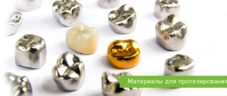
Composites
Composites are substances consisting of several dissimilar components.
In dentistry, composites are usually called substances consisting of an organic polymer matrix, an inorganic filler and a bonding layer (silane). The fundamental difference between composites and plastics is the presence of a third component that combines substances of dissimilar chemical structure (matrix and filler) into one material. A special property of composites makes it possible to add new portions of material to already hardened ones. The polymerized composite is an inert substance and does not have toxicity (except for first-generation composites). Fillings made from modern composites are applied without insulating gaskets, even in deep cavities. According to the requirement of the International Standards Organization (ISO), filling materials used to seal the chewing surface of teeth must be radio-opaque. Composites intended for filling only anterior teeth may not be radiopaque. Almost all modern composites are used in combination with adhesive systems, a description of which is given in the corresponding section.
Structure.
Organic polymer matrix.
The spread of composites became possible after the introduction of bisphenol glycidyl methacrylate (Bis-GMA) into practice. This monomer has a high molecular weight and is capable of forming very long chains that “envelop” filler particles. It hardens at room temperature and in the presence of a catalyst in just 3 minutes. Polymerization shrinkage is 5%. Bis-GMA forms the basis of almost all modern dental filling composites. To impart certain properties to composites, modifications of Bis-GMA are also used, such as urethane dimethacrylate, triethylene glycol dimethacrylate, etc. Some manufacturers use oligomethacrylates as the basis of the organic matrix. The organic matrix also includes polymerization initiators and inhibitors, catalysts, ultraviolet ray absorbers, and some other substances. The organic matrix determines the plasticity of the composite, its adhesive properties, and biocompatibility; influences the strength, color stability, and degree of polymerization of the composite.
Filler.
Determines such properties of composites as strength, shrinkage, water absorption, abrasion resistance, radiopacity, color stability. Fused and crystalline quartz, aluminosilicate and borosilicate glass, various modifications of silicon dioxide, aerosil, pre-polymerized crushed composite and other substances are used as fillers.
There is a fundamental difference in determining the amount of filler by mass and by volume. The inorganic filler is heavier than the liquid monomer, so its mass fraction always exceeds the volume fraction by 10-15%. The physical properties of the composite are better characterized by the volume ratio of the matrix and filler. The amount of shrinkage and other characteristics depend on the volume of organic matter. When comparing materials, it is necessary to take into account similar indicators.
The filler particle size can vary from 0.01 to 45 microns. The larger these particles are, the more of it can be introduced into the composition of the composite, the higher the strength of the material, the less shrinkage with unchanged ductility. However, large particles form a rough, lacking shine surface and contribute to increased abrasion of the filling. Small particles make the composite polishable and more resistant to abrasion. It is impossible to introduce a large amount of fine filler into the material, since small particles have a large surface area. In materials with small filler particles, basic physical indicators such as strength, water absorption, and color stability also deteriorate. To maintain ductility and strength, all filler particles must be “wrapped” in an organic matrix.
The shape of the filler particles also has a huge impact on the properties of the composite. As in amalgam, the needle-shaped, irregular filler becomes the basis for high strength, and the rounded, round filler allows the composite to be better polished and makes it more ductile.
Bonding layer.
Most often it is represented by silane, which is applied to the surface of the inorganic filler in the factory before mixing with the organic part. Silane is an organosilicon compound, a bipolar coupling agent. It forms a chemical bond, on the one hand, with the inorganic filler, and on the other, with the organic matrix. Due to this connection, the structure of the composite becomes homogeneous, its strength and wear resistance increase, and water absorption decreases.
All composites polymerize according to the free radical type. Free radical formation and curing occurs as a result of a thermal, chemical or photochemical reaction. Thermal polymerization is used only in laboratory conditions, since heating the composite to a high temperature in the oral cavity is impossible. The most widely used are composites of chemical and photochemical (light) activation.
Polymerization of composites never occurs 100%, which ensures a layer-by-layer connection, as well as the possibility of restoring old restorations.
When in contact with air, the surface of the composites interacts with oxygen, which leads to the cessation (inhibition) of the polymerization reaction. Thus, the surface of all composites cured in air is covered with an oxygen-inhibited layer. This layer promotes better bonding of the composite layers to each other. However, if there is an excess of the oxygen-inhibited layer, the process of joining the composite layers is disrupted, which can cause weakening of the structure and a change in its properties. The technique of plastic processing of the composite when laying the next portion allows you to correctly use the properties of the inhibited layer.
Not only atmospheric oxygen can block the polymerization reaction, but also oxygen released during the decomposition of hydrogen peroxide. Therefore, you should not treat the tooth cavity with hydrogen peroxide before using polymer filling materials. Tooth tissues are also saturated with oxygen during the process of chemical teeth whitening using peroxide compounds. After the last teeth whitening session using peroxide compounds, you should wait several days before restorative procedures to reduce the saturation of tooth tissue with oxygen. Eugenol can also block the curing of polymers. Therefore, it is not recommended to use eugenol-based cushioning materials or canal filling pastes before using polymer filling materials.
The polymerization shrinkage of composites varies, depending on the content of inorganic filler, from 1.8 to 5%. For light-curing materials, the shrinkage process is influenced by the intensity of the light flux at the beginning of polymerization. To reduce it, it is recommended to use a lower light intensity in the first few seconds (the so-called “soft start”).
Chemical activation composites
(chemical, self-curing). They are usually presented in paste-paste or powder-liquid systems. One of the components contains a chemical activator, the other a polymerization initiator. When the two components are mixed, free radicals are formed, which initiate a polymerization reaction. The quality of the composite in this case will depend on the accuracy of the dosage of the components and the thoroughness of their mixing. The colors of the catalytic and base pastes differ. The creation of a uniform color by mixing them indicates the readiness of the composite for introduction into the tooth cavity.
Some substances, usually in the catalytic paste, may spontaneously decompose when the temperature increases or is stored for a long time. The operating time of such materials is always limited and decreases with increasing temperature, and increases with decreasing temperature.
Polymerization of chemical composites occurs simultaneously throughout the entire volume. Consequently, the shrinkage of self-curing composites should be directed towards the “center” of polymerization. However, the last statement is controversial, since the polymerization reaction is accelerated upon contact with the warmer walls of the tooth, which are also covered with hardened adhesive.
Examples of composites in this group include “Evicrol”, Dental Spofa; Consise, 3M; "Adaptic", Dentsply; Epacryl, "Stoma".
Light activated composites
(light-curing, photopolymers, solar materials). They are one-component pastes, manufactured and packaged in a factory. The polymerization reaction is initiated by visible blue light with a wavelength of 450-550 nm. Under the influence of light of a certain wavelength, the polymerization initiator disintegrates, causing a set of reactions leading to the formation of free radicals and the formation of polymer chains. For proper polymerization of such materials, you should strictly adhere to the manufacturer’s instructions, both regarding the polymerization time and the type of device recommended for working with this composite. The depth of polymerization for different composites can range from 2 to 10 mm. It depends on the opacity and color of the material.
The shrinkage of photopolymers is theoretically directed towards the light source. However, taking into account the speed of propagation of the light flux, we can say that small portions of the photocomposite (within 2 mm of thickness) are polymerized simultaneously throughout the entire mass, similar to self-curing ones. Polymerization shrinkage of a light-curing composite can be reduced by a smooth start of polymerization, a decrease in the volume of the cured material, and directional polymerization.
Light-curing composites have significant advantages over chemically curing ones:
one-component; high strength; “command” polymerization; ease of work, lack of haste; high color stability; efficiency: the doctor takes as much material as he needs; high aesthetics and color accuracy; the ability to recreate many shades and several degrees of transparency.
A special feature of light-activated composites is the presence of pastes of varying transparency (or opacity, opacity). Similar to the structure of the tooth, there are 3 types of material based on this characteristic: an analogue of dentin - opaque tones; analogue of enamel - enamel tones; analogue of the cutting edge - the tone of the cutting edge. In terms of transparency, they differ from each other by an average of 20-30%. By placing different types of material in one restoration in color and transparency, it is possible to achieve a complete imitation of the tooth structure. Opaque tones serve to camouflage stains and create a “reflective” environment, similar to tooth dentin, enamel tones primarily color and diffuse light, incisal tones only refract and lightly diffuse light, creating a “vitality” of the restoration.
To activate the polymerization reaction of light-curing materials, an external source of blue light is required. Such a device is called a polymerization device, or lamp. To obtain blue light with a wavelength of 470-550 nm, special installations are used: halogen, diode, plasma, laser. They usually consist of the light source itself, a control unit and a light guide. For proper operation, a minimum luminous flux power of 300 mW/cm2 is required (for devices with a halogen lamp). The light guide should be located as close as possible to the surface of the material during polymerization. Removing it by 5 mm reduces the luminous flux power by 30%. In addition to light, polymerization plants can generate heat. The heat flow power should not exceed 50 mW/sq.cm. Curing devices from different manufacturers meet common standards and can be used to cure materials from different manufacturers. Due to the high intensity of light required for polymerization, avoid exposure to direct and reflected light by wearing safety glasses or shields. This light does not contain ultraviolet rays. Before using a specific device, you should carefully read the operating instructions.
The disadvantages of light-curing materials are the complex technology of their use, the need to use additional equipment (polymerization device, safety glasses, screen), and high cost.
Features of fiberglass pins
- High strength (1000 MPa), comparable to titanium.
- The elastic modulus of 35 GPa is close to that of natural dentin.
Thanks to a natural combination with hard tissues and modern composite materials for fixation, such pins are characterized by a long service life and high strength. Another reason for this is the uniform distribution of the chewing load throughout the entire tooth root. Due to the elasticity of the fiberglass pin, comparable to tooth tissue, the risk of mechanical destruction is minimal.
Advantages of fiberglass pins
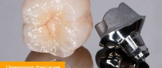
During fixation, the fiberglass pin does not need to be treated chemically, and there is no need to use specific “branded” materials. Fixation is possible using any conventional adhesive technique and composite materials - the pin is simply fixed in the root canal.
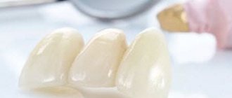
Such pins have excellent aesthetic characteristics - their color is close to the shade of natural tissues, so they are used, among other things, to restore front teeth. The material is easy to process. Using a diamond tool, the shape and length of the product can be adjusted. Light-curing composite materials are used to model the stump part of the structure.
Color perception and color reproduction in aesthetic dental restorations
High quality dental restoration in therapeutic dentistry is impossible today without accurate transmission of the shape, color, internal and external structure of the tooth and its optical characteristics. If we paid attention to issues of anatomical shape in our previous publications, then the issue of recreating color remains largely open.
Determining tooth color is a very complex and important process. Unfortunately, at a clinical appointment, the restoration of the color composition often occurs, just as during the reconstruction of the form, taking into account only the dentist’s own intuition and subjective perception of color. But if in the first case, doctors with good natural manual abilities are able to solve the problem of “form” in aesthetic restorations, then the solution to the problem of “color” sometimes occurs unconsciously and with great difficulty. Often doctors do not know even the most basic basics of “color science”, for example, what is color, hue and saturation? What is the difference between warm and cool tones of color? Which colors are called complementary and which are primary and what are their characteristics? And if we add to all this different methods and different colors from manufacturers of dental materials with identical characteristics, then even the most advanced dentists may experience great confusion in the matter of color selection.
Studying the topic of color in dentistry cannot be superficial, as many manufacturers, for example, call for, offering certain technologies and methods for using the materials they have developed, and not only because this topic is quite relevant today and dentists are concerned about it almost every day, but primarily because that any problem must be solved on the basis of solid knowledge and natural laws, and not separately developed skills and technologies.
On the pages of this publication, we decided to touch upon the topic of color in dentistry and begin its solution not with superficial practical methods taking into account the specifically advertised material, but from the original sources: the physics of color, the psychology of human color perception, the laws of color science and the optical properties of natural tissues. And only after this, on the basis of this information, we will determine their practical application in dental practice and give clinical examples that are already understandable to the reader, on which we conduct practical classes and seminars today.
The psychological effects of color were noticed by people at the same moment that color itself was noticed.
In primitive times and in antiquity, coloring served to highlight certain things, thereby focusing attention on them. For a long time, humanity has created various theories of color physics. Among ancient peoples, the question of color classification was resolved in close connection with the question of the structure of the cosmos, the world of gods and people. Later, during the Renaissance in Europe, both ancient (Alberti) and medieval color classifications were used. The artist Leonardo da Vinci introduces a “practically painterly” color system based on the painter’s minimal palette
In the 17th century, the physical characteristics of color perception changed. I. Newton introduces a natural scientific (physical) basis for the classification of colors, namely the white spectrum, in which there are seven simple spectral colors and one purple, formed by mixing the extreme colors of the spectrum. A curious fact is that this discovery was made completely by accident. At one of the English markets, the scientist bought a glass prism and was surprised to notice how the incident light from one surface was reflected onto another, emitting different colors. For quite a long time, the English scientist could not convince his colleagues of the multicolored color of the sun. Even his follower Goethe argued that Newton’s assumptions about the spectral composition of sunlight are more reminiscent of “children’s fairy tales.”
Based on the spectrum, a color wheel was constructed, which turned out to be a very convenient system for technical and scientific purposes.
A little later, at the end of the 18th century, Goethe proposed a new way of classifying colors. The color wheel he constructed consists of three pairs of contrasting colors. The basis of the circle was a triangle of the main colors of red, blue and yellow, thereby revealing the phenomenon of color induction. Later, thanks to the works of Philip Otto Runge, the color system acquired a third dimension and entered space. The German artist built a “color ball” that combined spectral and achromatic colors, whitened, blackened and broken. In the 19th century, thanks to the works of G. Helmholtz, the question of primary colors was clarified; they turned out to be red, green, and blue, which in mixtures give all the other colors of the spectrum of any saturation. These colors have found their practical application in printing, pointillist painting, and television.
Very important research on this topic was made by the English physician Thomas Young. By experimenting with color projectors during elimination, he found that for the same spectrum, the component colors could be reduced to three primary colors. Daylight can not only be divided into spectral colors, but also reconstructed in reverse order from the three primary colors (red, green and blue). As a result of the experiments, T. Jung made an important conclusion: nature strives to minimize (towards the optimum) when mixing three colors: blue, red and green, you can recreate any shade. A physiologically normal person is able to distinguish about 150 colors, dividing them by tone and brightness, in total this is about 1 million shades, and for this it is necessary to use a large number of visual cells. Hermann von Helmholtz, based on key discoveries of Jung, hypothesized that in certain areas of the eye there are structures that respond to three primary colors: blue, green and red. Numerous visual cells respond to light stimuli. In this case, each group of cells perceives the wavelength that corresponds to their sensitivity. Cone cells are responsible for color (daytime) color perception; there are three types of them, acting together, the cells form a common color spectrum. Special visual rod cells are responsible for black and white vision (night vision). Night vision is less clear, since the cells are located over the entire surface of the fiber and have less density
Thus, on the basis of the scientific discoveries of many artists, physicists and philosophers, the physical basis of color was determined, which is today relevant not only for technical and industrial human activities, but also for dentistry in particular. An important task facing dentists today is the ability to correctly use the above-mentioned scientific research in the field of color in daily clinical practice.
The essence of human psychophysical perception of color is as follows: We see all objects in reflected color. The spectrum of white color consists of seven primary colors and ranges from violet through blue, cyan, green, yellow, orange, to red (Fig. 1).
Any painted object, from the general spectrum of colors, reflects a single color (shade) while absorbing all the others (absorption and reflection). The human eye reads the beam reflected from the object, which is focused on the retina, analyzed by the brain and perceived by the person as the color of the given object or object. It should be noted that the human eye is capable of perceiving only a certain wavelength range, unlike other living beings, whose wavelength range is sometimes much larger than that of humans. Radiation in the wavelength spectrum that a person is on average ready to perceive is from 380 nm to 760 nm. (1 nm - 1/1000000 mm) each wavelength corresponds to a certain color of the spectrum.
Colored objects vary in color, saturation and hue. Each of the colors is close to warm (red tones) or cold tones (blue tones), lighter tones (close to white) or darker (close to black)
When merging with each other, they create various combinations and shades, dividing into primary red, yellow, blue and secondary green, orange and purple, which can be formed from the fusion of the first (Fig. 2)
Colors in their general range can either complement or suppress/destroy/ each other with their color activity. If you form a color wheel from the main colors of the spectrum, then for each of the colors you can find an additional one, in combination with which the first one will be more saturated than in combination with other colors. (Figure 3.).
By violating the basic laws of color construction, we may encounter color compositions that will create great confusion in our perception, since the same color, in combination with another color, can behave completely differently.
In Figure 4 you can see how the same colors can change when surrounded by others.
In dentistry, the concept of complementary colors is especially important. Its essence lies in the fact that when some colors are combined, the overall gamut becomes more saturated and natural, and when others are combined, the colors will turn out to be unnatural and dull. At a clinical appointment, we sometimes do not notice those factors that can have a negative effect on our color perception of objects, this could be the rich color of the walls in the office where the doctor is seeing, bright clothes or makeup of the patient, or simply the color of the coffee dam, which can dramatically change change the color of the tooth being restored.
Experimenting with VITA shades, we photographed teeth against different backgrounds. The result surprised us very much; the same tooth color against the background of a base wax plate and light paper was perceived completely differently. Agree, during a clinical appointment we don’t have time to think about the relationship between primary and secondary colors.
The generally accepted color scale contains a more limited number of colors, but their combination also occurs on the principle of mutual complementarity and contrasts. (Fig. 
You should also pay attention to the fact that when determining the color, the dentist is influenced by a certain number of negative factors.
Before studying the correct restoration of colors in aesthetic restoration, it is necessary to determine some of them:
1. The light source plays an important role in determining color. There are two light sources: warm and cold. In a natural light source, cool colors predominate (colors close to the blue range), in contrast to this, ordinary incandescent lamps have warm colors (colors close to the red range). Therefore, when determining natural color, it is necessary to pay attention to the fact that the light source is natural lighting (cold tones), otherwise the color determination will not be accurate.
In very low light conditions, the perception of color by the human eye becomes almost impossible, since the rods of the retina are not sensitive to color. With enough light, more intense signals enter the receptors. The cones are involved in the process of vision and the ability to perceive color becomes clearer. When the lighting is too bright, partial blinding occurs and the human eye is unable to correctly perceive not only the color, but often the shape of objects for quite a long time. It is very important to pay attention to this when the doctor works with a light curing lamp or under very bright artificial lighting. The doctor's direct or lateral vision is also very sensitive to an excessively bright light source.
A natural light source, at different periods of time and in different regions, also has a different composition. For example, in the northwestern regions of our country, on a sunny day, warm pink shades predominate in nature in the morning, bright silver in the afternoon, and orange-red in the evening. On a cloudy day, neutral silver tones predominate. To determine color, the time of day is also important. For optimal perception, neutral daylight falling from the north side of the building from 11 a.m. to 1 p.m. is best.
2. The perception of color is also greatly influenced by age-related and physiological changes in a person.
The human visual system is especially sensitive to age-related changes. After 30-35 years, a large number of people experience an accumulation of macular pigment covering the central part of the retina, which leads to yellowing of the lens and disruption of the correct determination of color assessment. Light in this situation changes its spectral composition even before it hits the light-sensitive receptor cells and the human eye receives a distorted perception of the light wave. A decrease in the mobility of the eyeball, the development of farsightedness with age and other physiological processes lead to a subjective assessment of color, and therefore to the lack of an objective assessment of the color and optical properties of teeth.
It should also be noted that approximately 1.5% of women and 7.5% of men experience partial color blindness, which leads to the inability to correctly determine color.
3. In addition to the direct light source, the surface of the teeth is greatly influenced by the reflected light reflex, which has a very large influence on determining the color of both one individual tooth and the entire dentition as a whole. If, for example, the clothes of a doctor or patient, the color of the walls in the office, the chair of a dental unit, or the floor have a very bright color scheme, then determining the color under these conditions will be practically impossible. Falling and reflecting from surrounding objects, the light will be changed in its spectral composition.
Natural light, which, as we have already said, has a complex color spectrum, creates a three-dimensional shape of any illuminated object, forming on its surface a highlight (direct reflection of a ray of light), light, penumbra, shadow and reflex (reflection from a nearby surface). If an object has a uniform color, then the visual size of the object can be changed using light and shadow.
Teeth (for example, the anterior group), due to their anatomical shape (equator line, medial and distal edges, line of primary inclination, etc.) also have a pronounced gradation in tone, with the help of which we can change the shape of a smile by creating from a narrow shape face, a wide grin of a smile, lightening the tone of the lateral part of the teeth and, conversely, lightening the tone of the front teeth, from a wide face shape, a more “narrowed” shape of the dentition, and therefore the visual perception of the smile of a patient with a wide face. In practical classes on color, we analyze such situations using the example of constructing a dentition in a tonal solution.
4. In addition to light sources, color perception is greatly influenced by a number of other factors. One of them is the phenomenon of metomery. Its essence is as follows: if the composition of the material is not homogeneous, then with one light source two materials with different compositions can have the same color, and with another source the colors of two objects can be different. The internal composition of the two materials manifest themselves completely differently under different light sources. This phenomenon becomes especially relevant if you pay attention to the fact that composite materials of color and natural teeth are different in their structure and composition.
5.An important problem in determining the color of teeth can be caused by color adaptation. With prolonged concentration of attention, the human eye ceases to perceive certain nuances of the color scheme. Color adaptation is expressed in reduced sensitivity of the eye. At the end of the working day, or with prolonged attention, the color sensitivity of the eye's receptor system decreases and the doctor is unable to distinguish not only small deviations and nuances, but sometimes the most basic colors.
In aesthetic restorations, determining the tooth color, tone and natural transparency is one of the most difficult stages. This is due, first of all, to the rather complex structure of natural teeth. According to the anatomical shape, the tooth is built from several layers of dentin (peripulpar dentin, predentin, interglobular dentin), dentin-enamel space (protein layer), several layers of enamel (inner, surface enamel). The color of the tooth crown is formed not only by the color composition of the three above-mentioned layers, but also by their optical property of light refraction. The color of the restoration is determined using special colors offered by manufacturers of dental materials. VITA shade is considered one of the closest to the natural color of teeth. According to this shade, teeth have four main groups of colors, each of which is distributed by color:
-brown colors (by color tonality A1, A2, A3, A3, 5, A4).
-yellow colors. (by color tone B1, B2, B3, B4.)
- gray colors (C1, C2, C3, C4)
- warm gray colors (D2, D3, D4)
If a color scale of four primary colors is decomposed by color tone, then they can be arranged in the following sequence: B1, A1, B2, D2, A2, C1, C2. D4, A3, D3, B3, A3.5, B4, C3, A4, C4.
In nature, the natural colors of teeth are mainly close to two groups: group A (brown colors), group B (yellow colors). Using a certain thickness of dentin and enamel, proper distribution of transparent layers, only from group A and group B, it is possible to recreate the color of teeth as close as possible to natural. But if the restoration is built not according to the banal scheme of “matching the color,” but taking into account the subtle transitions and reflexes of natural teeth, then the restoration will take place according to a slightly different scheme.
Let's look at this in a little more detail and examine two main issues in more detail: 1- The structure of the construction of complementary colors and 2- optical characteristics.
The structure of constructing complementary colors.
The color volume of the tooth crown works according to the law of warm-cold combination of primary and secondary colors and this happens in approximately the same way as it happens in the structure of a sound chord. Back in the 17th century, the famous physicist I. Newton expressed a version about the correspondence between the structure of musical sounds and colors. The seven sounds and seven primary colors of the color spectrum exist in clear correspondence. Understanding this pattern provides an answer to the question of why, during the restoration of a direct veneer in the three main zones of the upper middle and lower third of the crown, the effect of an unnatural plastic volume occurs.
In the sound chord A minor, in addition to the main sound A, there are a number of additional sounds that create a “live” harmony of the chord in the overall sound. The same laws of primary and secondary colors work on the vestibular surface of the crown.
Many modern manufacturers of light-curing materials deliberately try to introduce the so-called “chameleon” effect to create a feeling of natural play of color, but most often this effect is perceived as a half-measure.
Five color and a certain number (depending on age) light zones are formed on the surface of the crown, which we will consider a little later in the section on the optical laws of light refraction. Basic and additional color combinations according to
VITA coloring occurs in the following combination: group B (yellow colors) in the general color combination of colors refers to warm colors and clearly manifests itself against the cold background of group C (gray colors), group A (brown colors) also
belong to warm colors and will be able to express themselves to the maximum against the background of cold colors of groups C and D. The combination of similar colors in a warm-cold combination, for example, group A and group B, or group C with group D will lead to the destruction of the color composition. If in painting we combine yellow colors against a background of brown colors, then the latter will have a depressing effect on the first, and if yellow is depicted against a background of blue or gray colors, then it will begin to manifest itself with maximum activity. This is precisely the not complicated, but important law of primary and secondary colors that we need to apply in dentistry. (Figure 10)
At the practical seminar, we analyze in detail the main algorithms for the task of restoring the primary color of groups A, B, C and D, and we also analyze the age characteristics of the same color groups up to 20 and after 40 years of age.
The structure of optical refraction of light.
The most important thing in recreating the natural nature of teeth is the ability to reproduce the optical refraction of light. According to the anatomical structure, the natural shape of the tooth forms an internal glow of dentin, with pronounced mamilon lines, which is expressed in the overall transparency of the crown. It can be quite difficult to recreate this, especially if we do not pay attention to the laws of optical refraction of light.
To create certain optical effects in the formation of a crown, we must not only know well the basic laws of optics, but also skillfully use them.
1 The angle of incidence is equal to the angle of refraction. This pattern is applicable in all layers of the tooth crown (dentin, enamel, dentin-enamel junction) and in all materials that we use (composite, ceramics, etc.). The only difference will be that in different layers of the tooth crown we will use this law to varying degrees; for surface enamels, the angle of reflection will be from a smooth straight line, since the enamel must transmit light (absorption), and in the dentinal layers, the angle of reflection should be from textured (broken) lines, dentin concentrates the light source and subsequently reflects it (reflection)
If we analyze the presented drawings, we can understand why in restorations the dentine layers need to be made textured, and the enamel layers as smooth as possible, and the less we manage to get into the true color of the tooth crown, the more we must mask the inner layers of dentin, creating textured volumes of mamilons.
2 For a general idea of the refraction of light in volume, let us consider the movement of light in a well-known optical crystal. In cutting a diamond crystal, the optical laws of light refraction also work. The angle of incidence reflects the angle of refraction of the beam in a huge number of facets, creating a stunning crystal play effect. There are a few details to pay attention to. Firstly, this form was invented by jewelers over many centuries, and secondly, the height of the upper, let’s say, trapezoid and lower triangle is an exact mathematical calculation and a violation of the proportions of the two above volumes will disrupt the overall composition of the reflection of light. Another point that can lead to a violation of the optical refraction of light is a violation of the transparency of the bottom wall. If we cover the surface of the triangle with an opaque layer, then the light reflection in the crystal will stop (Fig. 12).
But returning to the optical properties of the crowns of the front teeth, it should be noted that while people have been experimentally developing the proportions of diamond cuts for centuries, nature has long had it. If we look closely at the structure of the construction of the volumes of the anterior group of teeth (view from the cutting edge). We can pay attention to a certain similarity in the proportions and structure of the construction of these two volumes (Figure 12.4). The optical laws of light refraction in the volume of the tooth crown occur according to the same laws as in the crystal, with the exception of the absence of light refraction in the opaque layers (dentin volume) of the crown (Fig. 12.2)
Taking into account the above, it is possible to show and explain the main stages of restoration using the example of reconstruction of the crown of a central incisor.
Initially, any destroyed shape of the crown, which may not suit us to one degree or another, must be restored. At a clinical appointment, we restore the lost shape of the cutting edge with a composite material, or using modeling wax on plaster models, after first taking an impression from the patient. Next, after taking an impression from the silicone mass, we begin to create a “silicone key”, which, taking the form of a matrix, should have a counter-volume shape of the palatal surface and the plane of the cutting edge of the crown, which we previously formed.
In the reconstruction of the crown of the central incisor, an important place is occupied by the stages of crown preparation. At this stage of recreating the vestibular surface of the crown of the central incisor, we will have to solve not only the problem of aesthetics, but also the full, functional load of the future restoration. At practical seminars, we pay special attention to this issue, analyzing the stages of preparation of various clinical situations using phantom models (grades 3 and 4). In the scope of this article, we do not consider this issue, but we will definitely examine it in the microprosthetics section.
We need to create a “silicone key” for two important reasons: 1) to form a previously restored tooth shape and 2) to create an algorithm for color construction of a crown with the effect of optical refraction of light.
In order for the volume of the restored crown to have the ability to transmit and then reflect a light wave, we need to restore the palatal surface of the crown with light-conducting layers of material (in this situation, surface enamel) using a silicone key (Fig).
At the next stage, we restore the opaque layers of dentin (forming in this case the volume of three mamilons) (Fig. .).
We complete the construction of the color volume of the crown with internal and external enamels in accordance with the combination of primary and additional colors.
If this algorithm for constructing transparent and opaque layers is violated and instead of transparent layers of surface enamel, opaque layers are formed on the palatal surface of the “silicone key,” then the volume of the tooth crown will acquire the effect of a “dead” plastic tooth. since the law of optical refraction of the tooth will be violated. After final polishing, on the labial surface of the crown we see all the inner layers of dentin layers. The crown has a transparent line of the cutting edge, which is more typical for the teeth of young patients. (Fig.) Using this technique, it is possible to recreate a tooth with different age characteristics.
In the next publication, we will definitely analyze the possibilities of practical application of the color restoration technique, clinical examples of restoring the crowns of anterior teeth in the 3rd, 4th class and the direct method of restoring the vestibular surface of the crown in direct veneers.
At practical seminars conducted on this topic, the stages of preparation of the anterior and lateral groups and restoration algorithms using “silicone keys” and individual phantoms are discussed in detail. Possibilities for restoring both young, bright-colored teeth and methods for simulating internal cracks, “chalky” inclusions, using dyes and intensives.
Information of interest can be found in Anton Vetchinkin’s studio