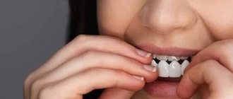
Are you looking for a dentistry that will provide high-quality prosthetics for your upper teeth? The PRESIDENT complex offers its services! Experienced doctors, progressive techniques, reasonable prices - make an appointment by phone.
In orthopedic dentistry, one of the main tasks is prosthetics of the upper teeth. Their loss is not uncommon, especially when it comes to elderly patients. Restoring lost teeth is of great importance for a number of good reasons.
Why is missing teeth dangerous?
Losing a tooth is not just a cosmetic defect. It is capable of triggering irreversible changes in the human body that only progress over time.
If even one upper tooth is lost, its pair on the lower jaw is almost not involved in the process of chewing food. As a result, the adjacent teeth take on the load. The result is a violation of their ligamentous apparatus, due to which the risk of loss and periodontal development becomes extremely high.
The loss of several dental units increases the likelihood that the height of the bite will change. As a result, the lower third of the face becomes smaller. This not only leads to an aesthetic problem - the appearance of early wrinkles. Due to the unevenly distributed load, the temporomandibular joint suffers - inflammatory processes and degenerative changes begin.
The patient may experience pain while chewing food or talking. Improper chewing of food leads to diseases of the stomach and other gastrointestinal tract organs.
Medical Internet conferences
Purpose: Selecting the most suitable method for preparing hard dental tissues for permanent restorations in clinical practice.
Objectives: 1) Familiarize yourself with the goals and principles of preparation.
2) Consider methods for preparing tooth surfaces.
3) Consider methods for forming a ledge.
4) Compare preparation methods to determine which is most suitable for use in clinical practice.
1.Purposes of preparation:
1) Preparation of all necrotic and damaged tooth tissues.
2) Maximum preservation of healthy tissues
3) Creation of conditions for retention of a fixed structure
4) Providing functional occlusion
5) Minimizing injuries to the oral mucosa.
Principles:
Preservation of hard tissues. Preparation of a tooth for a crown is accompanied by reduction of a sufficient amount of hard tooth tissue in order to provide space for a sufficient amount of restoration material, especially on the occlusal surface, where tissue reduction can reach 1.5 mm; it is necessary to bevel the functional tubercle (palatal in the upper jaw and buccal in the lower jaw) . It is important to preserve the anatomical shape of the prepared tooth and its group affiliation.
Retention of the restoration is ensured mainly by the shape of the tooth stump, such as: stump height (must be at least 2/3 of the initial crown height), stump area and taper.
The taper of the stump should be on average 6-10*, mainly for the anterior group of teeth. Retention reaches its maximum strength at a taper of 3*. As for the stability of the restoration, the following pattern can be identified: the greater the height of the stump and the smaller the radius, the more stable the restoration will be.
Tooth preparation has a number of contraindications:
1) General: previous myocardial infarction, hypertension, allergies to anesthetics, mental illness, etc.
2) Local contraindications are associated with the possibility of preserving vital dental pulp and are a reason for devitalization.
Preparation principles:
1) Biological aspect - maximum preservation of tooth tissue, elimination of excessive grinding, creation of a supragingival ledge and harmonious occlusion.
2) Aesthetic aspect – to achieve minimal visualization of the metal and maximum thickness of the ceramic coating.
3) Mechanical aspect - creating reliable fixation, stability, eliminating the possibility of deformation of the structure.
2. Preparation methods consist of separate stages, with the help of which in clinical practice the doctor maintains the biological width of the tooth and creates an adequate prosthetic space for restoration.
1) Oblique preparation method according to Martignoni and Schonenberg.
The method involves programming the depth of preparation of the hard tissues of the tooth by means of a saw cut beveled at an angle of 45* to the vertical axis along the cutting/chewing surface; later, the ledge is marked at the same angle, followed by excision of the remaining surfaces.
Pros:
— control of the depth of preparation in the cervical area.
— creation of sufficient occlusal space due to the formation of a beveled surface.
Minuses:
— The line of the ledge on the plaster model can only be determined with sufficient magnification.
— The method requires high manual skills of the doctor and great concentration during preparation.
2) Two-plane preparation method according to Kuwata based on the theory of three planes of the tooth.
The essence of the method is to prepare tissues while maintaining the inclination of the natural planes of the tooth and creating a ledge of 50° to the long axis of the tooth.
Pros:
— Manipulation does not require high manual skills.
— Rational direction of preparation, taking into account the individual topographical characteristics of the tooth.
— Preventive “protection” of the pulp of vital teeth.
Minuses:
— inability to control the depth of preparation.
— Relative consideration of tooth safety zones in terms of its position in the dentition.
3) Method of guiding grooves according to Stein.
The method is based on applying furrows of a certain depth to the prepared surfaces through the use of various calibration burs and further preparing the surfaces to a given depth.
Pros:
— Controlled preparation depth.
— the ability to more accurately take into account safety zones and individual topographical features of teeth.
Minuses:
— The need for accurate calibration of burs and its evaluation with a special measuring tool.
— The position of the tooth in the dentition is not taken into account.
— The impossibility of adequately assessing the depth of reduction of the tooth surface in case of dentition deformations.
4) Gürel's method.
The method is based on the preliminary fixation of a temporary composite restoration on non-prepared teeth, made on the basis of wax modeling. Without removing the composite restoration, we mark the depth of the preparation and then form the core.
Pros:
— Preparation is carried out taking into account the individual characteristics of the tooth.
— Visualization of the thickness of the surface preparation, taking into account the position of the tooth in the dentition.
— Minimization of tooth reduction during preparation.
Minuses:
— Requires additional time and economic costs, equipment and materials.
— Limited use, mainly when using adhesive indirect restorations.
— the difficulty of using the method in the distal parts of the jaw and with pronounced vestibular inclination of the teeth.
5) Method of preparation by tooth segments according to McLean.
The essence of the method is to divide the tooth into segments, the number of which depends on the functional surface of the tooth. First, one half is prepared from the vestibular, oral and occlusal surfaces. Later the second half. Subsequently, the cervical border is formed and finishing is performed.
Pros:
- Ease of manipulation.
— visualization of the thickness of the preparation.
— preparation taking into account the individual topographical characteristics of the tooth.
Minuses:
— relative control of the volume of preparation.
- difficulty of preparation in case of deformation of the dentition.
6) Two-stage preparation according to Massironi.
The essence of this method assumes that at the first stage the tooth is prepared for a certain structure with the location of the border of the closing line above the gum level and the subsequent fixation of provisional crowns. At the second stage, gum retraction is carried out with the final formation of a circular ledge and finishing of the tooth.
Pros:
— Controlled preparation taking into account individual topographic features.
— Protection of the epithelial attachment of the tooth.
Minuses:
— The need for a high level of manual skills, materials, tools, as well as time.
— Difficulty taking into account the peculiarities of the morphometric parameters of the biological zone of the teeth when forming a circular ledge.
3. Formation of a ledge is the most difficult stage in the preparation process, both its localization and choice of shape. The shape of the ledge determines: the amount of restoration material, marginal fit, the possibility of correction and constant care for it.
Principles of ledge formation:
Only with the correct connection and adaptation of the boundaries of the ledge and the orthopedic structure is its durability, functionality and aesthetics possible.
The ledge options are determined by the choice of design:
1) The groove-shaped ledge is used for MC and ceramics.
2) Ledge 120-135* for MK.
3) Shoulder shoulder 90* with an internal right angle for MK and ceramics.
4) Shoulder shoulder 90* with rounded internal corner for ceramics.
Also, the ledge is selected depending on the periodontium and hard tissues of the tooth.
There are two types of ledge arrangement:
1) Subgingival. Indications are aesthetic requirements, carious lesions and hyperesthesia in the area of the necks of teeth, low crown part, subgingival tooth fracture, repeated prosthetics.
2) Supragingival and at the level with the gingival margin. They are used in cases of low lip line, absence of visual necks of teeth in the smile, likelihood of complications from periodontal tissues, high clinical crown, generalized periodontitis of moderate severity, absence of previous sublingual preparation.
In order to minimize complications from periodontal tissues, the preparation of the ledge requires compliance with the biological width.
Biological width is a complex of tissues located above the alveolar ridge, including connective tissue and tooth-attached epithelium, which fill the space between the bottom of the sulcus and the alveolar ridge.
The edge of the subgingival preparation should end in the area of the middle of the periodontal sulcus.
The formation of the final ledge is carried out after installing the retraction thread with an increasing tip at low speeds.
Conclusion: Having considered all the above preparation methods, we can draw the following conclusion: due to the variety of clinical cases, indications and contraindications, it is impossible to choose one best preparation method. You should also take into account the huge variety of materials, manufacturing technologies for fixed dentures and the different technical capabilities of medical institutions. Based on this, in practice it is permissible to use any of the above methods, which is most convenient for the attending physician to use and allows one to obtain the most positive result for the patient’s health.
Methods of upper teeth prosthetics
Modern orthopedic dentistry uses many effective technologies for dental restoration. Various types of prostheses are used:
- bridge-like. They are a non-removable structure that is installed when 1-2 units of dentition are lost. They attach it to supporting units;
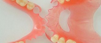
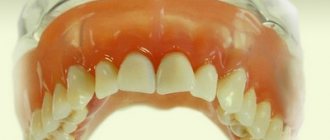
- removable plate. Inexpensive removable structures installed in place of lost teeth. They can be partial (restoring several units of the dentition) and complete (restoring the entire dentition);
- clasp (arc). They are a removable structure that uses a light metal arc. A durable, convenient solution that can restore chewing function well;
- conditionally removable. Can only be removed by a dentist. A system of locks (attachments) or a telescopic system without support from the sky can be used as fastening;
- crowns and bridges on implants. Today, the installation of implant systems is a priority method of prosthetics in European countries. Their installation is possible even with complete edentia.
The main difficulty when replacing defects in the lateral group is the proximity of the Maxillary sinus. It is extremely important that it be longer than the future implant. This is only possible if there is sufficient bone tissue. In some cases, a special operation is required in which bone tissue or a synthetic substrate is implanted.
Prosthetics for a completely toothless upper jaw
With complete edentia, it is possible to use various types of prostheses - plastic plates, lying on the oral mucosa, conditionally removable, which are fixed with implants.
Complete restoration from a functional and aesthetic point of view is possible if a minimum of four intraosseous implants are installed. The use of basal, mucosal implants is also considered acceptable, but such methods are not widespread.
Acrylic removable dentures are distinguished by their characteristics. The plate almost completely covers the hard palate. The valve area needs to be enlarged to improve fixation of the prosthesis. However, this method of prosthetics has a significant drawback - patients often note an increased gag reflex, as well as general discomfort.
However, it is worth noting that prosthetics for an edentulous upper jaw are simpler than for the lower jaw. This is due to the fact that the base of the prosthesis will not come into contact with the moving mucosa, thereby improving fixation and reducing discomfort when eating.
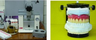
How are removable dentures made?
Prosthetics of the upper teeth using removable structures is a very common technique. It is carried out in several stages:
- inspection. At this stage, anamnesis is collected and a treatment plan is developed;
- taking impressions. For this, alginate material is used, as well as a standard dental instrument - a metal spoon;
- determination of jaw ratio. This is required so that the future prosthesis fits comfortably relative to the position of the jaw joint. Samples are carried out using an individual spoon;
- taking an impression. The spoon used during functional tests is used, and the doctor takes an impression. Based on it, a wax frame is made so that the patient can try it on;
- installation of a prosthesis. A prosthesis with a plastic base is placed in the oral cavity. After a few days of using it, correction is carried out.
PRESIDENT specialists have extensive experience in prosthetics of upper teeth. They will select the optimal type of prosthesis with which you will feel comfortable and can significantly improve your quality of life!
Prices
| Dental prosthetics | Price |
| Dental prosthetics using an implant: installation of a zirconium dioxide bridge supported by ASTRA-TECH implants (Sweden) | from 36,000 rub. |
| Prosthetics with partial removable plate dentures | from 28350 rub. |
| Prosthetics with partial removable lamellar dentures (immediate denture) | from 19600 rub. |
| Prosthetics with removable clasp dentures | from 49350 rub. |
| Dental prosthetics with complete removable plate dentures (immediate denture) | from 23,700 rub. |
| Dental prosthetics with complete removable plate dentures (immediate denture) in the area of the 1st tooth | from 15,000 rub. |
| Dental prosthetics with complete removable plate dentures (immediate denture) up to 3 teeth | from 12600 rub. |
| Restoring the integrity of the dentition with fixed bridges (tooth) | from 15500 rub. |
| Restoring the integrity of the dentition with fixed bridges (crown) | from 19350 rub. |
To avoid possible misunderstandings, please clarify the cost of services in clinics with the administrator or during a consultation with a doctor. Prices on the website are not a public offer.
