A number of dental procedures are impossible without laboratory diagnostic results.
Sometimes it is difficult to establish a diagnosis through examination of the patient by a dentist and using hardware diagnostic tools. In case of planned surgery, tests are also mandatory.
Laboratory diagnostics in dentistry can include both general clinical tests and complex morphological and biochemical screening.
This allows you to assess the work and condition of individual systems of the patient’s body, as well as predict the effectiveness of therapy, the approximate time of treatment and recovery.
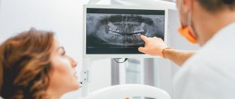
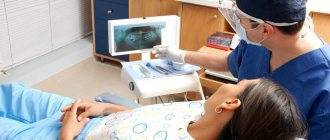
At the first stage of making a diagnosis, the dentist examines and interviews the patient about problems with the dental system, listens to his complaints and the history of the development of the disease. In some cases, this is enough to make a diagnosis. But most often, the dentist refers the patient for an additional X-ray examination or orthopantomogram (second stage).
In 90% of cases, the data obtained is enough for a correct diagnosis and development of a treatment plan. In difficult cases, the doctor prescribes laboratory diagnostics, as well as hardware examination methods - MRI, CT, ultrasound. Popular diagnostic measures are general clinical blood and urine tests, as well as serological and microscopic examination.
Basic examination methods
Patient interview. This is done at every appointment. The doctor collects information about complaints, existing symptoms, past illnesses, medications taken, etc.
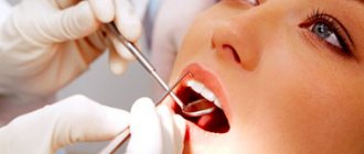
Inspection. The dentist examines the mucous membranes, teeth, evaluates the structure of the face, the closure of the dentition, the presence of swelling, edema, and other signs of inflammation. After a general examination, the condition of each of the teeth in the upper and lower jaw is assessed.
Crowns are examined using a dental mirror (allows you to see hard-to-reach areas and direct light to them). A sharp probe is used to assess the condition of the enamel. If there is a possibility of inflammation, percussion is performed, tapping the tooth (along the cutting edge or chewing surface). Normally it should be painless.
To assess the condition of the periodontium, the gums are examined, the presence of swelling, reddened, swollen, and injured areas is checked. Shallow probing may be performed to detect bleeding. If there is swelling or swelling, palpation (feeling) is performed. It is also used to assess tooth mobility.
Tests before implantation
Dental implantation, like any other procedure involving surgery, requires careful preparation. Neglecting the implantation protocol is fraught with its rejection and inflammatory processes. Therefore, it is important to pass all the necessary tests before implantation. This will allow:
- Identify hidden problems with gum tissue;
- Eliminate reactions to anesthesia and the use of medications;
- Determine the condition of the patient’s bone tissue;
- Assess the patient's general condition before surgery.
A complete list of tests that can be prescribed by a dentist before implantation:
- Clinical general blood test;
- Analysis of glucose levels in the patient's blood serum;
- Syphilis test RPR HBsAg;
- APTT;
- Fibrinogen test;
- Determination of prothrombin;
- INR;
- Determination for Antithrombin III;
- Thrombin time analysis;
- Determination of the presence of antibodies to HIV 1 and 2;
- Determination of the presence of HIV antigen 1 and 2;
- Qualitative test Anti-HCV-total;
- Urine analysis for protein and creatinine content.
Tests for periodontal disease
Periodontal disease is an extremely dangerous disease, which has a pronounced systemic-dystrophic nature in relation to periodontal tissues. Often, to get rid of a disease, it is necessary to jointly diagnose and draw up a treatment plan from specialists in several fields - a general dentist, periodontist, and therapist. Therefore, in the process of preparing for therapy, the doctor can prescribe the widest possible list of laboratory tests. The results of the study make it possible to identify which system of the patient’s body has weakened, which led to the development of periodontal disease.
Radiography
An image is taken of a small area of the jaw, more often to diagnose the condition of one tooth. It allows you to evaluate the characteristics of bone tissue, the tooth itself, the tissues around it and its root (gums, bone, ligaments).
X-rays in dentistry are used to:
- diagnose hidden caries, pulpitis, inflammation or cracks of the root canals;
- collect information about the dental system before implantation, prosthetics or monitor their results;
- assess the volume of bone tissue during sinus lift;
- check the quality of root canal treatment (must be filled without voids or pores);
- assess the structure of the dental system before and after orthodontic treatment;
- perform an accurate diagnosis if the patient’s complaints correspond to several diseases.
There are several types of X-rays used in dentistry.
Intraoral, dental. Performed to obtain an image of a single tooth or small area. The picture is taken using a radiovisiograph - a digital camera that captures an x-ray signal. The technology allows you to obtain high-resolution images: the doctor can see the condition of the canals, hard tissues, etc., and identify diseases at the initial stage. Radiation exposure with digital radiovisiography is minimal, which allows the method to be used for primary and intermediate diagnostics, when assessing the results and quality of treatment. The image is taken in a few minutes: a sensor is placed in the patient’s mouth on the side of the tooth being examined, and a pulsed radiation source is placed near the face. The image is available for viewing immediately.
Panoramic. It is performed to assess the structure and condition of the jaw as a whole, allows you to identify foci of inflammation or infection, pathology or disease of the TMJ, and assess the condition of the maxillary sinus and periodontium. Panoramic photographs are needed in preparation for implantation, prosthetics, and orthodontic treatment. They are performed using a special apparatus. The radiation dose is higher, but remains safe.
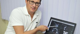
CT scan. It is used in planning sinus lifting, bone grafting, implantation, and other surgical interventions, and in diagnosing periodontal diseases. The photographs are taken in certain projections and used to build a three-dimensional model of the jaw. Computed tomography is very informative. The study provides information about the localization of inflammation (including intraosseous), the presence of tumor-like formations, their structure, size, features of the location of the root canals, etc.
Diagnostics in dentistry
The primary task for a dentist is to make the correct diagnosis for the patient. The diagnosis is the starting point for planning the treatment process, and the achievement of a successful result depends on its accuracy.
Why is accurate comprehensive diagnosis so important?
Nowadays, some patients perceive dentistry in a distorted way. Among the reasons we can highlight competition between clinics that, in search of patients, are ready to promise everything at once. In this case, insufficient time is devoted to diagnosis, and the correctness of the diagnosis remains in question.
Doctors are also contributing to this negative trend. Some of them quickly make a diagnosis based only on images or a simple examination, and are ready to immediately begin treatment. This practice is common not only in therapeutic dentistry, but also in prosthetics and implantation. One can only imagine what risks exist for the patient without a full diagnosis, and what complications may arise as a result of such an approach.
When contacting a general practitioner at a clinic, we all understand the appointment of many tests and images, as well as referrals to other specialists. Why shouldn’t dentists adopt this approach? We know well that everything in the human body is interconnected. The dental system also interacts with other systems of the body, so simple dental treatment can affect them as well. It can be difficult to establish the root cause of dental diseases without additional diagnostics, because by the time the patient comes to the clinic, his condition may worsen under the influence of psychosomatics.
Thus, thanks to the diagnostics carried out in each specific case, the correct diagnosis can be made. This is the only way to confidently exclude diseases of the respiratory system and spine, as well as neurological diseases. An individual approach to each patient includes personal communication with the dentist to establish contact and clarify the medical history. Thanks to this, the upcoming treatment will take place in an atmosphere of mutual trust and cooperation.
Palpation
One of the standard diagnostic methods is muscle palpation. Using this method, you can analyze the work of the muscles of the head and neck. When comparing muscle function with bilateral palpation, dysfunctions in the functionality of the muscular system can be identified. Thus, having basic anatomical knowledge, the dentist diagnoses problems of the dentofacial apparatus as a whole.
Bite analysis
Wax plates are used to record occlusion and articulation indicators in dentistry. They are used to determine the movements of the lower jaw and the contact of teeth at rest. Using plates is a simple and effective way to evaluate the closure process. However, in addition to this, a general examination is also important, during which you can find out about the condition of the periodontium and teeth, the presence of removable structures and restorations. Thanks to this, the dentist can draw conclusions about oral hygiene and the condition of the mucous membrane. This information is extremely important because malocclusions are often accompanied by periodontal problems.
Photo protocol
Currently, photography is widely used in diagnostics. To optimize the process, a protocol has been developed that the specialist follows.
For a complete diagnosis, it is necessary to take two groups of images – extraoral and intraoral.
Extraoral photographs record the position of the patient's head in individual positions. Based on these images, one can judge the characteristic features of appearance, asymmetry and facial features, muscle tension in the neck and head.
Intraoral photographs can reveal the position of the jaws, the condition of the dentition and other features.
If you have a problem similar to that described in this article, be sure to contact our specialists. Don't diagnose yourself!
Why you should call us now:
- We will answer all your questions in 3 minutes
- Free consultation
- The average work experience of doctors is 12 years
- Convenient location of clinics
Single contact phone number: +7
Make an appointment
Diagnostic models
One of the most important components of the diagnostic process is the production of models based on casts. At this stage, an articulator is used, which shows the position of the upper and lower jaws with maximum accuracy. The articulator is adjusted individually in each specific case, thanks to which the dentist receives comprehensive information about the dental system.
When diagnosing, an important role is played by the analysis of information about the patient’s bite. Maximum contact between teeth is not informative enough for the doctor. The primary task is to calculate the unforced posterior position of the mandible. Thanks to this, it is possible to determine the real relationship of the jaws and identify the source of the detected pathology.
Condylography
An effective method for diagnosing TMJ is condylography. It allows you to calculate the characteristics of movement in a joint. By analyzing the information obtained during condylography, the dentist can determine the presence of pathologies and make the correct diagnosis. Of course, data from other diagnostic methods are also taken into account.
Due to its non-invasiveness, condylography is an easy-to-use method. However, it requires special knowledge and skills from the dentist, without which it is impossible to obtain accurate data.
It is important to mention that condylography data is used both to clarify the diagnosis and for subsequent orthopedic work.
Other diagnostic methods
Currently, the following types of diagnostic methods are used in dental practice:
- Computed tomography;
- Magnetic resonance imaging of the central nervous system;
- Orthopantomogram;
- Teleradiogram, etc.
Let's talk briefly about some of these methods.
Teleradiogram
This method allows you to obtain accurate data on the structure of the skeleton and soft tissues. It is carried out according to a standard scheme, which increases the efficiency and effectiveness of treatment. In reconstructive dentistry, the importance of TRG analysis cannot be overestimated.
Magnetic resonance imaging of the HFNS
MRI is one of the most important methods for diagnosing dysfunctions of the HFNS. It helps to analyze the condition of the joint and soft tissues with maximum accuracy. However, the effectiveness of tomography directly depends on the qualifications of the doctor and his ability to interpret the image.
CT scan
Computed tomography is another common diagnostic method. It is used when making an initial diagnosis and in preparation for dental implantation.
Saliva examination
To make a diagnosis, it is sometimes necessary to determine the amount of saliva and the rate of its secretion, calculate the pH value, buffer capacity, and check the mineral composition. For example, a deficiency or excess of one or another element can provoke unfavorable conditions for enamel.
No less important are the biochemical parameters of the oral fluid:
- indicators of anti- and pro-oxidant systems;
- free radical oxidation values;
- the amount of enzymes in saliva, etc.
Biochemical methods
This is mainly a biochemical blood test. It is performed to detect signs of inflammation and changes in metabolic processes. Periodontal disease, especially in people over 40 years of age, is often accompanied by changes in lipid levels. A short (several indicators) or extensive biochemical analysis can be performed. Close attention is also paid to the composition of cells, their color, the presence of microbes, cell adhesion, etc. As a result of research, it is possible to establish the intensity of the development of inflammation and the direction of development of the pathological process.
Up to contents
Types of laboratory tests
Cytological
They help determine the nature of the cellular material, detect caries pathogens or specific cells that cause a pathological process in the oral cavity. Exfoliative material is obtained from the surfaces of ulcers, areas of leukoplakia, and from surgical discharge. Aspiration – from tumors, lymph nodes, foci of inflammation. In addition, swabs are used for research. The material is fixed, stained and examined under a light microscope for its constituent components.
Mycological and microbiological
Biomaterial is taken from areas of damage to the tooth or mucous membrane. The objective of the study is to obtain the most detailed information about the microecology of the oral cavity, salivary gland duct, periodontal pocket or other biological niche. These methods help to identify the causative agents of the pathological process and determine the sensitivity of microorganisms to antibacterial agents.
Virological
This group of methods is aimed at identifying traditional pathogens that cause the most common diseases of the oral mucosa. The presence of specific IgG and IgM antibodies in certain reactions will indicate that the disease is viral in nature. During acute infection and during relapse, they manifest themselves differently, which makes it possible to differentiate the stage of the pathological process.
Immunochemical
This includes various research methods, including the indirect enzyme immunoassay method. It is used to determine the titers of specific antibodies to fungi, viruses and bacteria in saliva and blood serum.
DNA polymerase
The material being studied is tested using high-precision methods for the presence of DNA pathogens.
Immunological
The methods are focused on checking the level of general (systemic) and local immunity of the patient and allow further adjustment, for example, of the treatment of pulpitis of primary teeth depending on the immune response. Instead of skin tests, today laboratories often resort to enzyme immunoassay methods.
General clinical laboratory tests are also carried out to diagnose dental diseases. They study the physicochemical and microscopic properties of blood, urine, feces and other fluids and separated substrates from the body.
Computed tomography (CT)
Computed tomography is used to examine bone structures in more detail .
Most often, the need for CT arises for intra-articular fractures and to clarify the structure of bone tumors. The accuracy of the study is achieved through a series of layer-by-layer X-ray images of the examined area, obtained by the doctor after computer processing.
Pros of CT:
- High information content in the study of bone structures;
- Ability to evaluate soft tissues;
- Possibility of computer 3D reconstruction of the studied area.
Cons of CT:
- High radiation exposure;
- Inability to use frequently;
- The impossibility of conducting research in patients with fear of closed spaces (claustrophobia).
Microbiological studies
This is a determination of the composition of the oral microflora and its sensitivity to antibacterial drugs. The reason for the importance of the study is not only the huge role of the microbial factor in the development of inflammation, but also the large number of cases where the disease is particularly resistant to traditional therapeutic interventions. Microbiological tests make it possible to determine the causative agent of inflammation, determine which antibiotic will be most effective in eliminating it, and also determine the treatment tactics for a specific form of periodontitis. Microbiological studies are also important because new microbial associations are emerging and their resistance to common antibiotics is increasing. Analysis of oral samples is usually performed before and after treatment to control its quality.
Up to contents