From this article you will learn:
- what to do if a child has an abscess on his gum,
- Do I need to remove a baby tooth?
- how the treatment is carried out (video),
- how to distinguish a purulent lump from a cyst on a child’s gum.
The article was written by a dentist with more than 20 years of experience.
An abscess on the gum in children usually looks like a red or yellowish lump located in the projection of the root of one of the teeth, which caused the inflammation. The inside of such a lump is filled with pus, and it has a yellowish color if the mucous membrane is thin and then the pus shines through it. The appearance of such a lump on the gum above a tooth in a child is always evidence of the development of purulent inflammation at the apex of the roots of a baby or permanent tooth, and indicates the development of apical periodontitis.
Apical periodontitis of primary and permanent teeth in children is most often associated with the lack of timely treatment of caries and pulpitis, as well as with mechanical trauma to the front teeth, for example, due to a fall. Therefore, caries or an old filling can always be found on the causative tooth, and if we are talking about an injury, there may be a chip of the enamel or a fracture of the tooth crown. In cases where the traumatic impact does not lead to damage to the hard tissues of the tooth, the crown of the tooth may be stained bluish or gray.
Abscess on a child’s gum: photo
Immediately before an abscess appears on the gum, children usually complain of pain or discomfort in one of the teeth, which is associated with the formation of pus at the apex of the tooth root. But after a pus-filled lump forms on the gum, children in most cases immediately feel relief (only pressing on the lump with your fingers can remain painful). Next, the lump may spontaneously open with the formation of a fistula, from which pus will leak into the oral cavity, or this may require an incision in the gums.
Important: however, not every lump on the gum indicates the development of purulent inflammation. There are also so-called eruption cysts. For example, even 2-3 weeks before the eruption of a baby or permanent tooth, a cyst may form on the child’s gum, which will have a bluish color (Fig. 7-8). We will also talk about such cysts at the end of the article.
What does a purulent abscess on a child’s gum look like (video) –
Causes of stomatitis
Inflammation of the oral mucosa can be caused by various reasons.
- Mucosal injury6.
- Viral, bacterial or fungal infection6.
- Allergy2.
- Immunity disorders (autoallergy, that is, a reaction to one’s own altered tissues2).
- Diseases of the blood, digestive, cardiovascular, nervous and endocrine systems2.
- Hypovitaminosis - lack of vitamins C, B1, B12, E, D32.
- Common infectious processes: measles, rubella, scarlet fever and diphtheria, infectious mononucleosis, tuberculosis and others1.
Only a specialist can understand the causes of stomatitis. Therefore, if symptoms of the disease appear, you should definitely contact your dentist.
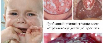
Inflammatory diseases of the oral cavity can occur in a child of any age6. Immaturity of the body is the primary factor predisposing to the occurrence of stomatitis in children under one year of age. The mucous membrane of the oral cavity during this period of a child’s life is very vulnerable, its natural protective properties are reduced. The immunity received from the mother during fetal development and which protected the baby immediately after birth weakens every day. Therefore, the infection easily penetrates the mucous membrane and causes its inflammation. Most often at this age, candidal stomatitis occurs, commonly called thrush1,6.
From the age of one to 3 years, children actively develop immune mechanisms to protect the oral mucosa (local immunity), but its permeability to viruses remains extremely high. Therefore, in a 2-3 year old child, stomatitis is more likely to be viral, in the vast majority of cases - herpetic1,3,4.
From 4 to 12 years of age, stomatitis in children is more often caused by allergic and autoimmune reactions. In particular, chronic aphthous forms of the disease occur during this period1.
Up to contents
A child has a lump on his gum - causes and treatment
This section is very important because... will show how untimely dental treatment, parents ignoring changes in the color of baby teeth or their superficial destruction - leads to the rapid development of periodontitis of the baby tooth, against the background of which a purulent abscess forms on the gum. Usually parents believe that they cannot do anything about it, because... The child still won’t let his teeth be treated. But modern dentistry allows even infants to have their teeth treated under sedation, allowing them to sanitize all their teeth in one visit without fear.
Reasons for the appearance of an abscess/bump on the gum –
- Untreated caries of one of the teeth is the main reason for the appearance of such ulcers. Caries in children occurs against the background of irregular hygiene and constant snacking between main meals. Caries sooner or later turns into pulpitis (at this stage, infection from the carious cavity penetrates into the tooth pulp, causing inflammation in it). Inflammation of the pulp leads to the gradual death of the neurovascular bundle inside the tooth - as a result of which a focus of purulent inflammation develops at the apex of the tooth root (apical periodontitis).
- Poorly treated teeth - in public dental clinics, usually no one will bother with your child, persuading him to be patient while the doctor carefully drills out the carious tissue and puts a filling.
Usually, the maximum that dentists offer to children under 3-5 years of age is the method of silvering teeth. But this method only slows down the development of caries and is not a complete treatment method. In addition, silvering of the same tooth must be carried out repeatedly (after a certain time). As a result, in most teeth treated with the silvering method, first pulpitis (inflammation of the nerve in the tooth) always develops, and then periodontitis.
- Traumatic damage to a tooth is the second most common reason why a lump appears on a child’s gum.
A strong mechanical impact on the tooth (fall, bruise, blow) can lead to the death of the nerve in the tooth, and even to a fracture of the crown or root of the tooth. Dental injuries do not always lead to these consequences; for example, if the blow was not very strong, then everything may well end well. If an abscess has nevertheless formed, and there is no caries or filling on the tooth (in the projection of the abscess), then the obvious reason for the development of purulent inflammation will be mechanical trauma to the tooth. This reason will also be supported if you find mobility of the tooth crown (in the projection of the abscess that has formed on the gum), or a change in the color of the tooth crown - to bluish or gray.
How does a purulent abscess form further?
The formation of a purulent abscess on the gum of a child occurs as follows... In general, periodontitis of the tooth for a long time in a child can be asymptomatic, but with hypothermia or decreased immunity, chronic inflammation at the apex of the root worsens - with the formation of pus at the site of inflammation. The pus tends to come out and therefore it gradually “makes” its way through the bone tissue, first falling under the periosteum of the jaw, where a subperiosteal abscess is formed.
Let us recall that the surface of the jaw bone tissue is covered with periosteum, and only on top of the periosteum is the mucous membrane located. When a purulent abscess is located between the surface of the bone and the periosteum, dentists call such inflammation the term “periostitis.” With periostitis, there will be a characteristic swelling on the gum that is dense to the touch, touching which will be very painful (but there will not yet be a clearly contoured lump with pus).
Next, purulent melting of the periosteum may occur, and then pus enters the submucosal membrane, forming a so-called “submucosal abscess.” The mucous membrane has high elasticity, and therefore a “bump”, soft to the touch, with clear contours, filled with pus (usually painless when touched) is formed on the gum. It should also be noted that in parallel with inflammation of the gums, swelling of the soft tissues of the face, as well as lymphadenitis of the submandibular lymph nodes, can also be observed.
Traumatic stomatitis
Inflammation of the mucous membrane in the mouth can be preceded by trauma: mechanical, thermal, chemical, radiation1,6.
Small children who put everything in their mouths can injure the mucous membranes with the sharp edges of toys or household items. Injuries often occur due to inept use of cutlery as a result of falls. In older children, stomatitis may be associated with a thermal burn, blows to the teeth, or the bad habit of chewing a pencil or pen. Sometimes the cause of inflammation is dental diseases and their treatment: sharp edges of damaged teeth and fillings, braces and aligners1.
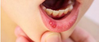
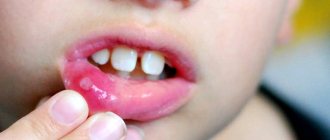
When stomatitis develops in a child on the gum, on the inner surface of the cheek or in another place, the mucous membrane becomes red and swollen, and a painful erosion or ulcer may appear in the area of damage. Since there are many microorganisms in the mouth, there is always a risk of a bacterial or fungal infection1.
If the mucous membrane is constantly injured, painless whitish or whitish-gray layers may form on it.
In children under 1 year of age, stomatitis can be the result of using the wrong nipples: long, tight, irregularly shaped. The resulting Bednar aphthae are located in the area of the transition of the soft palate to the hard palate and are round erosions or ulcers1.
Treatment of traumatic stomatitis in children includes:
- elimination of traumatic factors;
- rinsing the mouth, irrigating the mucous membranes and treating wounds with drugs with an analgesic and antiseptic effect1.
Up to contents
Abscess on the gum of a child: treatment
Thus, if a lump or fistula appears on a child’s gum, you should immediately go to the dentist. The strategy for temporary and permanent teeth in this case will be very different, and below we will describe in detail when such teeth can be treated and when they must be removed. If you find it difficult and cannot decide if it is a temporary or permanent tooth, use the tables with the timing of teething in primary and permanent dentition in children.
Viral stomatitis
About 80% of all stomatitis in children is caused by herpes viruses3,5. In approximately 70% of cases, the disease develops in children aged 1 to 3 years3,4.
The disease can occur in mild, moderate and severe forms.
With mild herpetic stomatitis, the general condition is practically not disturbed. Body temperature does not exceed 37-37.50 C. Symptoms of inflammation are limited to swelling of the gums and usually the simultaneous appearance of single painful herpetic blisters and erosions on the mucous membrane (no more than 6). After 1-2 days, the contours of the lesions are blurred, the rashes turn pale, and the erosions heal without scarring3.
With moderate stomatitis, the child’s temperature reaches 38-390 C and lasts until rashes continue to appear. General intoxication manifests itself in the form of weakness, headache, nausea. The child becomes capricious, lethargic, refuses to eat and play3.
As the temperature rises, the oral mucosa becomes red and swollen, and the gums begin to bleed. The number of herpetic elements reaches 20-25, repeated rashes are accompanied by fever3.
Severe stomatitis in children looks like a common acute infectious disease and is accompanied by severe intoxication: fever 39-400 C, chills, headache, aching muscles and joints, heart rhythm disturbances, nosebleeds, nausea and vomiting. The number of herpetic elements can reach 100; they are located not only in the mouth, but also on the skin of the face, on the eyelids and conjunctiva of the eyes, and earlobes. In addition, upper respiratory tract symptoms may be present.3
Treatment includes treating the affected area with painkillers and antiseptic drugs, antiviral therapy, drinking plenty of fluids, a balanced diet and proper nutrition3,5.
Up to contents
How to distinguish stomatitis from other diseases
Since stomatitis can appear for a variety of reasons, and the forms of its manifestation can be very different, it is highly not recommended to self-medicate, since in this case you can only aggravate the course of the disease and cause the appearance of new symptoms. Due to such diversity, methods of treatment and prevention of this disease can also be very different.
Manifestations of stomatitis may be similar to a number of diseases, from each of which a doctor, when examining a patient, will definitely be able to distinguish this ailment:
- Sore throat or stomatitis: you can determine a sore throat, which may be similar to stomatitis, by damage not only to the mucous membranes of the mouth, but also to the pharynx. It is very painful for the patient to swallow.
- Herpes or stomatitis: In the case of herpes, the rashes first look like blisters and appear later.
- Cancer or stomatitis: the first disease is distinguished by the fact that ulcers appear, which grow together over time. As a result, one ulcer forms and begins to grow.
- Thrush or stomatitis: when examining the mouth, if there is a disease such as candidiasis, a yellow or white coating is detected, redness, the wounds are round in shape, and the ulcers are small.
- Syphilis or stomatitis: the first disease is distinguished by the nature of the ulcers. They appear in the form of eosis and do not hurt. Sometimes there is a depression in the center of the wound.
The doctor selects the treatment for stomatitis based on the cause of the disease, the complexity of its course, the form of manifestation, the individual characteristics of the body, and so on - in general, you will have to take into account a lot of things in order to choose the correct and most effective treatment. A specialist usually easily determines that it is stomatitis and not another disease.
Candidal stomatitis
In 80% of healthy children, Candida fungi can be found in the oral cavity. They get there during childbirth, from nipples and pacifiers, from care products, in contact with the mother's skin, with food during eating and usually do not cause any problems. Candidal stomatitis, or thrush, occurs when immunity decreases1.
Predispose to the disease:
- prematurity and postmaturity;
- developmental defects and concomitant diseases;
- treatment with antibiotics and hormones;
- artificial feeding;
- poor care and poor feeding hygiene;
- Using the wrong nipples1.
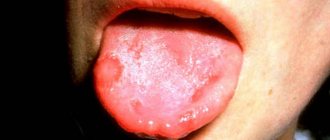
What does candidal stomatitis look like? In children's mouths, whitish or whitish-gray dotted formations appear on the reddened mucous membrane. They merge into films of a cheesy nature; when the films are rejected, bright red painful erosions are formed. Because of the pain, the baby becomes restless, cries often, sleeps poorly, and refuses to eat1.
In severe cases of candidiasis, a cheesy coating may appear on the palate and on the lateral surfaces of the tongue, on the tonsils and the back wall of the pharynx - candidal tonsillitis and pharyngitis develop1.
If your child has a fungal infection, you should always consult a doctor; he will tell you how and what to treat the mucous membrane with candidal stomatitis, so that the child does not have complications and recovers faster. Treatment, as a rule, involves treating the oral cavity with drugs with antifungal activity (chlorhexidine, hexethidine). In severe cases of the disease, antifungal agents, probiotics and immunomodulators are prescribed1.
Up to contents
Allergic stomatitis
Allergic damage to the mucous membrane most often occurs in the form of contact stomatitis and chronic aphthous form of the disease1.
Allergens can include medications, food products, varnishes and paints that coat toys, toothpastes, mouth rinses, chewing gum, and dental metals included in braces1,2.
With allergic stomatitis in children, erosions and ulcers may appear in the mouth, but more often the matter is limited to redness and swelling of the mucous membrane1.
Treatment is avoiding contact with the allergen, rinsing or irrigating the mouth with antiseptic solutions to prevent infection. If necessary, doctors recommend taking antihistamines1,2.
Chronic recurrent aphthous stomatitis often occurs in schoolchildren and adolescents due to allergies2. In addition, its development can be provoked by diseases of the gastrointestinal tract, upper respiratory tract infections, disorders of the nervous system, and hypovitaminosis1,2.
With aphthous stomatitis in children, itching and burning first appear in the oral cavity. The mucous membrane at the site of the lesion becomes red and swollen, then an aphtha forms on it - a round or oval erosion 0.5-1 cm in diameter rising above the surrounding tissues with a red rim along the periphery and a bottom covered with a grayish-white coating. Aphthae are extremely painful, and if many of them form, they cause significant distress to the child1,2.
How many days does aphthous stomatitis last in children? With a mild course of the disease, the elements of inflammation persist for up to 5-7 days, then they heal without scar formation2 and do not appear for quite a long time. However, in severe cases, aphthae can occur constantly, and then many elements of inflammation can be found on the mucosa at different stages of development1,2.
To treat aphthous stomatitis, doctors use:
- antihistamines;
- immunomodulatory drugs;
- vitamins;
- probiotics2.
As a local therapy, it is recommended to treat the mucous membrane with drugs with analgesic, antiseptic, proteolytic (protein-breaking), anti-inflammatory and regenerating effects2.
Up to contents
How to treat
If a child has a white sore on his gum, you can resort to drug treatment to achieve a quick therapeutic effect. It is recommended to give preference to the following medicinal formulations:
- Metrogyl Denta is a dental gel that has an antimicrobial effect. Allowed for use in children over six years of age. The drug is prescribed for the treatment of gingivitis, periodontitis, bacterial aphthous stomatitis, inflammation of the hood under the wisdom tooth, and periodontal abscess. Sores should be treated twice a day after meals and subsequent treatment of the oral cavity with antiseptic compounds.
- Cholisal is a dental gel that has antimicrobial, anti-inflammatory and analgesic effects. This drug is perfect for children, because it has no age restrictions. The ulcers should be treated with the drug twice a day for a duration of treatment of about 10 days.
- Kamistad is a combined drug for topical use that has a local anesthetic, antimicrobial, and anti-inflammatory effect. For children over the age of three months, it is recommended to apply the composition to the sores three times a day, keeping the strip length to 5 mm.
- Chlorhexidine is an antiseptic solution used for hygienic treatment of the oral cavity. Has a detrimental effect on bacteria and other harmful microorganisms.
- Acyclovir is a drug prescribed for the treatment of formations on the oral mucosa associated with herpes infection. The medicinal composition helps accelerate the healing of formations, relieve pain, and provide an immunostimulating effect. It is recommended to treat ulcers with cream about 5 times a day for a treatment duration of 5 to 10 days.
- Benzydamine is a medicinal non-steroidal anti-inflammatory composition that has local antiseptic, analgesic and antipyretic effects. Available in the form of lozenges, creams, and solutions. As part of complex therapy, the drug is indicated for stomatitis, pharyngitis, aphthous ulcers, tonsillitis, and gingivitis. The medicine can only be used after reaching 12 years of age.
Chlorhexidine solution.
Hexoral in the treatment of stomatitis
For local treatment of stomatitis in children, drugs from the HEXORAL® line can be used.
For irrigation of the oral cavity, the doctor may recommend aerosol HEXORAL® based on hexethidine, which has antiseptic properties7. Thanks to the fine spray, the drug is evenly distributed over the entire surface of the mucosa7. A solution of HEXORAL®8, similar in composition, is intended for rinsing the mouth. Both medications can be used to treat children 3 years of age and older.
Mint-flavored lozenges HEXORAL® TABS based on chlorhexidine and benzocaine may be suitable for boys and girls over 4 years of age9.
For patients with stomatitis over 6 years of age - HEXORAL® TABS CLASSIC based on the antiseptic amylmetacresol. The assortment includes tablets with lemon, orange, black currant, lemon and honey flavors10.
HEXORAL® TABS EXTRA may be suitable for adolescents aged 12 years and older. The lidocaine it contains can relieve even severe pain11.
The information in this article is for reference only and does not replace professional advice from a doctor. To make a diagnosis and prescribe treatment, consult a qualified specialist.
Up to contents
Literature
- Khomenko L. A. Therapeutic dentistry of children. Textbook for university / ed. 2007 – pp. 643-722.
- Ismailova G. T. Chronic recurrent aphthous stomatitis // Bulletin of surgery of Kazakhstan. - 2011. - No. 4. — P. 124-125.
- Drobotko L.N., Strakhova S.Yu. Acute stomatitis in children // Issues of modern pediatrics. - 2010. - T. 9. - No. 2. - P. 146-149.
- Suerkulov E. S., Yuldashev I. M., Mamyraliev A. B., Toktosunova S. A., Tsepeleva A. S., Sooronbaev A. A. Prevalence and structure of the incidence of stomatitis in children // Bulletin of Science and Practice. 2022. T. 4. No. 11. — P. 91-96.
- Suerkulov E. S., Yuldashev I. M., Mamyraliev A. B., Zhumashova N. K., Yuldasheva G. I. Complex therapy of inflammatory diseases of the oral mucosa in children // Bulletin of Science and Practice. 2022. T. 5. No. 5. — P. 96-104.
- Pankrusheva T.A., Maravina I.N., Chekmareva M.S. Research on the development of the composition and technology of tablets for the treatment of stomatitis // Scientific result. Medicine and pharmacy. – T.4, No. 1, 2022. – P. 78-87.
- Instructions for medical use of the drug HEXORAL® aerosol: , .
- Instructions for medical use of the drug HEXORAL® solution: , .
- Instructions for medical use of the drug HEXORAL® TABS: , .
- Instructions for medical use of the drug HEXORAL® TABS CLASSIC: , .
- Instructions for medical use of the drug HEXORAL® TABS EXTRA: , .
Up to contents