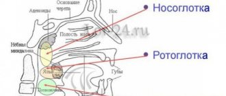
CLINICAL ANATOMY OF THE LARYNX.
CLINICAL ANATOMY OF THE LARYNX
The larynx
(Larynx)
is a hollow organ whose upper part opens into the laryngopharynx and the lower part passes into the trachea. The larynx is located under the hyoid bone on the front surface of the neck. The inside of the larynx is lined with a mucous membrane and consists of a cartilaginous skeleton connected by ligaments, joints and muscles. The upper edge of the larynx is located on the border of the IV and V cervical vertebrae, and the lower edge corresponds to the VI cervical vertebra. The outside of the larynx is covered with muscles, subcutaneous tissue and skin, which is easily displaced, allowing it to be palpated. The larynx makes active movements up and down when speaking, singing, breathing and swallowing. In addition to active movements, it passively shifts to the right and left, and the so-called crepitus of the laryngeal cartilages is noted. In the case of a malignant tumor, the active mobility of the larynx decreases, as well as its passive displacement.
In men, in the upper part of the thyroid cartilage, a protrusion or elevation is clearly visible and palpable - the Adam's apple, or Adam's apple (prominentia laryngea, s. pomum Adami).
In women and children it is less pronounced, soft and its palpation determination is difficult.
In the lower part of the larynx in front, between the thyroid and cricoid cartilages, you can easily feel the area of the conical ligament (lig. Conicum, s. cricothyreoideum),
which is dissected (conicotomy) if it is necessary to urgently restore breathing in case of asphyxia.
Laryngeal cartilages.
The skeleton of the larynx is made up of cartilages
(cartilagines laryngis),
connected by ligaments (Fig. 4.1 a, b). There are three single and three paired cartilages of the larynx:
• Three singles:
1) cricoid cartilage (cartilago cricoidea)
;
2) thyroid cartilage (cartilago thyreoidea)
;
3) epiglottic cartilage (cartilago epiglotica)
or epiglottis
(epiglottis).
Rice. 4.1.
Skeleton of the larynx:
a – front view; b – rear view: 1 – thyroid cartilage; 2 - cricoid cartilage; 3 – epiglottis; 4 – arytenoid cartilage; 5 – tracheal rings; b – hyoid bone
• Three doubles:
1) arytenoid cartilages (cartilagines arytaenoidea);
2) corniculate cartilages (cartilagines corniculatae);
3) wedge-shaped cartilages (cartilagines cuneiformes, s. Wrisbergi).
Cricoid cartilage (cartilago cricoidea)
is the basis of the skeleton of the larynx.
In shape it really resembles a signet ring facing backwards. The narrow part facing forward is called the arc (arcus),
and the widened rear part is called the signet or plate
(lamina).
The lateral surfaces of the cricoid cartilage have superior and inferior articular platforms for articulation with the arytenoid and thyroid cartilages, respectively.
Thyroid cartilage (cartilago thyreoidea),
the largest cartilage of the larynx, located above the cricoid cartilage (Fig. 4.2). Thyroid cartilage confirms its name both by its appearance and its role in protecting the internal part of the organ. Two irregularly shaped quadrangular plates that make up the cartilage at the site of fusion
Rice. 4.2.
Thyroid cartilage
in front along the midline they form a ridge, at the upper edge of which there is a notch (tasura thyreoidea).
On the inner surface of the angle formed by the plates of the thyroid cartilage, there is an elevation to which the vocal folds are attached. At both sides
the posterior sections of the plates of the thyroid cartilage have processes extending upward and downward - the upper and lower horns (cornila). The lower ones - shorter ones - serve for articulation with the cricoid cartilage, and the upper ones are directed towards the hyoid bone, where they are connected to its large horns by the thyrohyoid membrane. On the outer surface of the plates of the thyroid cartilage there is an oblique line (linea oblique),
running from back to front and from top to bottom, to which part of the external muscles of the larynx is attached.
Epiglottic cartilage (cartilago epiglottica),
or epiglottis, is a leaf-shaped plate resembling a flower petal.
Its wide part stands freely above the thyroid cartilage, is located behind the root of the tongue and is called the petal. The narrow lower part - the stalk (petiolus epiglottis)
- is attached to the inner surface of the angle of the thyroid cartilage by means of a ligament. The shape of the lobe of the epiglottis varies depending on how much it is thrown back, elongated or curled, which is sometimes associated with errors during tracheal intubation.
Arytenoid cartilages (cartilagines arythenoideae)
have the shape of triangular pyramids, the tops of which are directed upward, somewhat posteriorly and medially.
The base of the pyramid articulates with the articular surface of the signet of the cricoid cartilage. to the anterior internal corner of the base of the arytenoid cartilage - the vocal process (processus vocalis)
, and the
- to the anterior external angle (processus muscularis)
. The second part of the vocal muscle is fixed to the lateral surface of the pyramid of the arytenoid cartilage in the region of its anteroinferior third, where the oblong fossa is located.
Wedge-shaped cartilages (cartilagines cuneiformes, s. Wrisbergi)
located in the thickness of the aryepiglottic fold.
Horn-shaped cartilages (cartilagines corniculatae)
located above the apex of the arytenoid cartilages. Wedge-shaped and corniculate cartilages are small-sized sesamoid cartilages, not constant in shape and size.
Joints of the larynx.
The larynx has two paired joints.
1. Cricothyroid joint (articulatio cricothyreoidea)
formed by the lateral surface of the cricoid cartilage and the lower horn of the thyroid cartilage. By bending forward or backward at this joint, the thyroid cartilage thereby increases or decreases the tension of the vocal folds, changing the pitch of the voice.
2. Cricoarytenoid joint (articulatio cricoarytenoidea)
formed by the lower surface of the arytenoid cartilage and the upper articular platform of the cricoid cartilage plate. Movements in the cricoarytenoid joint (forward, backward, medial and lateral) determine the width of the glottis.
Laryngeal ligaments
(Fig. 4.3). The main ligaments of the larynx include:
Rice. 4.3.
Laryngeal ligaments:
a – front view; b – posterior view: 1 – lateral thyrohyoid, 2 – cricotracheal, 3 – cricothyroid, 4 – aryepiglottic fold
• thyrohyoid medial and lateral (tig. hyothyreoideum medium et lateralis);
• thyroid epiglottis (tig. thyreoepigtotticum);
• sublingual-epiglottic (tig. hyoepigtotticum);
• cricotracheal (tig. cricotracheate);
• cricothyroid (tig. cricothyroideum);
• vocal fold (ptica vocate);
• aryepiglottic (tig. aryepigtotticum);
• lingual-epiglottic middle and lateral (tig. gtossoepigtotticum medium et tateratis).
Thyrohyoid median and lateral ligaments
are parts of the thyrohyoid membrane
(membrana thyrohyoidea),
with the help of which the larynx is suspended from the hyoid bone. The median thyrohyoid ligament connects the upper edge of the thyroid cartilage with the body of the hyoid bone, and the lateral ligament connects with the greater horns of the hyoid bone. The neurovascular bundle of the larynx passes through the hole in the outer part of the thyrohyoid membrane.
Thyroglottic ligament
connects the epiglottis with the thyroid cartilage in the area of its upper edge.
Hypoepiglottic ligament
connects the epiglottis to the body of the hyoid bone.
Cricotracheal ligament
connects the larynx with the trachea; located between the cricoid cartilage and the first ring of the larynx.
Cricoid or conical ligament
connects the upper edge of the cricoid cartilage arch and the lower edge of the thyroid cartilage.
The cricothyroid ligament is a continuation of the elastic membrane of the larynx (conus etasticus),
which begins on the inner surface of the plates of the thyroid cartilage in the region of its angle. From here, the elastic bundles fan out vertically downwards towards the upper edge of the arch of the cricoid cartilage in the form of a cone, forming a conical ligament. The elastic membrane forms a layer between the inner surface of the cartilage and the mucous membrane of the larynx.
Vocal fold
is the superior posterior bundle of the elastic cone;
covers the vocal muscle, which is stretched between the inner surface of the angle of the thyroid cartilage in front and the vocal process (processus vocatis)
of the arytenoid cartilage in the back.
aryepiglottic ligament
located between the lateral edge of the epiglottis and the inner edge of the arytenoid cartilage.
Glossoepiglottic median and lateral ligaments
They connect the middle and lateral parts of the root of the tongue with the anterior surface of the epiglottis; between them there are depressions - the right and left fossae of the epiglottis (valecula).
Muscles of the larynx
(Fig. 4.4). All muscles of the larynx can be divided into two large groups:
1) external muscles involved in the movement of the entire larynx as a whole;
2) internal muscles that cause the movement of the cartilages of the larynx relative to each other; these muscles are involved in the functions of breathing, sound production and swallowing.
External muscles
Depending on the place of attachment, they can be divided into two more groups:
Rice. 4.4.
Muscles of the larynx:
a – external muscles: 1 – sternohyoid, 2 – geniohyoid, 3 – stylohyoid, 4 – digastric, 5 – sternothyroid, 6 – thyrohyoid, 7 – sternocleidomastoid, 8 – cricothyroid, 9 – omohyoid ; b – internal muscles: 1 – oblique arytenoid muscle, 2 – aryepiglottic, 3 – transverse arytenoid, 4 – posterior cricoarytenoid, 5 – cricothyroid
1. To the first group
There are two paired muscles, one end of which is attached to the thyroid cartilage, and the other to the bones of the skeleton:
• sternothyroid (m. sternothyroideus);
• thyrohyoid (m. thyrohyodeus).
2. Muscles of the second group
attached to the hyoid bone and to the bones of the skeleton:
• sternohyoid (m. sternohyoideus);
• scapular-hyoid (m. omohyoideus);
• stylohyoid (m. stylohyoideus)
;
• digastric (m. digastricus)
;
• geniohyoid (m. geniohyoideus). Internal muscles
the larynx performs two main functions in the larynx
Features:
1. Change the position of the epiglottis during the act of swallowing and inhalation, performing a valve function.
2. Change the tension of the vocal folds and the width of the glottis between them (voice function).
The position of the epiglottis is changed by two pairs of antagonist muscles.
Arepiglottic muscle (m. aryepiglotticus)
located between the apex of the arytenoid cartilage and the lateral edges of the epiglottis. Being covered with mucous membrane, this muscle forms the aryepiglottic fold in the area of the lateral entrance to the larynx. During the act of swallowing, contraction of the aryepiglottic muscle leads to the retraction of the epiglottis backward and downward, due to which the entrance to the larynx is covered and food is displaced laterally into the pyriform fossa towards the entrance to the esophagus.
Thyroepiglotticus muscle (m. thyroepiglotticus)
stretched on the sides of the thyroid epiglottis between the inner surface of the angle of the thyroid cartilage and the lateral edge of the epiglottis. When the thyroepiglottic muscle contracts, the epiglottis rises and the entrance to the larynx opens.
The muscle group that performs the function of the vocal apparatus is more numerous than the previous one and is divided into muscle groups that constrict, dilate, tense and relax.
glottis.
Lateral cricoarytenoid muscle (m. cricoarytenoideus lateralis)
(paired) begins on the lateral surface of the cricoid
th cartilage and is attached to the muscular process of the arytenoid cartilage. When it contracts, the muscle processes move forward and down, and the vocal processes move closer together, narrowing the glottis.
Transverse arytenoid muscle (m. arytenoideus transverses)
connects the posterior surfaces of the arytenoid cartilages with each other, which, when it contracts, come closer together, narrowing the glottis mainly in the posterior third.
Oblique arytenoid muscle (m. arytenoideus obliqus)
(paired) begins on the posterior surface of the muscular process of one arytenoid cartilage and is attached to the apex of the arytenoid cartilage of the opposite side. Both oblique arytenoid muscles enhance the function of the transverse arytenoid muscle, located directly behind it, crossing each other at an acute angle.
Posterior cricoarytenoid muscle (m. cricoarytenoideus post. s. posticus)
begins on the posterior surface of the cricoid cartilage and attaches to the muscular process of the arytenoid cartilage. When inhaling, it contracts, the muscular processes of the arytenoid cartilages rotate posteriorly, and the vocal processes, together with the vocal folds, move to the sides, expanding the lumen of the larynx. This is the only muscle that opens the glottis. When it is paralyzed, the lumen of the larynx closes and breathing becomes impossible.
Thyroarytenoid muscle (m. thyreoarytaenoides)
begins on the inner surface of the plates of the thyroid cartilage. Going posteriorly and upward, it attaches to the lateral edge of the arytenoid cartilage. During contraction, the arytenoid cartilage rotates outward around its longitudinal axis and moves anteriorly.
Cricothyroid muscle (m. cricothyroideus)
It is attached at one end to the anterior surface of the cricoid cartilage arch on the side of the midline, and at the other to the lower edge of the thyroid cartilage. When this muscle contracts, the thyroid cartilage bends forward, the vocal folds become tense, and the glottis narrows.
Vocal muscle (m. vocalis)
- triceps, makes up the bulk of the vocal fold; begins in the region of the lower third of the angle formed by the inner surfaces of the plates of the thyroid cartilage, and is attached to the vocal process of the arytenoid cartilage.
A narrow strip of elastic connective tissue runs along the medial edge of the muscle; it plays a significant role in the formation of sound. When this muscle contracts, the vocal folds thicken and shorten, the elasticity, shape and tension of its individual sections change, which plays an important role in voice formation.
TOPOGRAPHY OF THE LARYNX
Larynx
suspended from the hyoid bone by the thyrohyoid membrane;
downwards it passes into the trachea, attached to it by the cricotracheal ligament. In front, the larynx is covered with skin, subcutaneous tissue, superficial fascia of the neck, and muscles. The fascia of the thyroid gland is attached to the lower part of the cricoid cartilage in front, the lateral parts of which cover the muscles (m. sternothyroideus et m. sternohyoideus).
The anterolateral surface of the larynx is covered by the sternohyoid muscle, and under it are the sternothyroid and thyrohyoid muscles. At the back, the larynx borders the laryngeal part of the pharynx and the entrance to the esophagus. On the sides of the larynx lie neurovascular bundles.
Blood supply to the larynx
carried out by two arteries:
• superior laryngeal (a. laryngea superior);
• lower laryngeal (a. laryngea inferior).
Superior laryngeal artery
is a branch of the superior thyroid artery
(a. thyreoidea superior),
which, in turn, arises from the external carotid artery.
The superior laryngeal artery is larger than the inferior one. As part of the neurovascular bundle of the larynx (a. laryngea superior, v. laryngea superior, ramus internus n. laryngei superior),
the artery penetrates the larynx through an opening in the outer part of the thyrohyoid membrane.
Inside the larynx, the superior laryngeal artery is divided into smaller branches, where another branch departs from it - the middle laryngeal artery (a. laryngea media),
which anastomoses with the artery of the same name on the opposite side in front of the conical ligament.
Inferior laryngeal artery
is a branch of the inferior thyroid artery
(a. thyreoidea inferior),
which originates from the thyroid trunk
(truncus thyreocervicalis).
Venous drainage
is provided cranially through the superior thyroid vein
(v. laryngea superior)
into the internal jugular vein
(v. jugularis interna),
caudally through the inferior thyroid vein
(v. laryngea inferior)
into the brachiocephalic vein
(v. brachiocephalica).
Lymphatic system
The larynx is divided into:
• upper section;
• lower section, which are separated by vocal folds. The lymphatic network of the upper section is more developed ,
especially in
areas of the vestibular folds and laryngeal ventricles. From here, the lymph, converging with other lymphatic vessels, is directed along the neurovascular bundle of the larynx to the deep cervical lymph nodes located along the deep jugular vein.
Lymphatic vessels of the lower section
pass under and above the cricoid cartilage, collecting in the preepiglottic lymph nodes. In addition, there is a connection with the deep cervical lymph nodes located along the deep jugular vein. Contralateral metastasis is possible here due to the existence of a connection with the pre- and paratracheal lymph nodes. The connection between the lymphatic system of the lower larynx and the mediastinal lymph nodes is of great clinical importance.
Innervation of the muscles of the larynx
provided by two branches of the vagus nerve:
• superior laryngeal nerve (n. laryngeus superior);
• lower laryngeal nerve (n. laryngeus inferior sn recurrens).
Superior laryngeal nerve
is mixed and departs from the vagus nerve in the region of the lower part of the vagus nerve ganglion
(ganglion nodosum n. vagi).
Behind the greater horn of the hyoid bone, the superior laryngeal nerve is divided into two branches: the external branch
(r. externus),
motor, innervating the cricothyroid muscle, and the internal branch
(r. internus),
penetrating through the hole in the thyrohyoid membrane; it gives off sensitive branches to the mucous membrane of the larynx.
Inferior laryngeal nerve (n. recurens)
mixed, innervates all the internal muscles of the larynx with the exception of the cricothyroid muscle and provides sensitive innervation to the mucous membrane of the lower floor of the larynx, including the area of the vocal folds. The lower laryngeal nerves on different sides are continuations of the right and left recurrent nerves, which arise from the vagus nerve in the chest cavity at different levels. The right recurrent nerve departs from the vagus nerve at the level of the subclavian artery, the left - at the place where the vagus nerve bends around the aortic arch. Next, the recurrent nerves of both sides rise up to the larynx,
giving numerous branches to the trachea and esophagus on its way, with the right one located laterally between the trachea and esophagus, and the left one lying on the anterior surface of the esophagus on the left.
The sympathetic nerves arise from the superior cervical sympathetic cervicothoracic (stellate) ganglion (ganglion stellatum).
The cavity of the larynx
(cavitas laryngis),
shaped like an hourglass, is narrowed in the middle section and expanded upward and downward. According to clinical and anatomical characteristics, it is divided into three floors (Fig. 4.5):
• upper
- vestibule of the larynx
(vestibulum laryngis)
- located between the entrance to the larynx and the vestibular folds, has the appearance of a cone-shaped cavity, tapering downwards;
• average
- glottis
(rima vocalis)
- the space between the vocal folds through which communication occurs with the lower floor of the larynx;
• lower
- subglottic cavity, extending from the vocal folds to the trachea, having the appearance of a cone-shaped cavity, expanding downwards.
Entrance to the larynx
in front is limited by the epiglottis, behind - by the apices of the arytenoid cartilages and on the sides - by the aryepiglottis.
mi folds, in the lower part of which lie corniculate and wedge-shaped cartilages, forming tubercles of the same name. Between the aryepiglottic folds and the walls of the pharynx there are pear-shaped pockets (recessus piriformes),
which behind the larynx pass into the esophagus. At the bottom of the pyriform sinus there is a fold of mucous membrane running posteriorly and downward, formed by the internal branch of the superior laryngeal nerve and the superior laryngeal
Rice. 4.5.
Floors of the larynx: 1 – upper; 2 – average; 3 – lower
artery. The recesses between the median and lateral lingual epiglottic folds, which connect the anterior surface of the epiglottis with the root of the tongue, are called lingual epiglottic recesses.
or
valleculae (valleculae epiglotticae).
At the level of the middle and lower third of the thyroid cartilage in the laryngeal cavity, on either side of the midline there are two pairs of horizontal folds of the mucous membrane.
The upper pair are called vestibule folds (plica vestibularis),
the lower ones are called
vocal folds (plica vocalis).
The length of the vocal folds in newborns is 0.7 cm;
in women - 1.6-2 cm; in men - 2-2.4 cm. On each side between the vocal and vestibular folds there are depressions - laryngeal
(Morgani)
ventricles (ventriculi laryngis),
in which there is a pocket outward and anteriorly, ascending upward. In the thickness of the mucous membrane of the laryngeal ventricles there is an accumulation of lymphadenoid tissue, which is sometimes called laryngeal tonsils, and when they are inflamed, accordingly, laryngeal tonsillitis. The width of the lumen (the glottis between the vocal folds in the posterior third) of the larynx in men is about 15-22 mm, in women – 13-18 mm, in a 10-year-old child – 8-11 mm.
The mucous membrane of the larynx is a continuation of the mucous membrane of the nasal cavity and pharynx and is covered mainly by multirow cylindrical ciliated epithelium. The vocal folds, the upper part of the epiglottis, the arytenoid folds and the laryngeal surface of the arytenoid cartilages are lined with stratified squamous epithelium, which is important to consider in the diagnosis of tumor diseases.
4.2. CLINICAL ANATOMY OF TRACHEA AND ESOPHAGUS
The trachea
(tracheae)
is a hollow cylindrical tube that is a direct continuation of the larynx (Fig. 4.6). The trachea begins at the level of the body of the VII cervical vertebra and extends to the level of the bodies of the IV-V thoracic vertebrae, where it ends in a branching (bifurcation) into two main bronchi. The level of bifurcation is higher in young people. The length of the trachea is on average 10-13 cm. The wall of the trachea consists of 16-20 hyaline cartilages, shaped like a horseshoe, the arc of which faces forward, and the rear open ends are connected by a connective tissue membrane - the membranous part of the wall.
Rice. 4.6.
Trachea skeleton
ki of the trachea (paries membranaceus tracheae).
This membrane contains elastic and collagen fibers, and in deeper layers - longitudinal and transverse smooth muscle fibers.
The width of the membranous wall ranges from 10 to 22 mm. The hyaline cartilages of the trachea (cartilagines trachealis)
are connected to each other by means of annular ligaments
(lig. annularia).
The inner surface of the trachea is lined with mucous membrane,
covered with columnar ciliated epithelium. In the submucosal layer there are mixed glands that produce a protein-mucosal secretion. On the inside of the trachea, at the site of its division into two main bronchi, a semilunar-shaped protrusion is formed - the junction of the medial walls of the main bronchi - the tracheal spur (carina trachea).
The right bronchus is wider, extends from the trachea at an angle of 15°, its length is 3 cm; the left one is at an angle of 45°, its length is 5 cm. Thus, the right bronchus is practically a continuation of the trachea, and therefore foreign bodies more often enter it.
TOPOGRAPHY OF THE TRACHEA
From above, the trachea is attached to the cricoid cartilage by the cricotracheal ligament (lig. cricotrachaele).
In the cervical part, the isthmus of the thyroid gland is adjacent to the anterior surface of the trachea, and its lobes are adjacent to the sides. Posteriorly, the trachea is adjacent to the esophagus. To the right of the trachea is the brachiocephalic trunk, to the left is the left common carotid artery (Fig. 4.7).
In the thoracic region in front of the trachea is the aortic arch. To the right of the trachea are the right pleural sac and the right vagus nerve, to the left are the aortic arch, left carotid and subclavian nerves.
Rice. 4.7.
Topography of the trachea: 1 – thyroid gland; 2 – common carotid artery; 3 – aortic arch; 4 – thymus gland; 5 – vagus nerve
arteries, left recurrent nerve. In children under 16 years of age, the thymus gland is located in the thoracic region in front of the trachea.
Blood supply to the trachea
carried out using the inferior thyroid
(a. thyroidea inferior)
and internal thoracic arteries
(a. thoracica interna),
as well as
counting the bronchial branches of the thoracic aorta (rami bronchiales aortae thoracicae).
In the innervation of the trachea
(n. vagus)
and the tracheal branches of the lower laryngeal nerve
(n. laryngeus inferior)
take part The sympathetic influence is represented by nerves arising from the sympathetic trunk
(truncus sympathicus).
Lymph
The trachea flows mainly into the lymph nodes located on both sides on its sides. In addition, the lymphatic system of the trachea has connections with the lymph nodes of the larynx, upper deep cervical and anterior mediastinal nodes.
Esophagus
is a hollow organ in the form of a tube that connects the pharyngeal cavity with the stomach cavity. From above, the pharynx passes into the esophagus in the projection area of the VI cervical vertebra at the level of the lower edge of the cricoid cartilage. Below, the junction of the esophagus with the stomach corresponds to the level of the XI thoracic vertebra. The length of the esophagus in an adult is on average 23-25 cm, and the width is from 15 to 20 mm.
There are three sections in the esophagus:
• cervical;
• chest;
• abdominal.
Cervical region
extends from the level of the VI cervical vertebra to the thoracic vertebra, its length ranges from 5 to 8 cm. The anterior border with the thoracic region is the level of the jugular notch.
Thoracic region
has the greatest length - 15-18 cm and ends at the level of the X-XI thoracic vertebrae at the point of entry into the diaphragm through the esophageal opening
(hiatus esophageus).
The abdominal section is 1-3 cm in length and ends in a slight expansion at the junction with the stomach.
Extending in front of the spine, the esophagus along its path has four bends (two in the sagittal and two in the frontal planes) and three narrowings. First narrowing
located at the junction of the pharynx and the esophagus (15 cm from the upper edge of the incisors).
The pressure of the aorta and the left main bronchus determines the existence of a second narrowing
of the esophagus.
The third narrowing
is at the point of passage through
the hiatus esophageus
(Fig. 4.8).
In the cervical region, on the sides, the common carotid arteries and recurrent laryngeal nerves are close to the esophagus. In the thoracic region, at the level of the IV-V thoracic vertebrae, the esophagus passes next to the aortic arch. In the lower third, the esophagus touches the pericardium and passes into the abdominal part, which is covered in front by the left lobe of the liver.
The wall of the esophagus has three layers: internal (mucous), middle (muscular) and external (connective tissue).
Innervation
The esophagus is carried out by the esophageal plexus
(plexus esophagealis).
Blood supply
the esophagus in the cervical region is carried out by the lower thyroid gland
Rice. 4.8.
Physiological narrowing of the esophagus
no artery (a. thyroidea inferior),
in the thoracic region - by the esophageal and bronchial arteries
(aa. esophageae, bronchiales),
in the abdominal region - by the left gastric artery
(a. gastrica sinistra),
the lower left renal artery
(a. phrenica inferior sinistra).
4.3. CLINICAL PHYSIOLOGY OF THE LARRYNX, TRACHEA AND ESOPHAGUS
The larynx and trachea perform respiratory, protective and voice-forming functions.
Respiratory function
- The larynx conducts air to the lower sections - the trachea, bronchi and lungs. When inhaling, the glottis expands, and the size of the glottis varies depending on the needs of the body. With a deep breath, the glottis expands more, so that even the bifurcation of the trachea is often visible.
The opening of the glottis occurs reflexively. The inhaled air irritates numerous nerve endings in the mucous membrane, from which impulses are transmitted along the afferent fibers of the upper laryngeal nerve through the vagus nerve to the respiratory center at the bottom of the fourth stomach. From there, motor impulses travel through efferent fibers to the muscles that expand the glottis. Under the influence of this irritation, the function of other muscles involved in the respiratory act, the intercostal and diaphragm muscles, increases.
Protective function
larynx is associated with the presence of three reflexogenic zones of the mucous membrane of the larynx (Fig. 4.9):
• the first of them is located around the entrance to the larynx (the laryngeal surface of the epiglottis, the mucous membrane of the aryepiglottic folds);
• second zone – vocal folds;
• the third zone is located in the subglottic space on the inner surface of the cricoid cartilage. The receptors embedded in these areas have all types of sensitivity - tactile, temperature, chemical. When the mucous membrane of these areas is irritated, a spasm of the glottis occurs, due to which the underlying respiratory tract is protected from saliva, food and foreign objects.
Rice. 4.9.
Reflexogenic zones of the larynx (indicated by arrows)
An important manifestation of the protective function of the larynx is also a reflex cough that occurs when the reflexogenic zones of the larynx and subglottic space are irritated. A cough expels foreign objects that enter the respiratory tract with air.
Finally, at the level of the entrance to the larynx, the respiratory and digestive tracts are separated. Here, in the figurative expression of V.I. Vojacek,
there is a smoothly functioning railway switch mechanism. During the act of swallowing, the larynx rises upward and anteriorly to the root of the tongue, the epiglottis bends backward and closes the entrance to the larynx, approaching the posterior wall of the pharynx. Food masses flow around the epiglottis on both sides and enter the pyriform sinuses, and then into the mouth of the esophagus, which at this moment opens. In addition, during swallowing movements, the vestibular folds close and the arytenoid cartilages bend forward.
Vocal function
The larynx has social significance in human life, since it is directly involved in speech function.
All parts of the respiratory apparatus are involved in the mechanics of sound reproduction and speech formation: 1) lungs, bronchi and trachea (lower resonator);
2) vocal apparatus of the larynx;
3) the oral cavity, pharynx, nose and paranasal sinuses, in which sound resonates and which can change their shape by movements of the lower jaw, lips, palate and cheeks (upper resonator).
For sound to be produced, the glottis must be closed. Under the pressure of air from the lower resonator, the glottis opens due to the elasticity and elasticity of the vocal folds. Thanks to these forces, after stretching and upward deflection, the phase begins
return, and the glottis closes again. Then the cycle is repeated, during which the air stream vibrates over the vocal folds and at the same time the vocal folds themselves vibrate. They perform oscillatory movements in the transverse direction, inward and outward, perpendicular to the stream of exhaled air. The frequency of oscillatory movements of the vocal folds corresponds to the height of the emitted tone, i.e. sound is created. Wanting to pronounce a sound of a certain height, a person, contracting the laryngeal muscles in a certain way, reflexively gives the vocal folds the necessary length and tension, and the upper resonators a certain shape. The vibration pattern of the vocal folds is similar to the vibration of a steel plate in the form of a ruler, one end of which is clamped and the other is free. If you tilt and release its free end, it will vibrate and make a sound. In the larynx the same scheme occurs, only the force causing the oscillations (air pressure in the trachea) acts for an arbitrarily long time. All this relates to the normal formation of sound - the chest register.
The name comes from the fact that when pronouncing a sound, you can feel the trembling of the front wall of the chest with your hand.
In contrast, with falsetto
The glottis does not completely close; a narrow gap remains, through which air passes with increasing force, causing only the edges of the folds close together to vibrate. Thus, if in the chest register the vocal folds are tense, thickened and closed, then in falsetto they appear flat, very stretched and not completely closed, so the sound is high, but weaker than the chest one.
When whispering, the vocal folds do not close along their entire length, but only in the anterior 2/3. A triangular slit remains in the posterior region through which a stream of air passes, producing a noise called a whispering voice.
Sound has its own characteristics and varies in pitch, timbre and strength.
The pitch of the sound is related to the frequency of vibration of the vocal folds, and the frequency, in turn, is related to their length and tension. As a person grows, the size of the vocal folds changes, which leads to a change in voice. A change in voice, or its fracture (mutation), occurs during puberty (between 12 and 16 years). For boys, the voice changes from treble or alto to tenor, baritone or bass, for girls - to soprano or contralto. Oral and nasal cavities,
being an upper resonator, they enhance some overtones of the guttural sound, as a result of which it acquires a certain timbre. By changing the position of the cheeks, tongue, lips, you can arbitrarily change the timbre of sounds, but only within certain limits. The characteristics of the timbre of each person’s voice, although they depend on gender and age, are exceptionally individual, so we recognize the voices of familiar people.
Physiological role of the esophagus
- carrying food into the stomach. In the oral cavity, the food bolus is pre-crushed and moistened with saliva. The tongue pushes the prepared bolus of food to the root of the tongue, which causes the act of swallowing. At this time, the larynx rises upward. The entrance to the larynx is closed by the epiglottis, the return of food back to the oral cavity is blocked by the raised root of the tongue, and the food bolus, moving along the pyriform sinuses, enters the esophagus. The passage of food through the esophagus occurs as a result of its peristaltic movements: the section of the esophagus lying directly above the food bolus contracts, and the underlying section relaxes, the bolus is, as it were, pressed into the section of the esophagus that has opened in front of it. This passage of the lump through the esophagus to the stomach takes 4-5 seconds.
Swallowing is a complex reflex act. Contraction of the muscles of the swallowing apparatus is carried out reflexively with the participation of the cerebral cortex and the vagus nerve. A prerequisite for swallowing is stimulation of the receptors of the pharynx and the mucous membrane of the esophagus.
The topic is vast
The structure of the larynx, its role in voice formation
Slide 1
Voice formation Voice formation
Slide 2
2 Vocal folds and glottis Vocal folds (aka vocal cords) begin on either side of the arytenoid cartilages and are attached to the inner surface of the thyroid cartilage. Above the vocal folds there are more poorly developed folds of the vestibule. The void between the vocal folds and the folds of the vestibule is called the laryngeal ventricle. The glottis is the space between the vocal folds that remains for air to pass through. This gap is constantly changing - from a narrow gap during the utterance of sounds to the shape of a triangle during silence. Normally, during breathing, the lateral sides of the triangle of the vocal folds have a whitish color, and in the posterior section the medial surfaces of the arytenoid cartilages are visible. The base of the triangle is the posterior wall of the larynx, and the apex is called the anterior commissure. Like the vocal folds, the folds of the vestibule also form a fissure. All of the above together forms the vocal apparatus.
Slide 3
Location
Slide 4
The role of the larynx in voice formation 4 The voice is formed in the larynx. This process is called phonation. During quiet breathing, the vocal cords are relaxed and form a wide triangle that does not interfere with the passage of air. When a person is about to utter a sound, the vocal cords tense, move and form a narrow gap. A stream of air exhaled from the lungs passes through the closed vocal folds and causes them to vibrate. As a result, a weak sound is generated, which is amplified in the cavities of the nose, pharynx and mouth, which in this case act as a resonator. There, during the articulatory movements of the lips, tongue, cheeks and jaw, certain sounds are formed that make up coherent speech. The strength of the voice depends on the pressure of the air stream, the elasticity of the vocal cords and the amplitude of their vibrations; the length and frequency of these vibrations determine the pitch of the voice
Slide 5
References Samusev R.P. Atlas of human anatomy / R.P. Samusev, V.Ya. Lipchenko. - M., 2002. Gracheva M.S. Morphology and functional significance of the nervous apparatus of the larynx. - M., 1956. Palchun V.T., Magomedov M.M., Luchikhin L.A. Otorhinolaryngology. Textbook for universities. 2nd ed., rev. and additional M.: GEOTAR-Media, 2008 Palchun V.T., Kryukov A.I. Otorhinolaryngology: A guide for doctors. M.: Medicine, 2001. Vasilenko Yu.S. Voice. Phoniatric aspects / Yu.S. Vasilenko. – M.: Energoizdat, 2002. Babiyak V.I. Otorhinolaryngology. - RF: St. Petersburg, 2009 Yakovlev M., Drozdova M. Complete course in 3 days. Human anatomy Shimkevich V. M.,. Thyroid gland, anatomy / Encyclopedic Dictionary of Brockhaus and Efron Shimkevich V. M.,. Jugular veins / Encyclopedic Dictionary of Brockhaus and Efron Atlas of Human Anatomy Sinelnikov R.D. and others. Volume 3 Gellat P.P. Breathing and position of the larynx. 2 lectures on physiology Muzehold A. Acoustics and mechanics of the human vocal organ
content .. 121 122 123 124 125 126 127 128 129 130 ..Larynx (human anatomy)
Larynx
, larynx, a hollow organ of complex structure, which at the top is suspended from the hyoid bone, and at the bottom passes into the trachea. The upper part of the larynx opens into the oral part of the pharynx. Behind the larynx is the laryngeal part of the pharynx. Being an organ of voice formation, the larynx has: 1) a cartilaginous skeleton, consisting of cartilages that articulate with each other; 2) muscles that determine the movement of cartilage and tension of the vocal cord; 3) mucous membrane.
Laryngeal cartilages
. The cartilaginous skeleton of the larynx is represented by 3 unpaired cartilages - cricoid, thyroid and epiglottis and 3 paired - arytenoid, corniculate and wedge-shaped (Fig. 123).
Fig. 123. Cartilages, ligaments and joints of the larynx. a — front view: 1 — hyoid bone; 2 - granular cartilage; 3 - upper horn of the thyroid cartilage; 4 - left plate of the thyroid cartilage; 5 - lower horn of the thyroid cartilage; 6 - arch of cricoid cartilage; 7 - tracheal cartilage; 8 - annular ligaments of the trachea; 9 - cricothyroid joint; 10 - cricothyroid ligament; 11 - superior thyroid notch; 12 - thyroid-hyoid membrane; 13 - median thyroid-hyoid ligament; 14 - thyroid-hyoid ligament. b — rear view: 1 — epiglottis; 2 - greater horn of the hyoid bone; 3 - granular cartilage; 4 - upper horn of the thyroid cartilage; 5 - right plate of the thyroid cartilage; 6 - arytenoid cartilage; 7, 14 - right and left crico-arytenoid joints; 8, 12 - right and left cricothyroid joints; 9 - tracheal cartilage; 10 - membranous wall of the trachea; 11 — plate of the cricoid cartilage; 13 - lower horn of the thyroid cartilage; 15 - muscular process of the arytenoid cartilage; 16 - vocal process of the arytenoid cartilage; 17 - thyroid-epiglottic ligament; 18 - corniculate cartilage; 19 - thyroid-hyoid ligament; 20 - thyroid-hyoid membrane
1. Cricoid cartilage
, cartilage cricoidea, hyaline, forms the base of the larynx. It is similar in shape to a ring and consists of a plate, lamina cartilaginis cricoideae, facing posteriorly, and an arch, arcus cartilaginis cricoideae, facing anteriorly. On the upper outer corners of the plate there are arytenoid articular surfaces, fades articulares arytenoideae for articulation with the arytenoid cartilages, and on the posterolateral surfaces of the arch there are thyroid articular surfaces, fades articulares thyreoideae.
2. Thyroid cartilage
, cartilago thyreoidea, hyaline, the largest, consists of two plates - right and left, laminae dextra et sinistra, connecting in front at an angle of 60-70°. In the middle of the upper and lower edges of the cartilage there are notches: the upper one, incisura thyreoidea superior, and the lower one, incisura thyreoidea inferior. The thickened posterior edge of each plate continues up and down with the formation of protrusions - the upper and lower horns, cornua superiores et inferiores. The lower horns have articular surfaces from the inside for articulation with the cricoid cartilage. On the outer surface of both plates, an oblique line, linea obliqua, runs from the upper horn down and anteriorly, the place of attachment of the sternothyroid and hyoid muscles. The connection of the plates at the apex of the superior notch forms the laryngeal protrusion, prominentia laryngea, which is better expressed in men.
3. Epiglottis
, epiglottis, consists of elastic cartilage and has a leaf-like shape. Its anterior surface, facing the base of the tongue, is connected to the body and horns of the hyoid bone, and the lateral edges are connected to the arytenoid cartilages. The posterior surface faces the entrance to the larynx. Below, the epiglottis narrows into a stalk, petiolus epiglottidis, which is attached to the inner surface of the upper edge of the thyroid cartilage. The lower portion of the dorsal surface of the epiglottis forms a posterior projection called the tubercle, tuberculum epiglotticum.
4. Arytenoid cartilages
, cartilagines arytenoideae, elastic, paired, similar in shape to a triangular pyramid, the base of which, basis, is connected to the upper-posterior edge of the plate of the cricoid cartilage, and the apex, apex, is directed upward. There are three surfaces - anterolateral, medial and posterior. The medial surface is the smallest, the posterior one is concave, and the anterolateral surface is the widest. An arched ridge, starting from the mound, colliculus, runs along it, dividing this surface into two pits: the upper - triangular, fovea triangularis, and the lower - oblong, fovea oblonga, to which m. vocalis. At the base of the cartilage there are two processes: the lateral - muscular, processus muscularis, on which the muscles are attached, and the anterior - vocal, processus vocalis, where the vocal cord is attached.
5. Corniculate cartilages
, cartilagines corniculatae, elastic, paired, located on the tops of the arytenoid cartilages, which have a conical shape.
6. Wedge-shaped cartilages
, cartilagines cuneiformes, paired, rod-shaped, lie in the aryepiglottic ligament.
Articulations of cartilages and ligaments of the larynx. A number of joints are formed between the cartilages of the larynx, causing their mobility and, consequently, a change in the tension of the vocal cord (see Fig. 123).
1. Cricothyroid joint
, articulatio cricothyreoidea, paired, between the lower horns of the thyroid cartilage and the thyroid articular surfaces of the cricoid. Movements in the joint cause movement of the thyroid cartilage in relation to the arytenoids and tension or relaxation of the vocal cords.
2. Crico-arytenoid joint
, articulatio cricoarytenoidea, paired, between the articular surfaces of the arytenoid cartilages and the cricoid. Movements in the joint occur around a vertical axis, which is accompanied by rotation of the arytenoid cartilages, moving the vocal processes away or bringing them closer together. In addition, there may be sliding of the arytenoid cartilages towards each other and vice versa. In addition to the articulations, there are arycornoid synchondroses, synchondroses arycorniculatae, connections of the cornicular cartilages with the apices of the arytenoids.
The connection of cartilage, as well as the larynx with neighboring organs, is also accomplished with the help of the following membranes and ligaments.
1. Thyrohyoid membrane
, membrana thyreohyoidea, formed by the unpaired median thyrohyoid ligament, lig. thyreohyoideum medianum, stretching between the upper edge of the thyroid cartilage in the area of the superior notch and the body of the hyoid bone, and the paired lateral thyrohyoid ligaments, ligg. thyreohyoidei laterales, running between the upper edge of the plates of the thyroid cartilage, including the superior horns, and the greater horns of the hyoid bone. In their thickness lies granular cartilage, cartilago triticea.
2. Hypoepiglottic ligament
, lig. hyoepiglotticum. - between the middle of the anterior surface of the epiglottis, the body and horns of the hyoid bone.
3. Thyroepiglottic ligament
, lig. thyreoepiglotticum, - between the thyroid cartilage and the stem of the epiglottis.
4. Cricothyroid ligament
, lig. cricothyreoideum, - between the arch of the cricoid cartilage and the inferior notch of the thyroid cartilage. The ligament consists of elastic fibers.
5. Cricotracheal ligament
, lig. cricotracheale, - between the lower edge of the cricoid cartilage arch and the first cartilaginous ring of the trachea.
6. Cricopharyngeal ligament
, lig. cricopharyngeum, - between the lateral surface of the plate of the cricoid cartilage and the pharynx.
7. Posterior cricoarytenoid ligaments
, ligg. cricoarytenoidei posteriores, paired, - between the cricoid and arytenoid cartilages. It is a continuation in the lateral direction of the cricothyroid ligament.
8. Vocal cords
, ligg. vocales, paired, between the vocal processes of the arytenoid cartilages and the middle of the inner surface of the thyroid. Ligaments consist of elastic fibers. Both ligaments limit the glottis, rima glottidis.
9. vestibular ligaments
, ligg. vestibulares, paired, are located above the vocal cords in the thickness of the fold of the same name.
Muscles of the larynx. The muscles of the larynx are functionally divided into: 1) narrowing the laryngeal cavity or glottis (constrictors); 2) expanding the cavity and glottis (dilators); 3) changing the tension of the vocal cords (Fig. 124).
Rice. 124. Muscles of the larynx. a — rear view: 1 — epiglottis; 2 - greater horn of the hyoid bone; 3 - thyrohyoid ligament; 4 - hyoid-hyoid membrane; 5 - upper horn of the thyroid cartilage; 6, 8 - aryepiglottic muscle; 7 - arytenoid cartilage; 9 - transverse arytenoid muscle; 10 - muscular process of the arytenoid cartilage; 11 - cricoid cartilage; 12 - lower horn of the thyroid cartilage; 13 - posterior cricoarytenoid muscle; 14 - trachea. b — side view: 1 — epiglottis; 2 - thyroid cartilage (dissected); 3 - thyroid-arytenoid muscle; 4 - lateral cricoarytenoid muscle; 5 - cricothyroid ligament; 6 - cricoid cartilage; 7 - trachea; 8 - arytenoid articular surface; 9 - posterior cricoarytenoid muscle; 10 - muscular process of the arytenoid cartilage; 11 - aryepiglottic muscle; 12 - corniculate cartilage
Constrictor muscles
.
1. Lateral cricoarytenoid muscle
, m. cricoarytenoideus lateralis, paired, begins on the arch of the cricoid cartilage and attaches to the muscular process of the arytenoid. When contracted, it pulls the muscular process forward, turning the vocal process and bringing the vocal cords closer together.
2. Thyroarytenoid muscle
, m. thyreoarytenoideus, steam room, originates from the inner surface of the plates of the thyroid cartilage and stretches up and back to the muscular process of the arytenoid. With simultaneous muscle contraction, the cavity of the larynx above the vocal cords narrows.
3. Transverse arytenoid muscle
, m. arytenoideus transversus, unpaired, is located between the arytenoid cartilages and, when contracted, narrows the glottis at the back, bringing the arytenoid cartilages closer together.
4. Oblique arytenoid muscle
, m. arytenoideus obliquus, paired, begins on the muscular process of the arytenoid cartilage, goes obliquely upward and attaches to the apex of the opposite arytenoid cartilage. It functions simultaneously with the previous muscle, causing a narrowing of the glottis at the back.
5. Arepiglottis muscle
, m. aryepiglotticus, paired, originates at the apex of the arytenoid cartilage next to the previous one, goes up and forward in the thickness of the plica aryepiglottica and attaches to the lateral edge of the epiglottis. It narrows the entrance to the larynx and pulls the epiglottis down.
Dilator muscles
.
1. Thyroepiglottic muscle
, m. thyreoepiglotticus, paired, starts from the inner surface of the plate of the thyroid cartilage and is attached to the edge of the epiglottis, partially passing into the aryepiglottic fold. Expands the entrance to the larynx and its vestibule.
2. Posterior cricoarytenoid muscle
, m. cricoarytenoideus posterior, steam room, originates on the posterior surface of the plate of the cricoid cartilage and attaches to the muscular process of the arytenoid. During contraction, the muscular process moves posteriorly and medially, as a result of which the vocal process turns laterally and upward, which causes an expansion of the glottis.
Muscles that change vocal cord tension
.
1. Vocal muscle
, m. vocalis, paired, begins in front on the inner surface of the thyroid cartilage in the middle of the inferior notch and is attached to the vocal process. The medial edge of the vocal muscle is fused with the vocal cord, and the lateral edge is adjacent to the thyroarytenoid muscle. Relaxes the vocal cords and narrows the glottis.
2. Cricothyroid muscle
, m. cricothyreoideus, paired, originates from the middle of the cricoid cartilage arch, goes laterally and upward, attaches to the lower edge of the thyroid cartilage and its lower horn. During contraction, the thyroid cartilage is pulled forward, causing tension in the vocal cords and narrowing of the glottis.
Laryngeal wall
. The wall of the larynx is formed by: 1) its cartilage, united into a tube through ligaments and muscles; 2) fibrous-elastic membrane; 3) mucous membrane; 4) outer connective tissue membrane.
1. Cartilage and muscles
larynx are described above.
2. Fibrous-elastic membrane of the larynx
, membrana fibroelastica laryngis, is a layer of fibro-elastic connective tissue lying directly under the mucous membrane of the larynx. On the inner surface of the thyroid cartilage, between its inferior notch and the vocal processes of the arytenoids and the upper edge of the cricoid cartilage arch, there are dense bundles of elastic fibers that form an elastic cone, conus elasticus.
3. Mucous membrane
, is lined, with the exception of its vocal folds, by multirow ciliated epithelium. The vocal folds are covered with stratified squamous epithelium. The epiglottis is lined with multirow squamous epithelium, since here the mucous membrane of the larynx passes into the mucous membrane of the digestive tract.
The proper layer of the mucous membrane is represented by unformed connective tissue with many elastic fibers. It is tightly connected to the fibroelastic membranes of the larynx and contains mixed laryngeal glands, glandulae laryngeae, and laryngeal lymphatic follicles, folliculi lymphatici laryngei. The mucous membrane limits the cavity of the larynx.
4. Outer connective tissue membrane
, tunica adventitia, surrounds the cartilages of the larynx. It contains many elastic fibers and forms a fascial cover around the larynx (visceral layer of the fourth fascia of the neck (see section Elements of neck topography, this edition).
Laryngeal cavity
. The cavity of the larynx, cavum laryngis, is a tube with two expansions and one narrowing in the middle. At the top, the laryngeal cavity opens with the entrance to the larynx, aditus laryngis, which is limited in front by the epiglottis, behind by the tips of the arytenoid cartilages and on the sides by the aryepiglottic folds, plicae aryepiglotticae, formed by the mucous membrane.
The upper, expanded, section of the larynx forms its vestibule, vestibulum laryngis, which is located in the area from the entrance to the larynx to the vestibular folds of the mucous membrane, plicae vestibulares, limiting the fissure of the vestibule, rima vestibuli. The mucous membrane of the vestibule of the larynx is very sensitive and its irritation is accompanied by a reflex cough.
The middle, narrowed, section of the larynx extends from the vestibule fissure to the glottis, rima glottidis, formed by two vocal folds, plicae vocales. The vocal folds contain vocal cords and muscles. Between the vestibular and vocal folds, a depression is formed on each side - the ventricle of the larynx, ventriculus laryngis. The glottis is the narrowest part of the larynx. It has two sections, the intermembranous, pars intermembranacea, formed by the vocal folds, and the intercartilaginous, pars inter cartilaginea, limited by the vocal processes of the arytenoid cartilages. The vibration of the vocal cords during the passage of a stream of air during exhalation occurs under the influence of the muscles of the larynx, which tense and relax the vocal cords. Vibration of the ligaments causes the appearance of oscillatory waves of exhaled air, causing the appearance of sound. The sound arising in the larynx is amplified and acquires additional color under the influence of the resonator system, which includes the upper respiratory tract, oral cavity and paranasal sinuses.
The lower, expanded section of the larynx is the subglottic cavity, cavum infraglotticum, tapering downwards and passes into the trachea.
In a living person, the laryngeal cavity can be examined using a laryngoscope (laryngoscopy). During laryngoscopy, the vestibular and vocal folds, the mucous membrane of the larynx, and the condition of the glottis are visible. When breathing, the glottis is widened, and when sound is produced, it is narrowed or even closed. The vocal folds are pink, the vestibular folds are reddish. The surface of the mucous membrane is smooth and pink.
Topography of the larynx
. The larynx is located at the level of the IV-VI cervical vertebrae. Behind the larynx is the laryngeal part of the pharynx, on the sides are the neurovascular bundles of the neck and the lobes of the thyroid gland. The front of the larynx is covered with muscles starting on the hyoid bone.
Age-related features of the larynx
. In newborns, the larynx is short and wide. It is located 3 vertebrae higher than in adults, and reaches its final position by the age of 13. Corniculate cartilages are absent. The entrance to the larynx is wide. Thyrohyoid ligaments are absent. In subsequent years, the larynx increases in size. By the age of 7, all anatomical formations of the larynx appear. Boys aged 12-15 experience particularly significant growth of the larynx. Its cavity increases, the vocal cords lengthen, and therefore the voice changes (voice mutation). In girls, the growth of the larynx occurs more gradually.
X-ray anatomy of the larynx
. During an X-ray examination in the lateral projection, due to the presence of an air column, the contours of the anterior and posterior walls of the larynx and pharynx, the ventricles of the larynx, the epiglottis, the shadows of the vestibular and vocal cords, the upper and posterior contours of the cricoid cartilage, and the trachea are visible. The sagittal projection reveals the lateral walls of the larynx. A slightly contoured shadow of the epiglottis, shadows of the aryepiglottic folds, vestibular and vocal folds, and the ventricles of the larynx are visible.
Blood supply to the larynx occurs through the superior and inferior laryngeal arteries (from the corresponding thyroid arteries). Veins are formed from the venous plexuses of the mucous membrane and drain blood into the veins of the same name as the arteries, which flow into the thyroid.
Lymphatic vessels carry lymph to the deep cervical nodes.
The vagus nerves send the superior and recurrent laryngeal nerves to the larynx. Sympathetic fibers come from the cervical nodes of the sympathetic trunk.
content .. 121 122 123 124 125 126 127 128 129 130 ..