Home / Articles / What to do after tooth extraction
A tooth was removed: what to do now?
Unfortunately, the procedure for removing teeth is unpleasant, but there is no escape from it. How to avoid complications and negative reactions after tooth extraction?
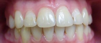
After a tooth is removed, a hole is formed in its place, which is gradually filled with blood (this is a protective function of the body). Over time, the blood coagulates and turns into a clot, which should completely cover the bone that has become unprotected after tooth extraction. Over the course of several months, the blood is absorbed and healthy bone tissue remains in its place. If the bone and gums sag a little at the site of the extracted tooth, this is normal. Most often, the healing process proceeds without complications, without causing any pain or discomfort to the patient. In some cases, fever, aching pain, prolonged healing, systematic visits to the dentist and other side effects are possible.
What do doctors recommend?
For several days, it is necessary to rinse the oral cavity with an antiseptic solution, which will prevent the entry of microbes from the external environment through the wound surfaces of the gums. The number and duration of rinses is determined by the attending physician. Rinse solutions should be no cooler than room temperature and no hotter than 37 degrees. The simplest and most inexpensive option is a solution of baking soda (1 teaspoon per glass of water). In addition, rinsing can be done using pharmaceutical products.
Rinses from the pharmacy
| Iodine-containing products | Iodinol, yox (spray) |
| Chlorine-containing | Chlorhexidine, miramistin, eludril, |
| Nitrofuran | Furacillin |
| Based on hexetidine | Stomatidin, hexoral |
| Vegetable | Chlorophyllipt, calendula tincture, |
| Antiseptic lozenges | Faringosept, septolete, grammidin |
| Means to accelerate the healing of mucous membranes | Rotocan, dental paste solcoseryl, stomatophyte, propolis spray, sea buckthorn oil. |
| Infection Control Products | Antibiotics, bacteriophages (staphylococcal bacteriophage, pyobacteriophage) |
Buildups and pins
Author: Simon Stone
Translation: Trichev Yuri
Buildups in vital teeth
This article consists of two parts: the first part examines buildups in vital teeth as a basis for overlay restorations. The second part examines the practical aspects of working with the increasing variety of restorative options for endodontically treated teeth, with an emphasis on the creation of buildups, the use of pins and inlays.
The Dictionary of Prosthetic Dentistry terms defines a buildup as “the center or basis of a structure”; the structure, of course, refers to the tooth being restored.
Thus, in vital teeth restored with indirect restorations, at least part of the buildup must be made up of the natural tooth structure. The other part of the buildup often contains a restorative material (eg, amalgam, composite, glass ionomers and their derivatives) to replace the missing part of the tooth. The purpose of creating a buildup is, firstly, to fill the space, especially in cases where it is necessary to cover undercuts (for example, with cavities of classes III and V). Secondly, in the absence of a large volume of tissue, direct restorative material can be used to give the buildup a shape similar to that of a prepared intact tooth. This thus provides additional support for the restoration. However, a chain is only as strong as its weakest link. In a buildup, not only the strength of the material is important, but also the connection (mechanical, chemical or both) between it and the tooth. Every dentist knows the unpleasant feeling that occurs when a patient returns with a build-up remaining in a fallen crown. We'll come back to choosing the right buildup materials and how they should be handled, but first we need to consider:
- The need to remove old restorations.
- Indications for tooth preservation.
- The choice between a buildup with or without restoration material.
The need to remove old restorations
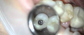
There is always a temptation to leave the existing restoration for a build-up (Fig. 1).
Figure 1 (a) Need for removal of old restorations. No sane dentist would use this amalgam filling as a build-up for an indirect restoration, but often seemingly sound restorations hide unwanted surprises.
Figure 1 (b) Pulp involvement is clearly visible here, but in some cases it is not so obvious.
However, if you are not sure about it, there are strong arguments for removing it:
- To check for caries underneath.
- To ensure that there are no marginal defects on the proximal surfaces.
- To determine whether there is enough tooth structure remaining to allow subsequent tooth preparation.
- To check the dentin for the presence of an open tooth cavity.
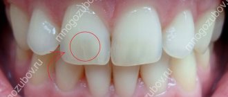
- To detect existing cracks in dentin and determine their extent (Fig. 2).
Figure 2 (a) Removal of the old restoration to determine the extent of the existing symptomatic crack at the distal edge of the tooth. The crack is located only within the dentin, but the cusps are significantly weakened. (b) A temporary restoration was made using amalgam to relieve symptoms - also
What to do after tooth extraction?
- According to the doctor's recommendation, after 15-20 minutes you need to spit out the gauze balls from your mouth. They are usually left if the bleeding from the socket has not completely stopped.
- You should not eat food in the next 2-3 hours. Food injures the hole.
- In addition, you should avoid eating too cold or hot food, steam rooms, saunas, and cold hardening. Sudden temperature changes provoke dilation and constriction of blood vessels, resulting in bleeding and pain. In the same way, alcohol can do this, which also needs to be excluded.
- You will temporarily have to give up aggressive tooth rinses, which can destroy the clot.
- You must try not to injure the blood clot in the socket to avoid inflammation. Most often this happens while brushing your teeth. Of course, it is necessary to brush your teeth after extraction, but only carefully and carefully.
- If your temperature rises after tooth extraction, you should immediately inform your doctor.
Tooth root removal
Tooth extraction can be of varying complexity. Sometimes this is a fairly simple process, but it can get complicated:
- after the start of the procedure for removing a heavily damaged tooth or a tooth with curved roots, a fracture occurs in the middle part of the root or its apex;
- It is not easy to remove the roots of a completely destroyed tooth, which are located deep in the alveolus.
Therefore, the removal method is selected individually, depending on the situation.
Removal with forceps
A part of the tooth protrudes above the edge of the hole from the outside and inside, which the dentist must grab with forceps. To do this, he carefully separates the gum from the root.
Sometimes the dentist has to peel off the mucous membrane and periosteum from the edge of the socket in order to properly grasp the tooth.
Even if, as a result of pathological changes, bone resorption has occurred, the cheeks of the forceps can be inserted deep enough to tightly grasp the root.
Upper jaw root removal
Depending on which tooth needs to be removed, the doctor uses special extraction forceps. For example, for large molars, bayonet-shaped forceps are used. Their cheeks may move deep under the gums. Incisors and canines are removed using an S-shaped instrument.
Basically, removal occurs using rocking movements. If the roots are located deep in the alveolus or are large, the dentist adds rotational movements.
If it is not possible to extract the root with forceps due to its deviation or the thick walls of the holes, then the doctor separates them with a bur.
If the root is deviated, the bottom of the tooth cavity is sawed where the buccal roots connect to the palatal roots. First, using a spherical bur, a hole is made in the interroot commissure, and then, using a fussur bur, the bottom of the tooth is sawed in the longitudinal direction. An elevator is inserted into the resulting cavity and the palatal root is dislocated. After this, it is removed with bayonet-shaped forceps.
Mandibular root removal
Typically, the roots of the lower jaw teeth are shorter and have thinner socket walls.
To remove the root of a lower jaw tooth, a dental surgeon mainly uses rib-curved forceps with thin and narrow cheeks.
To remove fangs, use forceps with wider cheeks. Sometimes difficulties arise when extracting the lower molars. Often it is not possible to insert the cheeks of the forceps deeply because they slip off. Since the alveolar process is quite thick at the edges of the socket, it is not possible to apply forceps to the cheeks. Therefore, when removing lower molars, an elevator is often used.
Tooth root removal using an elevator
If it is not possible to remove the roots of the teeth using forceps, an elevator is used. This situation may arise if they are deep in the hole. Using forceps can damage adjacent bone tissue and mucous membrane. Using an elevator is less traumatic.
Removal by direct elevator
It is used to remove the upper teeth and roots of teeth that are located outside the dentition, and sometimes when extracting the lower third molar.
The elevator is inserted between the wall of the hole and the root that needs to be removed. To do this, the convex part of the cheek is located towards the wall of the hole, and the concave part is facing the root. The dental surgeon presses on the handle and rotates it around the longitudinal axis in both directions. Periodontal fibers break and move to the opposite wall of the socket. After the cheek of the elevator enters approximately four millimeters, the tool rests on the edge of the hole and acts as a lever. The doctor’s force is transferred to the end of the elevator, and the root is squeezed out of the alveoli.
When removing the roots of molars that are welded together, they are first separated using a fissure bur and then removed one by one using bayonet-shaped forceps or an elevator.
Removal by corner elevator
Angled elevators are used to extract the roots of lower teeth, mainly molars. When removing them, hold the tool with your whole hand. In this case, the handle of the instrument should be on the cheek side.
The cheek of the corner elevator is inserted to the tooth to be removed with its concave part into the gap between the wall of the socket and the root or between the adjacent tooth and the root. In order for the cheek to move deeper, the thumb of the left hand presses on the intermediate part of the instrument at the point where it passes into the working part. At the same time, the elevator handle is moved forward and backward alternately by twenty degrees. After inserting approximately 0.6 centimeters into the periodontal fissure, rotational movements are made with the handle and the tooth is pulled out of the socket.
Treatment of the tooth socket
If the removal took place against the background of purulent inflammation, then treatment of the tooth socket is necessary. The hole must be washed with an antiseptic solution, and then an anti-inflammatory medicine is placed in it. It should be placed in the hole even if there is no inflammation. This is done for preventive purposes, so that after removal the wounds heal without inflammatory processes.
Stitching
To bring the edges of the wound closer together, they are sutured with special suture material.
After detachment with a trowel, the gums loosen a little, and a blood clot may fall out of the socket. To prevent this from happening, the wound is sutured. In addition, this will allow it to heal faster.
Thanks to the sutures, the size of the wound is reduced, and this reduces the possibility of inflammation developing in the hole.
Sutures are also used to stop bleeding. They help prevent bleeding that may occur some time after tooth extraction.
The stitches are removed after about five to ten days. If catgut was used as a suture material, then there is no need to remove the sutures, since catgut is absorbed.
After the extraction, the dental surgeon ends his appointment with care tips and, if necessary, prescription of medications.
The article used materials from the site: https://stoma.guru/lechenie/udalenie/osobennosti-udaleniya-kornya-zuba-kak-prohodit-operaciya.html#hcq=FmBUKCq
How to stop bleeding?
In some cases, if it is not possible to see a doctor, you can stop the bleeding yourself.
- A tight tampon twisted from cotton wool or gauze should be placed on the bleeding hole. Then squeeze your jaws tightly and do not loosen them for 15-20 minutes. If after such a procedure the tampon is not completely saturated with blood, then the bleeding has been stopped. A blood clot has formed at the site of the bleeding, which cannot be disturbed for at least a few hours.
- In case of a fiasco with the previous method, the procedure must be repeated again. Only the tampon now needs to be soaked in a 3% hydrogen peroxide solution.
- If the bleeding is too severe, then you should consult a doctor. At the dentist's appointment, electrocoagulation may be performed or stitches may be placed.
- If after all these manipulations the bleeding does not stop, you need to use medications that increase blood clotting. Prolonged bleeding (several days) entails urgent hospitalization.
How to relieve pain after tooth extraction?
When the effect of the anesthetic begins to wear off, the pain will return. In case of severe pain or a low pain threshold in the patient, it is necessary to take painkillers.
As a rule, non-steroidal anti-inflammatory drugs (diclofenac, voltaren, indamethacin, nimesulide, ibuprofen) are taken, which will also help relieve swelling and inflammation. It is worth remembering that long-term use of these drugs is contraindicated due to a decrease in the number of leukocytes in the blood and complications from the stomach. Most drugs in this group should be taken with omeprazole.
In addition, you can resort to using a topical drug in solution or lozenges: benzydamine hydrochloride (or tantum verde).
Emergency situations after tooth extraction
- If your cheek is swollen. The infection entered the soft tissue and caused periostitis, which led to swelling of the soft tissue. A doctor's examination is required.
- If your gums hurt. In any case, there will be swelling, but if it bothers the patient, it is necessary to visit a doctor.
- If bleeding resumes. When it continues for a long time, it can cause anemia. At the appointment, the doctor may cauterize the bleeding vessel, administer vitamin K preparations, or use a hemostatic sponge.
- If the stitches have clearly fallen apart. No one is immune from such a problem as cutting stitches, because putting them in the mouth is extremely inconvenient. Again, you need to urgently visit a doctor.
What and when can you do after tooth extraction?
Comprehensive hygiene for 2500 rubles! Air Flow 1000p for patients with braces!
A unique promotion for our regular patients! A set of hygienic procedures (removal of tartar and plaque using Air Flow and ultrasound , scaling, polishing, fluoridation) for regular patients once every 6 months for RUB 3,000!
Air Flow and polishing for only RUR 1,500 for patients with braces!
Oda Ferrul. When is it not worth saving your natural teeth?
This difficult topic came to light after the second interview with Ekaterina Kolb “Endodontic treatment: “complicated, expensive - why”?” After reading this article, the chief physician of the AldisDent clinic, Vyacheslav Bitno, made the following comment: “Sometimes people come to me with high-quality endodontics, and I upset them - it is better to remove the tooth and install an implant.”
The remark was worth considering this issue in more detail: money needs to be spent efficiently, and you wouldn’t want to suffer in vain! Therefore, we continue the conversation on the topic of preserving severely damaged teeth: when it is necessary to fight for preservation, and when it is useless.
Vyacheslav Leonidovich, please clarify your comment. Ekaterina so thoroughly and convincingly explained why a person needs to fight to preserve teeth, but it turns out that all this may be in vain?
My position has no contradictions with what Ekaterina Kolb said - high-quality endodontics helps preserve teeth, and rightly so. What I’m saying is that after endodontic treatment, proper restoration is needed, otherwise the efforts of the endodontist will be wasted.
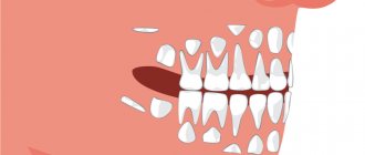
When a significant part of the tooth crown is lost, the remaining volume of hard tissues of the native tooth (ferrules) may not be enough to evenly distribute the load on the root during chewing, and it will simply collapse from overload.
I’ll explain it more clearly (I’ll even draw pictures).
In a healthy tooth, the tooth enamel takes the load and distributes it evenly to the root.
But then the tooth got sick, caries “ate” part of the dentin, maybe the tooth also chipped, the infection reached the pulp - I had to go to the endodontist to fill the canals. Next, if there is a small amount of dentin loss, the therapist makes a restoration with filling material or, if most of the tooth is lost, sends the patient to a prosthodontist for a crown.
How do further events most often develop?
An ordinary orthopedist looks: you can put a metal tab here and a crown on it. I decided - I did it, and provoked the following scenario: a person, while chewing, presses on this tab, which puts a load on the root, and the root breaks. Why? Because the load should be evenly distributed on the root, and not into the root. In a healthy tooth, this uniform load is ensured by the enamel. The tab puts the load directly into the center of the root, which is not functionally designed to withstand it.
What if the tooth is not restored with a metal pin, but rather with filling material?
A filling does not replace tooth enamel. It, like the pin, exerts concentrated pressure inside the root, as well as from the inside on the walls of the tooth, which leads to chipping and even greater reduction of the enamel.
Nature has provided reserves of health. A tooth can live without a nerve, as if without a heart, with sealed canals, but at the same time it cannot withstand the load directed from inside the tooth.
And what, nothing can be done to preserve the native roots so that they can also be chewed on?
To answer this question, let's go back to the beginning of the conversation. Do you need to fight to save your natural teeth? Of course it is necessary. Under what circumstances is preservation possible? There are two main conditions:
- No infection in the root canals (that is, high-quality endodontics).
- Presence of ferrules.
Even some doctors don’t know what ferrule is. A ferrule is a part of a tooth that can be subjected to external loads and through which the load is distributed to the root. The artificial crown must fit correctly on the tooth and distribute the load on the root, and for this you need to wisely use the residual ferrule. If this point is not understood, the doctor, when treating teeth for crowns, can further reduce the ferrule.
Ferrul is like a “formula of happiness”, a formula for a good prognosis. The more ferules, the better the treatment result. Hence the conclusion: if the doctor wants to make a ledge, it must be made in such a way as to preserve the ferule to the maximum.
And where it is impossible to saw, it is necessary to use the “vertiprep” technology - when the tooth is processed for a crown without a ledge, preserving this same ferrule as much as possible.
And if this ferrule is hopelessly small, does it mean removal and implantation?
There are two ways in which ferulus can be added. The first is orthodontic: we slightly pull the tooth out of the bone, lifting it from above. The root becomes smaller, but a ferrule appears on which you can place a crown and distribute the load. The second way is surgical: you can cut off part of the bone around the tooth and make a lower crown.
But this seems even more painful, and success is “fifty-fifty.”
Yes, and therefore a correct assessment of the situation and prospects is very important. But I want to emphasize: in many cases such radical measures are not required if what is left is used correctly.
How does a doctor determine “enough or not enough” - “by eye”?
Correct question! And very difficult. What height should the ferrules be: a millimeter, two, three? How much height of a tooth is needed to place a proper, load-distributing crown on top of it?
Here we again come up with the topic of bite, which has already been talked about a lot and, believe me, we talked only superficially. What determines the load that the patient will create on the tooth, and how to measure it? Again we need to talk about the bite, “but that’s a completely different story.”
To be continued…