Treatment of fibroids on the gum, lip and other soft tissues of the oral cavity, as a rule, involves surgical intervention - removal with a laser or radio wave method. However, in some cases it is possible to do without radical therapy, it depends on the causes of the formation and the ability to quickly eliminate provoking factors.
To make an accurate diagnosis, it is important to undergo a dental examination, consult with a doctor and, if necessary, take a photo of the pathological area. Timely visit to the clinic minimizes the likelihood of complications. In addition, the patient should adhere to preventive measures to prevent the disease and its consequences.
What is fibroma
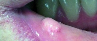
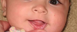
This is a benign neoplasm in the form of a small node on a stalk or broad base, which consists of fragments of connective tissue. In most cases, it is localized on the mucous membranes of the gums, lips, palate, on the inside of the cheeks, and a little less often on the tongue. The pathological process is more often encountered by primary schoolchildren and adolescents aged 6-15 years.
The patient is not in pain; at a very early stage he does not notice the pathology. As the thickening grows, it is easily felt and accidentally injured.
Can fibroids in the mouth resolve on their own, without medical intervention? In most cases, surgical excision of the tumor is required. However, do not worry about the complexity and duration of the operation. In less than an hour, an experienced doctor will eliminate the defect without subsequent complications and a long rehabilitation period. Currently, clinics use laser and radio wave techniques.
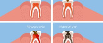
Despite the relative harmlessness of fibrous growths, they still need to be treated even in the absence of discomfort. If the seals increase in size and are constantly exposed to traumatic effects, it is highly likely that an infection will enter the wound. In this case, treatment will be very long, difficult and will not always lead to positive dynamics. In addition, the lack of therapy sometimes leads to the degeneration of a neoplasm into a malignant one.
Reasons for education
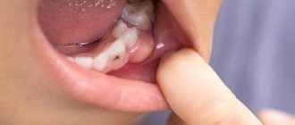
After conducting numerous studies, scientists have not been able to determine an exhaustive list of factors that provoke the development of pathology. However, some conditions and phenomena still occur most often when diagnosing this disease. For example, this is possible with a hereditary predisposition to fibroids on the gums (shown in the photo below), tongue, inside the cheeks and other areas of the mucous membranes. In this case, the disease develops in childhood in young patients from 1 to 10 years.
Often, in the presence of this diagnosis, a history of regular traumatic exposure is established. There may be constant biting of a certain area of soft tissue. They can also be injured by sharp edges of crowns, orthodontic structures, dentures, especially poorly fixed ones, and hard food.
Taking certain medications can also be a provoking factor. Some medications can cause the appearance of benign lumps in representatives of different age categories, not only in children and adolescents. Drugs that may cause fibromatosis after use:
- Cyclosporine. It is indicated to prevent organ rejection during transplantation.
- Valproate, used by epileptics.
- Verapamil and other calcium channel blockers.
- Estrogens of synthetic origin in oral contraceptives or other hormonal pills.
The culprits in the development of the disease may be inflammatory processes in the oral cavity. These include stomatitis, glossitis, periodontal disease, gingivitis, etc.
It is impossible to exclude a hereditary factor, but the risk of formations can be significantly reduced if you follow simple preventive rules:
- do not forget about hygiene;
- Carry out regular professional cleaning to remove thick plaque and tartar;
- protect soft tissues from traumatic effects (solid food, biting), and also avoid sudden temperature changes;
- treat dental diseases in a timely manner;
- consult with doctors before starting to take medications and avoid self-medication;
- visit the dentist for preventive maintenance at least once every six months.
Classification of fibroids
Pathology is divided into several types according to criteria. This includes the density of the benign neoplasm, the nature of its origin, as well as the severity of clinical manifestations.
Dense fibroma
Characterized by a solid consistency. This is due to the fact that the contents are quite hard fibers of connective tissue. They include a small number of cores. Formations of this type are most often found on the gingival surfaces and palate.
Soft fibroma
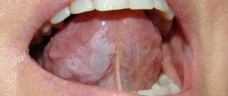
The fibrous structures are thin and freely located, so their clusters are characterized by a high degree of softness. This type is most often observed on the tongue and the inside of both cheeks. Benign neoplasms of a mixed type, combining the signs of all the varieties listed above, can be found on the sublingual part and on the mucous membranes of the floor of the oral cavity. For example it could be:
- Fibrolipoma. It is hard to the touch because it contains fibrous fibers. It can be eliminated surgically, with laser, or radio waves.
- Fibrohemangioma. As a rule, it is provoked by infectious processes occurring in the child’s body. It is extremely rare in adults. It never degenerates into malignant tumors. Most often it is treated surgically, in some cases it can resolve on its own.
Fibroma from irritation
This is not a tumor formation in its usual form, but the result of reactive hyperplasia. Chronic pathology is characterized by the development of focal lesions, which are caused by systematic mechanical action and subsequent injury.
The most common cause is installed crowns, fillings, and dentures. In the latter case, the disease is called prosthetic granuloma. The orthopedic structure exerts continuous pressure on the alveolar process, leads to its resorption, moves forward and contributes to the formation of compactions, which are associated with the inflammatory process.
The risk group includes not only patients who have undergone prosthetics, but also people with untimely cured caries, in adulthood, with foreign objects in the oral cavity (for example, with piercings). Studies have shown that women are more prone to such hypertrophic transformations than men.
Symmetrical fibromas
Doctors diagnose such tumors in the area of the third molars in the areas between the gums and the roof of the mouth. The tumor is hard to the touch and resembles a bean in shape.
It should be noted that this type of compaction does not apply to true fibromatosis. These are just overgrown tissues in the gingival membranes, accompanied by the process of scarring and other changes of a similar nature.
Lobular fibroma
It occurs as a result of reactive hyperplasia with systematic trauma to delicate sensory fibers with prostheses or other orthopedic structures. The main distinguishing feature of such formations is their rough, textured surface. When palpated, tubercles are felt.
Fibrous epulis
This is a dense growth of pinkish tissue that does not cause pain or other discomfort. The edges are often hyperemic, have clear boundaries and irregular shape. The base is quite wide.
The vestibular part of the gums is usually affected. There are cases of neoplasms occurring in the interdental spaces in the form of a saddle with spread to the intraoral surface.
Quite often, a dental unit located in a pathological area has a poorly fitted metal crown, extensive carious lesions, or a prosthetic clasp. It is these structures that are the provoking factor in the occurrence of a chronic inflammatory process with the formation of granules, which over time are transformed into mature connective fibrous fibers.
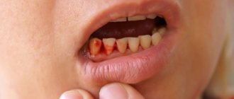
In dentistry, there are also angiomatous epulis. They are brighter in color, somewhat softer to the touch and bleed. In this case, blood appears not only at those moments when the surface is affected mechanically, but regardless of the presence or absence of a traumatic factor. When conducting diagnostic studies, many vascular branches are detected in the pathological area.
Symptoms
The tumor grows and develops quite slowly, so for a long time the patient may not even be aware of its presence in the mouth. Fibroma of the oral mucosa looks like a hemispherical growth rising above the plane, covered with pinkish tissue. If you press it, pain or other discomfort does not appear. The surface is smooth, there are no irregularities or roughness on it.
The appearance of ulcers with such a diagnosis is very rare. In such cases, an infection is usually associated with the subsequent development of the inflammatory process. Swelling, redness, erosion occur, and pain is felt. The pain persists even if you do not touch the pathological area.
If you do not injure the formation, it may not change its size for quite a long time and remain in a stable state. If it is exposed to constant traumatic effects, there is a high risk of malignant degeneration, which is dangerous to the life and health of the patient.
What does a wen look like on the lip?
White wen on the lips looks unsightly:
- A white hemispherical formation appears on the red border of the lips under thin skin.
- Sometimes a lipoma is located on the edge of the lips or under the nose, on the chin.
- Multiple appearances of small lipomas are possible. These are dots on the lips under the skin, scattered throughout the surface of the lips.
- Lipomas often form on the lips outside the red border, in the area around the mouth, most often on the upper lip, under the nose.
In any case, the owners experience serious psychological discomfort.
Diagnosis of fibroids of the upper and lower jaw
The doctor will not prescribe treatment until he is sure that the diagnosis is correct. To do this, diagnostic procedures are carried out, the results of which will confirm or refute the fears of doctors.
First, the patient is asked to describe the symptoms. The dentist examines and palpates the tumor. However, this is not enough to develop therapeutic tactics, since it is extremely important to determine the depth of tumor growth into soft tissue. For this purpose, an ultrasound examination is performed.
In difficult cases (ulcers, development of an inflammatory process in a pathological area of the gum, etc.), a biopsy is indicated. After surgical removal of the tumor, fragments are necessarily sent for histological analysis.
The examination is necessary not only to establish a diagnosis, but also to identify the factors that provoked the disease. A full dental examination is carried out to confirm the presence of inflammation. In addition, one cannot do without radiography, orthopantomogram and other images in different projections.
If a person has dentures, a consultation with an orthopedic dentist may be necessary. This is necessary to eliminate the possible traumatic effects of artificial elements on the mucous membranes.
Differential diagnosis
Biopsy remains one of the most informative methods for distinguishing fibroids from other benign neoplasms. The study is indicated for suspected papilloma, lipoma, epulis of various structures, neurofibroma, cyst, squamous cell carcinoma, wart, etc.
If the growth is localized on the tongue or sublingual part, it is extremely important to differentiate it from all existing seals. Timely diagnostic measures make it possible to detect cancer at the earliest stages and provide high-quality therapy with a short recovery period.
Preventive measures
If you have not had and do not have wen, then you are unlikely to think about their prevention. Once the problem has appeared, there is no need to talk about prevention. After lipoma removal, your doctor will advise you:
- give up bad habits - smoking, drinking alcohol;
- improve the functioning of the gastrointestinal tract, eliminate constipation and diarrhea;
- undergo an examination by an endocrinologist to check the body’s hormonal levels; correction may be necessary;
- give up unhealthy foods and switch to a rational, balanced diet.
Experience shows that simple preventive measures and a healthy approach to organizing your life can prevent many problems.
Treatment of oral fibroids
Surgery remains the most effective and most common therapeutic method. The seal is excised using a laser or radio waves. This procedure lasts about half an hour. If the tumor is very large, after its removal the wound is covered with a flap, which is formed by the doctor from the surrounding tissue.
When pathology is caused by taking certain medications, they should be completely eliminated and replaced with alternatives with similar properties. After discontinuation of drugs in such cases, the appearance of the mucous membranes is often restored without outside help, and the likelihood of relapse approaches zero. However, this does not apply to situations where the disease is advanced.
Surgery can also be avoided in case of traumatic effects of orthopedic structures. For example, when a crown, filling, or prosthesis puts pressure on the tissue. Elimination of the provoking factor often leads to a decrease or complete disappearance of a benign formation. Most likely, it will be necessary to dismantle the old structures and replace them with new ones.
On the Internet you can find stories of healing using home remedies. It is worth remembering that the disease cannot be treated with the help of folk recipes. Herbal decoctions and infusions, and other compositions are used only as an auxiliary element of complex therapy.
Types of lipoma removal
- classical surgical removal of a lipoma - excision with a scalpel, performed under local anesthesia (if the formation is small) or using general anesthesia;
- Laser removal of lipoma is the most effective alternative to classic surgical removal; it does not leave scars or scars, therefore it is most often prescribed when it is necessary to excise a formation on the face and neck;
- endoscopic removal of a lipoma - the incision is made at some distance from the tumor (in areas hidden from view, in natural folds of the skin), and not on the formation itself; through the access obtained, an endoscope is introduced into the removal area, broadcasting the progress of the operation on the screen, and surgical instruments;
- radio wave removal of lipoma is a method based on the use of a “radio knife” that affects the tumor with intense electromagnetic radiation;
- liposuction (puncture-aspiration method of lipoma removal) - a needle-cannula is inserted into the tumor, through which, using a special apparatus, the entire contents of the tumor are drawn out; The disadvantage of this method is the possibility of relapse.
View price
Let's sum it up
Mucous fibroids on the inside of the cheeks, gums, lips, and tongue grow very slowly and do not cause severe discomfort. They do not pose a danger to the patient, but the problem must be eliminated as early as possible. The fact is that a benign seal in the oral cavity with regular trauma can degenerate into a malignant one, and the oncological process requires serious therapy and is extremely dangerous to life and health.
It is impossible to get rid of the disease using traditional methods. It is important to undergo high-quality dental diagnostics and consult with specialists. In most cases, surgical excision is indicated, but with small growths, the absence of severe symptoms and the elimination of provoking factors, there is a possibility of rapid tissue restoration without external influence.
There is no need to worry if the doctor has decided to undergo surgery. Modern clinics practice laser or radio wave removal on an outpatient basis. These are the most gentle and highly effective methods. An extremely important point in a positive prognosis for the treatment of dental fibroids is an early visit to a doctor and proper excision by an experienced physician.
Is it possible to remove a wen yourself?
Removing a wen yourself is deliberately engaging in self-harm.
It’s better to consult a doctor, don’t skimp on your health.
This cannot be done categorically, and here’s why:
- There is a high risk of infection, then the whole procedure will end in an abscess.
- With your inept intervention, you can cause the degeneration of an ordinary lipoma into liposarcoma - a malignant neoplasm.
- By opening the wen and squeezing out the contents, you leave the capsule under the skin. Relapse is guaranteed.