FREE CONSULTATIONS!
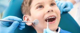
We are pleased to announce the start of free admission at the Konstanta Clinic for patients under 18 years of age by maxillofacial surgeon, pediatric surgeon L.A. Eremeyshvili. for questions:
- Congenital facial pathologies: cleft lip and palate;
- Benign neoplasms of the face and oral cavity;
- Diseases of the temporomandibular joints;
- Pathology of the frenulum of the oral cavity: lips and tongue.
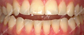
Correction (plasty) of tongue frenulum is provided free of charge for children under 1 year of age.
*Valid for residents of Yaroslavl and the Yaroslavl region.
You can make an appointment by phone or through the website (4852) 37-00-85 Daily from 8:00 to 20:00
Sign up for a consultation
Congenital cleft palate or cleft palate is a fairly common malformation of the facial region. This pathology is ranked 4th among all developmental defects, and it is diagnosed with a frequency of 1:700 births. Often, a cleft palate is not an independent deviation, but part of a hereditary syndrome.
Material and methods
Believing that the method of completing the operation should be determined depending on the extent of the pathological process, as well as on the individual anatomical features of the sacrococcygeal region, we proposed a classification of fistulous forms of the coccygeal tract according to the degree of complexity (see table). This classification facilitates a differentiated choice of the optimal method of surgical intervention and a comparable comparative analysis of treatment results. In the proposed classification, in addition to assessing the spread of the pathological process, the presence of unfavorable anatomical features of the sacrococcygeal region in the form of the so-called “high” buttocks was taken into account, the criterion of which was the depth of the intergluteal fold of more than 3 cm above the lowest located primary fistula opening with an angle of the intergluteal recess of less than 30 °.
Classification of fistula forms of the epithelial coccygeal tract by degree *The depth of the intergluteal fold is more than 3 cm above the lowest located primary fistula opening with an angle of the intergluteal recess of less than 30°.
This study included the results of treatment of 41 patients with a fistula form of the epithelial coccygeal tract of the III degree, which corresponded to the widespread prevalence of fistulas with external fistula openings outside the internal clivus of the buttocks, the presence of pronounced scars and infiltrates in the circumference of the fistulous tracts. There were 37 men, 4 women. The average age of the patients was 35.6±6.4 years. In 31 (75.6%) patients the disease was regarded as a relapse. Previously, they had made unsuccessful attempts to excise the coccygeal tract. The average duration of the disease was 7.4±2.1 years. The average body mass index of patients was 27.9±3.5 kg/m2. There were no severe concomitant diseases.
All operations were performed as planned in the absence of acute suppurative processes in the area of surgical intervention in the patient's Depage position under spinal anesthesia. After radical excision in one block of all fistula tracts, their external openings, inflammatory infiltrates, cavities and scar tissue, the wound was given a diamond shape (Fig. 1, a). At the same time, we tried to follow the classical recommendations of the ideal ratio of the angles of the rhomboid defect of 60 and 120°, although there were individual deviations depending on the prevalence of the process in the patient.
Rice. 1. Scheme of moving a diamond-shaped skin flap according to Limberg. Explanations in the text.
To replace the wound defect, a diamond-shaped flap was cut out in the adjacent gluteal region (see Fig. 1, a). According to the direction of the transverse axis of the diamond-shaped defect BD, a cut was made along the line DE, from which a cut was made parallel to the face DC along the line EF. The dimensions of the flap corresponded to the wound defect being replaced. This was easily determined by the correspondence of the axial dimensions, as well as the dimensions of the sides, i.e., the lengths of the cuts AB, AD, CD, BD, DF, FE were the same. The flap included the entire thickness of the subcutaneous fat separated from the underlying gluteal fascia. The degree of mobilization of the flap was determined by its free movement onto the wound defect without tension. After moving the flap, its edges were fixed to the edges of the wound defect with double-row sutures (see Fig. 1, b). The maximum tension of the sutures occurred at points F and D, which made it possible to eliminate tissue tension at the peripheral edges of the fixed flap.
Unlike smooth, flat areas of the skin surface, where this method of skin grafting can be easily performed, the sacrococcygeal region has a complex three-dimensional structure, which can directly affect the results of treatment.
Thus, the weak point of the classical Limberg plastic surgery when excision of the coccygeal tract is the location of the lowest part of the rhomboid defect in the depth of the intergluteal fold, which creates the precondition for complicated wound healing, especially in unfavorable anatomical conditions with “high” buttocks, practically closing with each other. It is in this place that a relapse usually occurs. In addition, if the use of the Limberg method on flat areas of the body surface does not require additional fixation of the flap to the underlying tissues, then the complexity of the three-dimensional geometry of the intergluteal region can cause incomplete adherence of the flap to the wound defect and the accumulation of exudate in the subflap space, which impairs the healing of the displaced tissues.
Taking this into account, we use a modified version of Limberg's plasty, the difference of which is the asymmetrical displacement of the lowest part of the excised rhomboid defect from the midline (Fig. 2). As a rule, the displacement was 2 cm. As a result, after moving the flap, the “critical point” of the wound was in the best conditions for healing.
Rice. 2. Scheme of modified Limberg plastic surgery.
The second feature of the modification is suturing the flap behind the subcutaneous tissue to the deepest part of the wound defect, namely to the bottom of the intergluteal fold (especially in its caudal part), for good fixation and to eliminate the possibility of accumulation of wound secretion in this area, which worsens healing conditions. This technique also made it possible to “straighten” the intergluteal angle, reducing the intergluteal depression, thereby leveling the basis for relapses of the disease. Peripheral zones of the subflap space with large defect sizes were drained, drainage tubes were connected to a device for constant evacuation of wound fluid.
If the child has a pathology
A diagnosis of “cleft palate” in a child is not a death sentence, and parents need to remember that the pathology responds well to treatment if it is started early. During the treatment period, the main role is given not only to surgical intervention, but also to the patience of parents, their responsibility and willingness to deal with the health of their own child. You must be prepared to carefully implement all recommendations.
Once a diagnosis of cleft palate is made, the child is taken to a surgeon who can create a treatment plan.
In our Clinic, such defects are dealt with by Sergey Nikolaevich Bessonov and Levan Avtandilovich Eremeyshvili, who are maxillofacial surgeons. Appointment with doctors S.N. Bessonov and L.A. Eremeyshvili takes place over the phone.
Thanks to the help of a charitable foundation, surgery to correct a cleft palate can be completely free. All you need to do is prepare the documents from the list and wait for an individual call to a specialist.
Introduction
Epithelial coccygeal tract is a fairly common congenital defect in the development of soft tissues of the sacrococcygeal region, occurring mainly in young people. This nosological form is benign and very rarely leads to serious complications, but it significantly worsens the quality of life and leads to disability in patients of the most active age group [1, 2, 7]. The results of surgical treatment remain not entirely satisfactory, which is due to the relatively large number of postoperative wound complications and frequent relapses of the disease [5, 6].
Many methods have been proposed for the treatment of the coccygeal tract, but none of them has found universal acceptance. The main controversy is caused by the choice of method for completing the surgical intervention, ranging from open wound management after excision of the fistula tracts to the use of complex plastic surgeries to close the wound defect.
The purpose of the work is to analyze the results of using the skin-plastic method of closing a wound defect after tissue excision in common fistulous forms of the coccygeal tract according to Limberg with some modifications.
Violations associated with vice
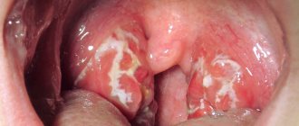
A cleft of the soft and hard palate leads to the appearance of a communication between the oral and nasal cavities. A cleft of the soft palate leads to improper attachment of muscle structures and deformities of the pharynx in the middle section. The soft palate is also significantly shortened.
A cleft palate leads to the fact that the child cannot normally perform sucking and swallowing movements, his breathing processes are disrupted, and his bite is formed incorrectly. With age, the deformation intensifies, and problems with speech appear.
Cleft palate
A comment
The article is undoubtedly of scientific interest and is devoted to a topic that is relevant for coloproctologists and surgeons - surgical treatment of complex recurrent forms of the epithelial coccygeal tract.
The author used a modified method of plasty of a wound defect with a diamond-shaped skin-subcutaneous flap according to Limberg. The described technique is quite popular abroad, but is extremely rarely used by domestic surgeons, which further increases interest in the results obtained in the study. To select patients, the author developed and used a special classification of fistulous forms of the coccygeal tract, with the help of which it is quite clearly possible to identify a group of patients for whom surgery using Limberg plastic surgery is indicated. This is undoubtedly important, since the rhomboid flap method should be used only according to strict indications, in the presence of a widespread scar-inflammatory process in the sacrococcygeal region. As a result of the work, the author obtained impressive results - recovery of patients in 97.3% of cases. For practical purposes, the article provides a number of recommendations aimed at reducing the incidence of complications and relapse of the disease, which significantly increases the practical significance of the work. Thus, the article is of scientific and practical interest and shows the possibilities of plastic closure of extensive wound defects after excision of complex recurrent forms of the epithelial coccygeal tract.
Prof.
Yu.A. Shelygin
Possible complications during treatment
Possible complications:
1. The danger of thrombosis is greatest within 20 minutes after restoration of blood flow through the anastomosed vessels. This time should be waited out, observing the pulsation of the stitched arteries.
2. A slow capillary reaction of the tissues of the transplanted flap indicates inadequate blood flow; its cyanosis indicates difficulty in venous outflow.
3. If the anastomosis is performed incorrectly, microvascular thrombosis cannot be prevented in any way, including the use of medications.
4. Vascular sutures, especially on vessels less than 5 mm in diameter, should not be attempted by a surgeon who has not been trained in microsurgery.
Prognosis after treatment method
Postoperative monitoring should be as close as operative monitoring. It is necessary to maintain body temperature and fluid balance. Pain relief is extremely important. The patient should not experience anxiety or malaise. A high level of observation and care for the patient is required, which is best achieved in the intensive care unit, where the patient should remain for the first 24-48 hours. In addition to monitoring the patient, it is necessary to carefully monitor the condition of the autograft. The surgeon or nurse on duty must constantly monitor the presence of a capillary reaction. In specialized microsurgery departments, monitoring of the patient and the condition of the autograft is carried out by a team on duty. If thrombosis is suspected, an urgent operation is required - thrombectomy. Complications that arise must be corrected as soon as possible, while the viability of the flap is still preserved. If everything goes smoothly during the flap transplant operation after removing the vascular clamps, the likelihood of complications is low. It increases in cases where there were difficulties in applying vascular anastomoses during surgery. A decrease in flap temperature compared to the patient's body temperature indicates arterial or venous insufficiency, or both complications. Blueness of the skin of the flap and accelerated capillary reaction indicate inadequacy of venous outflow, and vice versa: pallor, slower capillary reaction indicate arterial insufficiency. It is incorrect to assume that the success of a flap transplant depends only on the personal skill of the surgeon and on how skillfully he performed the anastomoses. Success, like failure, depends on the choice of the patient, the skill of the surgeons, anesthesiologist, and the staff of the operating room and recovery rooms.
Briefly about the treatment method
Elimination of skin defects using nearby tissues is called local plastic surgery. Local plastic surgery is used, in particular, in the presence of fresh injuries, cicatricial deformities after traumatic injuries, congenital defects, and also defects after removal of tumors in the skin and subcutaneous tissue. As defined by A.A. Limberg (1963), local plastic surgery is the main method of treating scar deformities or defects, as well as an additional method of plastic surgery after tissue transplantation from distant areas of the body.