From this article you will learn:
- how is tartar formed?
- Is it possible to remove tartar at home,
- which will help reduce the amount of dental plaque.
The article was written by a dentist with more than 19 years of experience.
The ability to remove plaque at home will depend on its type. For example, you can use special products, which we will discuss below, to remove not too massive pigment plaque that occurs in smokers and lovers of too strong tea or coffee. As for removing plaque, using a regular toothbrush and toothpaste only removes soft microbial plaque well. Removing partially mineralized plaque, and especially tartar, requires the use of special means.
With irregular oral hygiene, soft microbial plaque that is not removed from the teeth in time begins to gradually mineralize (absorbing calcium salts from saliva), turning into hard tartar. It will no longer be possible to remove tartar at home if it is completely mineralized or large in size. But if you have a smoker’s pigment plaque, or the microbial plaque is only partially mineralized and therefore has a loose consistency, then for these cases there is one fairly effective method.
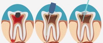
Hard dental plaque: photo
Such dental deposits (tartar) as in Fig. 1-3 can only be removed at the dentist by signing up for an ultrasonic teeth cleaning procedure. Only partially mineralized stone, which is small in size and has a loose consistency, can be removed at home - and all this applies only to supragingival dental plaque. Subgingival tartar, which is present in almost all patients with gum disease, can only be removed by a dentist.
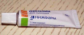
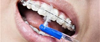
So what can replace a visit to the dentist if you have smoker plaque or a small amount of tartar. A combination of an Oral-b electric brush and a special teeth polisher will help us with this. Let us remind you that Oral-b toothbrushes are made on the principle of a dentist’s instrument, which the doctor uses to remove plaque and the remains of hard dental deposits. You will probably remember how, after the ultrasonic cleaning procedure, the doctor polished your teeth with a rotating attachment with bristles and polishing paste. And we will do the same.
So, for this we need:
- electric brush from Oral-b,
- special toothpaste.
What kind of remedy is this?
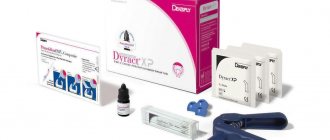
If ultrasound or mechanical teeth cleaning are contraindicated, a chemical method is used . Modern means of chemical treatment include Scaling gel. It is an alternative method if it is not possible to use medical instruments to remove hard deposits from teeth.
The drug is used to clean teeth of unnecessary stones in adults and to dissolve greenish plaque in children.
The effect of this dental gel is based on the fact that it is able to soften the hard tissue of dental plaque , which subsequently dissolves. The tooth enamel remains intact.
The drug consists of special particles that, when applied to teeth, stain damaged tissue. This makes diagnosis easier.
The dental product, in addition to all of the above, has good whitening properties. This effect can persist in patients with constant oral hygiene for about three months. The drug is very easy to use and has a very affordable price.
The drug contains the following components:
- silica;
- distilled water;
- iodine-containing substance “potassium iodide”;
- hydrochloric acid;
- polyhydric alcohols;
- special thickener;
Professional tartar removal
The most modern, effective and gentle way to remove tartar is ultrasonic cleaning. It is carried out in dental clinics. Hygienists or periodontists specialize in such procedures.
Before performing ultrasonic dental tartar removal, the doctor, guided by the main medical principle “Do no harm!”, assesses the condition of the patient’s oral cavity. Determines whether there are any contraindications. Selects a suitable nozzle for the ultrasonic cleaning device.
During ultrasonic exposure, the structure of the mineral plaque is destroyed. And in most cases this happens from within. Tartar simply falls off the surface of the enamel, falls out of periodontal pockets and interdental spaces.
After this, the enamel is polished. This is necessary in order to prevent the appearance of plaque for a long time. The result is not only noticeable - it is literally tangible. You can run your tongue over the surface of the enamel - it will be clean, smooth and tactilely pleasant.
If tartar has penetrated deep into periodontal pockets or has damaged the integrity of the enamel, causing increased sensitivity, the dentist can also use local anesthesia. This is necessary to make the procedure comfortable and painless for the patient.
Indications for use
The properties that this gel has determine its use for the purpose of cleansing hard plaque on teeth, even in the complex therapy of periodontal disease in adults.
Teeth affected by periodontal disease are most often mobile, so it is quite difficult to remove hard deposits from them. There is a risk of damage to the teeth themselves.
Products such as abrasive paste are not suitable, which is why they resort to using gel. It removes stones painlessly.
The coloring elements included in the gel can be used to find damage that is difficult to identify using other methods. They change the color of both healthy and damaged tissue for some time.
“Scaling” can also be used to remove stones in children. It is absolutely safe.
Toothpaste against plaque and tartar: what to look for
Nowadays, there is a huge selection of anti-plaque products. In most cases, such toothpaste may contain:
- pyrophosphates;
- fluorides;
- zinc compounds;
- citric acid salts;
- triclosan (as an antibacterial component).
Studies show that with the use of anti-tartar agents, the risk of plaque formation is reduced by approximately 50%. However, it is worth remembering that they will not solve existing problems and it is pointless to use such toothpastes to soften existing stone, especially if it is under the gum.
Contraindications
Contraindications include intolerance to any individual component included in the dental gel. For example, an allergic reaction to potassium iodides. And also contraindications common to all cleaning methods:
- infectious processes occurring in the gums;
- acute respiratory viral infections;
- special sensitivity of tooth enamel or damage to it;
- pregnancy in the last months.
All of the listed contraindications can be eliminated or adjusted.
The influence of scaling instruments on the surface of dental mini-implants
D. G. Kiparisova
postgraduate student of the Department of Orthopedic Dentistry and Orthodontics, State Budgetary Educational Institution of Higher Professional Education "Southern State Medical University" of the Ministry of Health of Russia (Chelyabinsk)
Yu. S. Kiparisov
postgraduate student of the Department of Orthopedic Dentistry and Orthodontics, State Budgetary Educational Institution of Higher Professional Education "Southern State Medical University" of the Ministry of Health of Russia (Chelyabinsk)
N. S. Nurieva
Doctor of Medicine, Professor of the Department of Orthopedic Dentistry and Orthodontics, State Budgetary Educational Institution of Higher Professional Education "Southern State Medical University" of the Ministry of Health of Russia (Chelyabinsk)
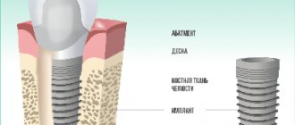
The protruding parts of the implant need daily thorough cleaning, since the lack of adequate hygienic care can lead to a decrease in the service life of implants and prostheses installed on them due to inflammation of the mucous membrane around the implant and the subsequent development of peri-implantitis - one of the most dangerous complications that arise during prosthetics. implants [1, 2].
To mitigate the risks associated with contamination of the implant surface with bacterial and food plaque, after completion of prosthetics, the patient is regularly scheduled for follow-up examinations [3].
One of the most important components of care after implantation is professional oral hygiene [4], which is usually carried out at least once every six months. It has a number of features compared to the standard procedure, such as the use of special attachments for ultrasonic scalers, powders and polishing pastes with rubber cups and polishing brushes, which do not have a damaging effect on the surface of the implant [5].
When carrying out professional hygiene, one should also take into account the loss of bone tissue around the implant (up to 1 mm during the first year after surgery and up to 0.1 mm per year in subsequent years) and, as a result, exposure of the implant neck. On the exposed part of the implant, rapid accumulation of dental plaque is possible due to the inaccessibility of this area and the surface topography [6]. This part of the implant needs to be thoroughly cleaned both during home hygiene care and during professional oral hygiene procedures [7].
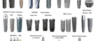
Currently on the dental materials market there are many attachments for ultrasonic scalers for removing hard dental plaque. In the context of a wide range of products, the question of choosing instruments that will ensure high-quality and safe professional hygiene for the implant remains relevant.
Purpose of the study
To evaluate the effect of ultrasonic scaling attachments on the surface of dental mini-implants.
Materials and methods
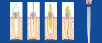
On the surface of a mini-implant, as on the surface of a natural tooth, the formation of dental plaque and tartar also occurs (Fig. 1). In order to study the influence of instruments for carrying out professional hygiene procedures on the surface of implants, we used dental mini-implants in the amount of four pieces.
According to in vitro protocols, the surfaces of mini-implants were treated with various ultrasonic scaling attachments for 20 seconds.
The surface of the first implant was treated with a stainless steel scaling nozzle - nozzle No. 1, the surface of the second implant - with a plastic scaling nozzle (made in Switzerland) - nozzle No. 2, the surface of the third implant - with a plastic scaling nozzle (made in France) - nozzle No. 3, the fourth implant was not processed and was used for control purposes (Fig. 2, 3).
Before the study, the implants were removed from the factory sterile packaging, after which the flange, microthread and macrothread were processed, then all implants were examined by scanning electron microscopy at an accelerating voltage of 20 kV at magnifications x30, x100, x300, x1000.
Results and discussion
When analyzing the results obtained, the effect of scaling attachments on the surface of mini-implants was established, which was expressed in the appearance of structural damage to the surface and in the form of trace elements of the attachment material on the surface of the studied samples.
Rice. 1. Deposition of tartar and plaque on the surface of the implant Fig. 2. In vitro study: mini-implants and surface treatment attachments. Rice. 3. Treatment of the mini-implant with a Swiss-made plastic attachment.
When processing the flange (the most polished part) of the mini-implant with metal nozzle No. 1, gross surface defects in the form of numerous scratches were revealed (Fig. 4). When processing the mini-implant flange with plastic nozzles No. 2 and 3, no surface damage was observed, but trace elements of the nozzle material were found on the surface of the samples: Figures 5-7, tables No. 1, 2.
When visually comparing the samples at this magnification, it was concluded that there was no damaging effect of plastic attachments.
When processing microthreads with a metal nozzle, gross surface defects in the form of scratches and smoothing of the specific relief of this area were also observed. When processing microthreads with plastic nozzles, no damaging effect was detected, but trace elements were detected.
Table No. 1. Damaging effect of scaling tips on the surface of mini-implants
Sample Average number of damages per 250,000 µ² surface Average longitudinal damage size (µm) Average transverse damage size (µm) Mini-implant treated with metal attachment (No. 1) 4 400 50 Mini-implant treated with plastic attachment (No. 2) not detected not detected not detected Mini-implant treated with plastic attachment (No. 3) not detected not detected not detected Control mini-implant not detected not detected not detected
Rice. 4. Mini-implant flange treated with metal attachment No. 1. Magnification x100. Rice. 5. Mini-implant flange treated with plastic attachment No. 2. Magnification x100. Rice. 6. Mini-implant flange treated with plastic attachment No. 3. Magnification x100. Rice. 7. Flange of the control mini-implant. Magnification x100.
Conclusion
From our in vitro study, we can conclude that when it is necessary to remove dental plaque from the implant surface, a safe instrumentation protocol should be followed.
Processing the polished part of the implant with stainless steel tools leads to scratches that affect the oxide layer of the surface, reducing the corrosion resistance of titanium implants. The appearance of scratches also enhances the attachment of bacterial plaque to the surface of the implant, which directly affects the service life of the implant.
Plastic scaling tips do not damage the surface of the implant, however, it becomes contaminated with trace amounts of the tip material, which can subsequently negatively affect the long-term success of implantation. Therefore, after scaling with a plastic nozzle, it is recommended to polish the implant superstructures with a brush using a low-abrasive polishing paste.
Table No. 2. Trace elements of scaling tips on the surface of mini-implants
Sample Average number of trace stripes per 250,000 µm² surface Average size of trace stripes (µm) Maximum size of trace stripes (µm) Mini-implant treated with metal attachment (No. 1) not detected not detected not detected Mini-implant treated with plastic attachment (No. 2) 8 90 200 Mini-implant treated with plastic attachment (No. 3) 10 145 300 Control mini-implant not detected not detected not detected
The main goal of the correct selection of instruments is the ability to gently but effectively remove dental plaque from the surface of the implant using the right scaler, without damaging the surface of the implant and without leaving hard fragments in the gingival sulcus. The data obtained are also consistent with the literature data of other authors [8–10].
Literature
1. Joyce M. The care and maintenance of dental implants. Dental Nursing 2013 9;3:138–142.
2. Kotsovilis S., Karoussis IK, Trianti M., Fourmousis I. Therapy of peri-implantitis: a
systematic review. J. Clin. periodontal. 2008; 35: 621–629.
3. Kracher CM, Smith WS Oral health maintenance dental implants. J.Dent. Assist.2010; 79 (2): 27–35.
4. Wingrove S. Professional hygiene in the field of implants and treatment of peri-implantitis. - M.: Tarkomm, 2014. - P. 167-199 .
5. Maximo MB, de Mendonca AC, Santos RV, Figueiredo LC, Feres M, Duarte PM. Short—term clinical and microbiological evaluations of peri-implant diseases before and after mechanical anti-infective therapies. J Clin Oral Implants Research 2009; 20: 99—108
6. Renvert S., Roos-Jansaker AM, Claffey N. Non-surgical treatment of peri-implant mucositis and peri-implantitis: a literature review. J. Clin. periodontal. 2008; 35: 305–315.
7. Todescan S., Lavigne S., Kelekis J., Cholakis A. Guidance for the maintenance care of dental implants: clinical review. Journal Can. Dent. Assoc. 2012; 78:107.
8. Dmytryk JJ, Moriatry JD The effects of scalling titanium surfaces with metal and plastic instruments on cell attachment. J. Periodontol. 1990; 61: 491–496.
9. Fox SC, Moriarty JD, Kusy RP The effects of scaling titanium implant surface with
metal and plastic instruments: an in vitro study. Journal Periodontal. 1990; 61: 485–490.
10. Ramaglia L., di Lauro AE, Mogrese F., Squillace A. Profilometric and standard
error of the mean analysis of rough implant surfaces treated with different instrumentations.
J. Implant. Dent. 2006; 15: 77-82.
Mode of application
- At the initial stage of the procedure for cleaning tartar, it is necessary to prevent the substance from getting on the gums.
- Then one of the syringes with the contents of the gel is taken from the package and the cap is removed from the instrument. A metal cannula, called an applicator, is placed on the syringe.
- Using applied force, the gel is squeezed onto the affected area with hard dental plaque that requires treatment. The product should be left to act on tartar for 35-40 seconds.
- Then the gel is removed from the surface to be treated with special tools and thoroughly washed with water.
- After the cleaning process, you need to remove the cannula from the syringe and close the instrument with a special cap.
In particularly difficult cases, it is recommended to perform dental procedures several times to achieve the desired effect. At the same time, do not forget to always protect the gums from getting the gel on it.
It must be remembered that Scaling gel is used exclusively in medical clinics. Do not use the drug at home.
You should know that after the plaque cleaning procedure, it is important to carefully follow strict oral care recommendations . They should include daily (morning and evening) brushing of teeth, gums and tongue, as one of the sources of decomposition of various microorganisms.
There is no need to be overzealous with eating foods that are colored or contain a lot of carbohydrates. This can leave pigmentation on the enamel and promote bacterial deposits. If, after scaling, bad breath appears again after several months, you must repeat this stone removal procedure again.
Why does tartar form on teeth?
There is always a certain amount of bacteria present in the oral cavity. This is fine. Moreover, bacteria help in digestion by starting the process of decomposition of food already during chewing.
Bacteria are found on the surface of all oral tissues, including teeth. The latter are protected from the harmful effects of microorganisms by enamel. Bacteria cannot penetrate through it.
But, nevertheless, nothing prevents bacteria from accumulating on the surface of the enamel. This biofilm is easily removed with everyday hygiene - brushing, flossing and irrigating. But in some cases - for example, due to untimely removal - the plaque begins to thicken. The reasons for this are:
- Excessive consumption of sweet foods, sugar, foods high in carbohydrates. All this creates a breeding ground for microorganisms;
- Use of low-quality hygiene products, untimely cleaning;
- Smoking. Tobacco smoke destroys the structure of the enamel, forming roughness, cavities and cavities on its surface;
- Irregularity of the dentition, malocclusion;
- Diseases of the digestive system, hereditary predisposition.
Bacterial plaque is easily removed. However, at some point it begins to petrify – that is, to become denser and acquire a mineral structure. And turns into tartar.
Tartar can no longer be removed with everyday hygiene products. It cannot be brushed or flossed off, and even a waterpik cannot destroy it. At the same time, bacteria in this colony continue to destroy both the tooth and the soft tissue located next to it. As a result, itching, swelling, bleeding begins, and increased sensitivity appears.
Cleaning your teeth at the dentist
Professional teeth cleaning at the dentist is a complex procedure. Today in dentistry the following techniques are used:
- Ultrasonic cleaning, in which the tooth enamel is cleaned by special sound vibrations. Cleaning is carried out in two stages, during which not only the stone is removed, but the enamel is also polished.
- Laser cleaning is recommended for subgingival calculus removal. The technique is atraumatic and precise.
- Cleaning using an Air Flow device is the most modern technology. A special air-water mixture with abrasive particles is applied to the tooth to clean it under pressure. The technique is suitable for working with children, pregnant women and patients with various types of braces.
Your dental hygienist will advise you on the best cleaning option. He will also talk about preventive measures to prevent relapses.
Kinds
Plaque is divided into two categories. First, supragingival stone appears, the cause of which is poor oral hygiene. Such deposits are located above the upper edge of the gingival crest, so they are easily diagnosed during a routine examination.
The characteristic features of supragingival tartar include:
- Localization on the inner surface of the tooth, from the side of the tongue;
- White or yellowish tint;
- The consistency is hard or clay-like.
The plaque is unstable, so it can be removed mechanically without any problems.
Subgingival calculus appears as a result of the fact that supragingival plaque descends down the root of the tooth and mineralizes. It forms in the gum pocket and is diagnosed only when using special dental instruments. Such deposits are considered age-related and most often appear in people over 40 years of age. If personal hygiene rules are not followed, it can appear at any age.
Signs of tartar:
- Dense and hard consistency;
- Light brown or greenish-black tint;
- Swelling and bleeding gums.
Why does it appear?
The main cause of any problem is insufficient hygiene. If you regularly cleaned your mouth poorly, did it with the wrong brush, or used a low-quality paste, then expect the formation of not only hardened plaque, but also destruction of hard tooth tissue, and other equally dangerous diseases. To avoid adverse effects, choose the right brush and toothpaste, also follow the correct technique for brushing your teeth and be sure to rinse and floss your mouth after eating.
The accelerated appearance of hardened plaque causes:
- Hobbies for confectionery, carbonated drinks, foods rich in simple carbohydrates;
- Frequent consumption of soft or liquid foods, food fillers and flavored sweeteners;
- Poor diet;
- Drinking alcoholic beverages and smoking;
- Heavy secretion of saliva;
- Taking a number of pharmacological drugs;
- Chronic inflammatory diseases.
Clinical picture
Signs of tartar appearing on teeth:
- bleeding gums;
- tooth mobility;
- toothache;
- constant unpleasant odor from the mouth;
- caries.
If such symptoms are detected, you should consult a dentist to find out how to get rid of tartar at home, or undergo the necessary procedures in the clinic.