As soon as you say “tooth canal,” people worry about toothache. Why does the mention of root canals scare people so much? Most patients believe that root canal treatment is a very painful procedure. While with the development of modern dental technologies, the treatment process has changed a lot.
There are many myths associated with root canal treatment, and in this article we will present real facts that will help people who are faced with a similar problem to find out the real state of affairs.
Root canal treatment is a very painful procedure.
This is not true. Root canal treatment does not cause pain. In fact, root canal treatment is performed to relieve pain from inflammation of the pulp chamber (where the nerve is located) or a dental abscess. The myth that root canal treatment causes severe pain is becoming a thing of the past. With the use of modern anesthesia methods, this procedure is no more painful than installing a filling.
If there is a serious abscess process, anesthesia may not work, and your doctor will prescribe antibiotics before root canal manipulation. If a root canal is difficult to access, your dentist may refer you to an endodontist, a doctor who specializes in treating dental cavities and root canals.
Method of lethal condensation of cold gutta-percha -
In Russia, 95% of all root canals are filled using this method.
The method of lethal condensation consists in the doctor performing a number of stages, the success of which largely depends solely on the high-quality implementation of the previous stages. We are talking about the correct measurement of the length of the root canal, as well as the quality of the instrumentation performed. Lateral condensation of gutta-percha (scheme) –
- Selection of the main gutta-percha pin - the pin is selected depending on how much the root canal was expanded during mechanical treatment)
- Filling the root canal with sealer – sealer is a special paste that is used to fill the voids between gutta-percha pins. After introducing the sealer, the main pin is inserted into the canal (Fig. 14a)
- Compaction of a gutta-percha pin with a spreader - the compaction process consists of making reciprocating movements with the spreader instrument, with its help the gutta-percha is pushed towards the canal wall and space is freed up for the insertion of new gutta-percha pins. The spreader itself is shown in Fig. 14(b), and the compaction process in Fig. 14(c).
- Insertion of smaller pins and their compaction (Fig. 14 d, e, f) - up to 8-12 gutta-percha pins can be “compacted” in just one root canal. As a result, if we look at an enlarged section of the tooth root, we should see a picture like this - Fig. 17.
- X-ray control of filling - if everything is OK - we proceed to the next stage... But if we see that the canal is not filled to the top, or the pins extend beyond the root into the surrounding tissues - it is necessary to remove all the pins and start filling the canals from the beginning.
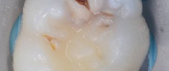
- Removing excess gutta-percha and sealer - after the canals are tightly obturated with gutta-percha and sealer, the tips of the gutta-percha pins protruding from the mouths of the root canals are cut off with a hot tool (Fig. 14 g, h). You can see what the mouths of the root canals look like before filling and after they have been cut, excess gutta-percha protruding above the mouths of the canals in Fig. 15 and 16, respectively.
- Temporary filling – After this, the tooth cavity is closed with a temporary filling. It is not allowed to fill the crown of a tooth in the same visit with filling the canals. Restoration of the tooth crown should be carried out at the next visit.
Root canal treatment is expensive
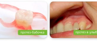
Right. Although root canal treatment is expensive, it allows you to preserve the tooth, as well as its functionality and chewing functions. If you have a root canal and install a crown on the tooth, the cost of treatment will be less than if you remove the tooth and replace it with a bridge or dental implant.
The cost of treatment will depend on how many root canals the tooth has, whether it is the first time it has been treated, and on the qualifications of the dentist (general dentist or specialist).
Endodontic instruments during cleaning
To effectively clean the canal, it is necessary to use several types of instruments, usually made of strong wire. Some of them are used in particularly difficult cases when the passage of channels is difficult. The specialist, taking into account the condition of the patient’s dental passages, can select rigid devices with a square cross-section, which are durable, but are less efficient at evacuating dentine filings. Another option is triangular-section reamers, which have increased flexibility but break more easily.
Groups of tools by purpose of use:
- channel expansion. The most commonly used options: Pro File 04, ProTaper, Rasp, K-flexofile, Rotary Instrument;
- increase in the diameter of the canal mouth. : Varieties: Largo and Gates Glidden;
- facilitating passage of the canal. Types: K-reamer, K-reamer farside;
- removal of tool remains. Instrument Removal System, Masseran Extractor Kit.
An auxiliary device is a microscope, which helps to visually zoom in on the channels and ensure high precision of the manipulations performed.
Advice! The safest instruments are considered to be nickel-titanium reamers, which bend easily and “remember” their shape after penetration into the canal.
Toothache disappears immediately after the procedure
Wrong. After completing the treatment procedure, the patient will feel a significant improvement. However, it is normal for the tooth to remain sensitive for a few days after the procedure - in this case, taking painkillers may help. Minor pain may be present, especially when chewing, and may persist for several weeks. After this period, the pain should completely disappear.
Is it possible not to experience any pain after root canal treatment? Yes, this is possible, and it depends on how difficult the treatment process was and whether the tooth was infected before the dental intervention.
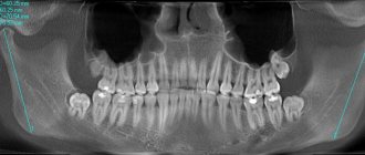
Microscope or X-ray
Currently, dentistry uses two types of control over the treatment process: x-ray and using a microscope. X-ray is inferior both in terms of diagnostic capabilities and from a safety point of view:
- Some types of pins and composite materials are not visible in the photographs, which blurs the objective picture of the condition of the dental canals;
- optical distortions and channel overlap are possible;
- if there is liquid in the cavity, then the x-ray will not show it;
- To diagnose and monitor the treatment of the canals, a series of images will be required, and this, although insignificant, is still radiation.
A dental microscope is capable of magnifying the image 30 times - the doctor will notice the smallest tissue defects. You can easily detect the mouths of additional canals, examine their most tortuous configurations, assess root damage and monitor the quality of cleaning.
The microscope allows for unsealing with ultrasound, thereby preserving a large volume of valuable healthy tissue. Optics has significantly expanded the possibilities of dentistry in terms of restoring teeth that were previously considered hopeless. Treatment under microscope control almost completely eliminates the development of recurrent inflammatory processes and ensures long-lasting results.
Patients of the IMEZA clinic can count on high-quality canal retreatment using high-precision optical equipment, technological instruments and modern materials. It is enough to make an appointment, during which experienced doctors will identify all potentially dangerous elements and determine the optimal therapeutic tactics.
The effect of treatment is short-lived
Wrong. Although nothing can completely replace a healthy tooth, quality root canal treatment followed by a suitable filling or crown can be very successful. In approximately 85% of cases, the effect of treatment is lifelong.
If a tooth becomes affected again several years after root canal treatment, it often needs to be re-treated. However, in some situations, such as a crack in the tooth, very severe tooth root decay, or severe loss of bone around the tooth, the dentist has no choice but to remove the affected tooth.
Mild but prolonged pain after treatment is normal
Wrong. It is not normal to experience constant pain for up to several months after root canal treatment. Causes of pain include hidden canals that were not cleaned during the procedure, or a crack in the tooth.
In these rare cases where pain persists, patients should be referred to an endodontist who specializes in root canal treatment for a diagnosis and appropriate treatment. In cases where the root or tooth is destroyed and no treatment method is suitable, the dentist has no choice but to remove the tooth.
A root canal “kills” the tooth.
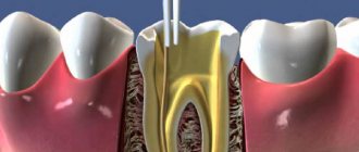
Wrong. During root canal treatment, the inside of the tooth is cleaned and disinfected, which helps heal it; it does not kill the tooth. The nerves and blood vessels located in the pulp chamber serve to develop the tooth if it is a child or teenager.
The function of the nerves is to signal pain when something is wrong with the tooth, be it decay, infection, inflammation or injury. Pain, therefore, is a defense mechanism that encourages a person to look for its cause and eliminate it.
The main stages on which the quality of root canal filling depends:
1) Determination of the working length of each root canal -
High-quality root canal treatment requires that the root canals be filled to the apex of the root. If the channel length is determined incorrectly, then there are 2 options :
- The root canal will be underfilled - this will lead to inflammatory complications, the development of periodontitis, cysts, and in the absence of retreatment of the tooth - to its removal.
- The root canal will be refilled - the filling material will be excessively removed beyond the apex of the root, and this can lead to long-term pain, neuralgia, and the development of inflammation.
Therefore, it is necessary to carefully measure the length of each canal in the tooth. This is done in good clinics as follows: after removing the pulp from the root canals, the doctor, using special thin hand instruments (for example, K-files - Fig. 7, 10) tries to go through each root canal to the apex.
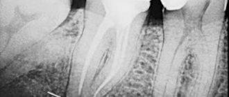
The advancement of the instrument deep into the canal is carried out under the control of a special device “apex locator” (Fig. 6), which is connected with the help of an electrode to the K-file located in the canal (Fig. 7-8). The display of the apex locator reflects the depth of the instrument's immersion, as well as the moment the instrument tip reaches the root apex.
Important: the apex locator only shows an approximately accurate picture. Therefore, after measuring the length of the canal with an apex locator, the K-file is left in the root canal and the patient is sent for an x-ray. These K-files are radiopaque, so the X-ray can clearly see whether the tip of the instrument has reached the apex of the root.
2) Mechanical treatment of root canals –
The purpose of mechanical treatment is to expand the root canal and make it suitable for filling.
Untreated canals in most cases are very narrow, have many imperceptible narrowings and expansions, which will not allow the canal to be properly filled with filling material along its entire length. Thus, mechanical treatment should remove all narrowings and irregularities throughout the root canal, and expand it to a certain size.
There are 2 methods of mechanical treatment of root canals:
- Hand instruments (Fig. 10) - the doctor rotates such instruments in the root canal with his fingertips. Below you can see photos and videos of this method.
- Using an endodontic tip (Fig. 11) - special Pro-files made of nickel-titanium are inserted into such tips (Fig. 12). The endodontic tip rotates the profile in the root canal, as a result of which the sharp edges of the profile remove chips from the walls of the canal, expanding it. Due to the fact that the metal of the profiles has a shape memory effect, they do not break during rotation even in highly curved root canals.
The advantages of processing canals with such machine Pro-files over hand tools:
- The quality of canal treatment is many times higher - the surface of the root canal walls after such treatment is very smooth, as if polished, and this facilitates the insertion of gutta-percha pins for filling the canal. In addition, the use of an endodontic tip can significantly reduce the processing time of canals (compared to manual processing).
- Safety - in most cases, the endodontic handpiece comes complete with a smart micromotor (Fig. 11), which controls the movement of the file in the canal.
If a certain load is exceeded, which threatens to break the profile in the channel, the micromotor stops the rotation of the profile and turns on auto reverse. Therefore, the threat of the tool tip breaking off in this case is minimal, which cannot be said about manual files. After all, the doctor’s fingers, with which he rotates the “manual” file, feel quite weakly the extreme resistance to the movement of the instrument, after which the instrument breaks off in the canal. If your clinic performs mechanical treatment of canals using such smart devices, this is a big plus. However, it is worth remembering that in any case such treatment is performed by a doctor, and even the smartest and best device in the wrong hands can cause more harm than good (24stoma.ru).
Mechanical treatment of canals with hand instruments and endodontic handpiece: video
3) Filling root canals with gutta-percha –
After the root canals have been expanded and the root canals have been treated with medication, they need to be sealed. Root canal filling methods that can be used in dental clinics:
- One paste method - the canal lumen is filled with a plastic material, which then hardens. At the moment, there is no more terrible method of filling canals; complications develop in almost 99% of cases. If your doctor fills your canals in this way, then you need to run urgently! And if you can’t run, then you should at least crawl to the exit.
- Single pin method - after filling the root canal with paste (as in the previous version), one gutta-percha pin is inserted into the root canal. This method is slightly better than the first, however, the percentage of complications after such treatment is also close to 99%, and you also need to run.
- Method of lethal condensation of cold gutta-percha - the meaning of this method is to compact the pins of cold gutta-percha as tightly as possible along the entire length of each of the root canals. We will discuss this method in more detail below. The method has an affordable cost and high reliability.
- Vertical condensation of hot gutta-percha is the most effective method of filling canals, which is carried out using gutta-percha heated to a fluid state.
The latter then gradually cools and hardens. Due to the fact that at the beginning it is in a fluid state, gutta-percha flows even into the lateral microchannels, which cannot be sealed by any other method (even the lateral condensation method). There are many techniques for using this method and materials for it. However, the best system for filling canals with hot gutta-percha called Termafil. If you are willing to pay for quality and reliability, then this is your method. But be prepared for the fact that the cost of filling canals with Termafil will be high.
Video: filling canals with cold and hot gutta-percha
Need to take painkillers
Right. Relatively, this is true. The pain experienced after root canal treatment is usually caused by inflammation around the tooth and may only last for a limited period of time. This process can be successfully managed with the help of common anti-inflammatory drugs: ibuprofen (Advil, Motrin), acetaminophen (Tylenol).
If severe pain continues for several months after the procedures, you should consult a dentist or endodontist to avoid possible complications.
Expert opinion
Lyubov Ivanovna Kopylova
dentist-therapist
Experience: more than 10 years
If you have the opportunity to go to a dental clinic equipped with an electron microscope, take advantage of it! Even the most experienced dentist works with canals without a microscope by touch. This means that there is a risk of underfilling or, on the contrary, excessive deepening into the canal, leading to perforation. The use of a microscope, when the working field is clearly visualized and the doctor efficiently cleans and seals even the thinnest and most curved canals, will allow this situation to develop.
Teeth that have undergone root canal treatment often require a crown.
Right. Often, teeth undergoing root canal treatment have severe caries or large fillings. A tooth with a large filling is at high risk of destruction. For this reason, your dentist may recommend that you get a post and crown after root canal procedures.
Some dentists place a post and filling on the tooth immediately after the root canal has been treated. Although a post adds more strength than a filling alone, a crown is also recommended to improve strength characteristics.
Root canal treatment is a lengthy process that requires several visits to the doctor.
Wrong. Today, root canal treatment can take one to two hours if there are no complications. The number of visits to the dentist often depends on the condition of the tooth and the number of canals in it.
In cases where the damage is severe, your dentist or endodontist may place medication inside the canal to prepare (disinfect) the inside of the tooth and then finish treatment after a few days. But if there are no lesions or complications, all procedures can be performed in one visit.