Pulpitis and periodontitis: clinical picture of the development of caries and its complications
If we talk about the structure of a tooth in a simplified way, then it has a crown part that protrudes above the gum, and a root (or several roots) that hold the tooth in the bone tissue of the jaw.
Inside, under the enamel layer, the tooth is filled with dentin, in the thickness of which there is a small space. There are nerve endings and blood vessels that supply the tooth. They enter the root canal through the apex, fill it from the inside, and also occupy a small space in the crown of the tooth. The neurovascular bundle inside the tooth is the pulp. If you delay the treatment of caries, pathogenic microorganisms will begin to penetrate the pulp, causing its inflammation. This complication caused by caries is called pulpitis. With this disease, the pulp swells and puts pressure on the nerve. This is why the pain is so intense. The difficulty of pulpitis in root canal treatment sometimes lies in the fact that the teeth are connected by nerve endings and it can be difficult to immediately determine which of them is causing the pain.
When the pain does not go away, and also intensifies at night, this indicates that the inflammation has gone further - into the root canals of the tooth. If you do not treat the tooth canals during pulpitis, and try to drown out the pain with medications, then the infection from the root canals will spread to the bone tissue of the jaw.
The inflammatory process that begins in the bone around the apex of the root is called periodontitis. Treatment of canals for periodontitis and elimination of bone inflammation itself is much more difficult. Here you absolutely cannot delay contacting a specialist, otherwise the inflammation will quickly spread and intensify. Or it will become chronic, in which the infected area of the bone is replaced by granulomatous tissue or granuloma. As a result, the tooth most likely cannot be saved. This process is very dangerous: you can not only lose a tooth, but also provoke serious health problems.
Deep caries is a potential pulpitis, followed by periodontitis. A simple examination 1-2 times a year will preserve the health and beauty of your teeth, nerves and money. Since canal treatment for pulpitis and periodontitis is not the easiest, and also not cheap. In addition, there is a high risk of complications and tooth extraction in advanced cases.
Ways to restore sevens if they are lost
In dental practice, to replace a lost molar, the following is used:
Temporary prosthesis "butterfly"
“Butterfly” is a removable structure that imitates a living tooth and masks a defect in the dentition. Includes an artificial crown made of plastic and a gum made of nylon or acrylic. The denture is attached to adjacent molars without grinding.
The advantage of removable prosthetics is efficiency and efficiency. The disadvantages of the “butterfly” include rapid wear and the need to regularly take it off and put it on. If a breakdown occurs, the prosthesis is replaced with a new one. The design is too small and does not create a physiological load on the jaw. This leads to its atrophy, which further complicates implantation. Recommended as a temporary option for replacing a missing row unit.
Dental bridge (bridge prosthesis)
Bridges are a type of fixed prosthetics, a traditional way of restoring a molar after removal. Externally, the prosthesis resembles an arch with combined crowns. When restoring one tooth, the number of crowns is three - the two outer ones rest on adjacent teeth on both sides of the defect, the middle one replaces the missing seven. The design hides aesthetic defects and lasts for a long time - up to 10 years. Before installing a bridge, preparation of the supporting teeth is required - root canal treatment, grinding of tissue for crowns. If the preparation is poor, the orthopedic structure will have to be removed and the teeth re-treated. In addition, extensive preparation leads to loss of enamel, which makes adjacent teeth fragile. The bridge increases the load on them and accelerates destruction.
The advantage of a dental bridge is its reliable fastening and ability to withstand chewing loads. The patient does not need to remove the structure: only a doctor can remove it. Crowns imitate the color of natural enamel and are made of metal-ceramics, porcelain, and other materials. The disadvantages of the method include the inability to stop bone resorption. Atrophic processes continue in the defect area, because the chewing load falls only on the neighboring units of the row.
Implantation
Installing an implant is the most progressive method of restoring the seven. The treatment does not affect adjacent molars and preserves healthy tissue. The doctor implants a titanium root in place of the extracted tooth. With the classic two-stage technique, the patient is implanted with an implant and a plug is placed. The abutment is installed 4-6 months after the root has grown into the jaw, and a permanent crown made of metal ceramics or zirconium is attached to it.
Seven implants restore chewing functions and create a physiological load on the jaw. This is the only way that stops the process of bone resorption after tooth loss and prevents sagging of soft tissues. Bridges and butterflies lack this ability. In addition, in the absence of a figure eight (wisdom tooth), it is impossible to install dentures, since an end defect occurs and there is no support for fastening.
Root canal treatment
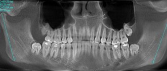
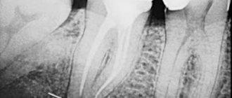
Before you begin root canal treatment, you need to take a photo. The fact is that in order to draw up an effective treatment regimen, the doctor needs to see the individual clinical picture of the disease: how deep the infection has spread and whether changes have begun in the bone tissue, how many roots the tooth has, perhaps the canals are too narrow and/or sclerotic. During treatment, it will be necessary to take more pictures to ensure the quality of the work done and the density of filling the canals with filling material. Only after making sure that everything is done correctly and efficiently will the doctor perform the final manipulations and restoration of the crown.
Treatment of root canal pulpitis
If no pathological changes in the bone tissue near the apexes of the roots are detected, then the canal treatment is performed according to the traditional scheme:
- Anesthesia with modern anesthetics. The drug is selected individually for each patient.
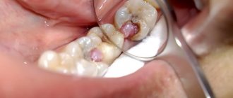
- Using a bur, the dentist removes tissue decay and opens the entrance to the pulp chamber.
- Then he needs to find the entrances to the channels, which is not always easy.
- Using special instruments and medications, the doctor removes the pulp from them, washes them, disinfects them from infection and dries them.
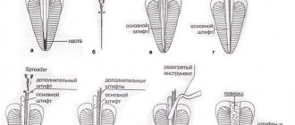
- The canals must be hermetically filled to the full depth with special filling materials - gutta-percha and polymer cement.
- Immediately after filling the canal, you must wait several days. Therefore, first the doctor places a temporary filling, and at a second visit he will restore the anatomical shape of the tooth with a modern photopolymer filling. Restoring the crown of a tooth after root canal treatment is a responsible task, the correct execution of which determines the reliability of the restoration and its service life. Often, the tooth is additionally strengthened with a pin, fixing it in the root canal.
Treatment of canals for periodontitis
It is not always possible to save a tooth if periodontitis has already developed. But with timely seeking help and proper treatment, the chances of a favorable outcome are quite high.
First of all, it is necessary to ensure the outflow of purulent mass from the inflamed area. Only after its elimination can treatment and filling of the root canals of the tooth be performed. Thanks to modern antiseptic drugs, it is possible to completely eliminate the infection from the affected area. However, in cases where the canal is too narrow to ensure the outflow of pus, it is not possible to save the tooth - it is immediately removed.
After the first stage of treatment, the patient is also given a temporary filling, with which he remains for about a week. You may need to undergo a course of antibiotic therapy. At the follow-up appointment, a thorough diagnosis is carried out. If no complications are identified, then the doctor installs a permanent filling and restores the tooth. Or prescribes further treatment based on diagnostic indications.
Features of endodontic treatment
Since childhood, for many, going to the dentist is inevitably associated with pain. Basically, this is just a psychological factor caused by the visual perception of the dental office almost like a hospital operating room. Most dental procedures are painless thanks to a combination of advanced treatment techniques and the use of effective local anesthesia. If we talk about endodontic dental treatment, it is somewhat painful, but this, firstly, is logically explainable, and secondly, the pain will be present for a short period. When a focus of infection appears in our body, pain is a natural reaction and a signal to action. This is inherent in nature itself. In the case of endodontics, we are talking about inflamed nervous tissue, which, of course, creates unpleasant sensations. However, an experienced dentist, using high-quality anesthetics, is able to carry out all the necessary actions with minimal discomfort for you. For some time after the end of treatment, increased sensitivity of the tooth to external influences cannot be ruled out. This feature does not appear for long and is easily eliminated with available medications.
“Clinic of Your Dentist”: root canal treatment with care for each patient
The prognosis for the quality of treatment of tooth root canals for pulpitis and periodontitis directly depends on their structure and the degree of development of the disease. Treatment of dental canals in complex cases especially requires good equipment, high-quality medicines and materials, as well as extensive practical experience from the doctor.
“Clinic of Your Dentist” fully meets even the most stringent requirements for the quality of services and the professionalism of its specialists. Conducting effective root canal treatment and saving the patient’s tooth is our main task, which we successfully cope with!
Features of implantation of the 7th upper tooth
Due to the anatomical features of the upper and lower jaws, the timing of implantation differs.
The upper jaw is subject to less stress when chewing. The bone tissue is looser compared to the lower row. Therefore, the implant takes 1-2 months longer than from below. Above are the maxillary sinuses, the infraorbital foramen and the facial nerve. If the implant is inserted incorrectly, there is a risk of damage, which increases the complexity of the procedure.
In case of sinusitis, sinus cyst, inflammation of the nasal mucosa, surgical intervention to implant a titanium root is not performed until the disease is eliminated.
If the upper molar is missing for a long time, the patient is indicated for a sinus lift. The procedure compensates for the lack of bone tissue by raising the bottom of the maxillary sinus and filling the space with synthetic material. The operation is performed open or closed.
In the first case, the procedure is carried out in two stages. The doctor builds up the jawbone, and after the osteoplastic material has fused with the tissues, an artificial root is implanted.
A closed sinus lift is combined with implant installation. The technique is less traumatic and reduces the duration of treatment. Loading of the artificial root with a crown is carried out after osseointegration.