Malignant tumors can form in almost all anatomical areas. Thus, oral cancer, the symptoms of which sometimes appear only in late stages, is not a rare form of oncology.
This disease is found among heavy smokers, chewing tobacco lovers and other categories of patients. Early treatment helps minimize the effects of tumor growth in the mouth.
What is oral cancer
Regular examination by a doctor - prevention
Oncological diseases of the oral cavity are caused by malignant cell growth in various tissues of this anatomical region. Since almost the entire oral cavity is lined with epithelial tissue, the likelihood of developing cancer is quite high.
This does not mean that anyone is susceptible to this disease. For tumor growth to occur, the influence of negative factors is necessary.
Malignant growth can affect the following parts of the oral cavity:
- Lips.
- Gums.
- Language.
- Buccal mucosa.
- Sky.
- Floor of the mouth.
The most common tumors are the tongue, the inner lining of the cheeks and the floor of the mouth. The clinical picture and the rate of spread of tumor cells depend on the location of the disease. When a tumor grows in the buccal area, the salivary glands may be affected.
Risk factors [5,6]
Science indicates the following reasons that can trigger the appearance of a malignant tumor of the oral cavity:
- weak immunity;
- poor nutrition;
- poor oral hygiene ;
- viral infections (primarily human papillomavirus);
- precancerous diseases: Bowen's disease, lupus erythematosus, lichen planus, leukoplakia, candidiasis;
- smoking, chewing tobacco, using tobacco mixtures;
- consumption of strong alcoholic drinks;
- deficiency of vitamins A, E and C;
- chronic injuries to the mucous membrane due to the sharp edge of a tooth or filling, incorrectly selected orthopedic structures, defects in the dentition;
- bad habit of biting your tongue, cheeks, lips;
- unfavorable labor factors: heavy metal salts, acid vapors, mineral fertilizers, petroleum products.
With the development of medicine, the list is supplemented and revised. Thus, if previously tobacco and alcohol were considered the main dangers, today more and more attention is paid to neglect of oral hygiene, poor nutrition, immunodeficiency and viral infections [5].
Causes
Oral cancer
The mechanism of development of oral cancer is generally similar to the stages of formation of any malignant tumor. Healthy epithelial cells, after dividing, perform their functions and are gradually destroyed.
Cell division is regulated by genetic information and special intracellular mechanisms. Under certain conditions, the division process can be disrupted, resulting in the formation of an abnormal cell mass called a malignant tumor.
How is the process distributed?
The process is distributed in several ways:
- The path is lymphogenous. Mutated cells spread through the lymph vessels along with the lymph. This variant is observed in most carcinomas.
- The route is hematogenous. Cancer cells choose blood vessels to spread throughout the body. This is how sarcomas and kidney cancer spread.
- The implantation path. The serous membrane is the channel for the spread of the disease.
- The path is portable. Associated with mechanical spread of tumor fragments during diagnostic and surgical procedures.
Malignancy
Symptoms and signs
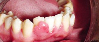
Poorly healing ulcers are a warning sign
Symptoms of any cancer can be variable. Often patients do not have any complaints until the tumor becomes large enough.
Symptoms depend on the location of the malignant growth and the individual body's response to the disease.
The most common symptoms include:
- The appearance of a lump, thickening, rough patch, or slight swelling in any area of the oral mucosa.
- Unexplained bleeding in the mouth.
- Numbness and loss of sensation in the face or neck.
- Constant appearance of sores in the mouth area.
- Painful sensations while eating food.
- Difficulties with chewing and swallowing. Speech dysfunction.
- Partial numbness of the tongue.
- Pain in the ear area.
- Gradual decrease in body weight.
The listed signs are not specific to oncology and may indicate a variety of diseases of the oral cavity. However, if such symptoms are detected, you should consult a doctor for a detailed diagnosis.
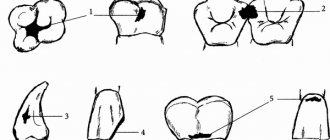
Malignant tumors of the upper jaw
Clinical manifestations of malignant neoplasms of the upper jaw are diverse, but not pathognomonic and depend on the initial location of the tumor, prevalence, shape and direction of preferential growth, and morphological structure.
Since epithelial malignant tumors originate from the mucous membrane of the maxillary sinus, in stages 1 - 2 they are usually an accidental finding (for example, during gairotomies), because tumors of such prevalence are not available for visual observation, symptoms may resemble ordinary sinusitis, or even absent. This explains the predominant detection of malignant tumors of the upper jaw in stages III-IV. In a significant proportion of patients, the first symptoms are pain of various types in the upper jaw, radiating to the head. Often the disease is manifested by pathological changes in the nasal cavity: one-sided difficulty or complete absence of nasal breathing, serous-purulent or bloody discharge. More rare first symptoms of malignant tumors of the upper jaw are unilateral profuse lacrimation, swelling of the eyelids or soft tissues of the face of the corresponding side, and impaired skin sensitivity. Some patients, as the first sign of disease, note a tumor in the upper jaw that is already visible to the naked eye.
To fully characterize the clinical manifestations of malignant tumors of the upper jaw and accurate topical diagnosis, in 1933 the Swedish oncologist Ohngren proposed a conditional division of the jaw into sectors. It is carried out by drawing an imaginary inclined plane starting from the inner corner of the orbit and going to the corner of the lower jaw. Two sectors or segments are obtained: superior posterior and inferior anterior. The second plane passes sagittally through the center of the maxillary sinus and divides each of the previous segments into lateral and medial parts.
Thus, two lateral and two medial segments arise:
- superoposterior external;
- superoposterior internal;
- inferoanteroexternal;
- inferoanterointernal.
Clinical observations confirm the feasibility and practical significance of the Ongren scheme, since it allows one to accurately determine the location of the tumor, since the lesion of each segment is characterized by certain clinical manifestations. Correct assessment of the symptoms of the disease allows you to timely suspect a malignant tumor and take the necessary measures. The danger hidden in delaying treatment for malignant tumors of the upper jaw is so great that it should be considered justified in all unclear and suspicious cases to resort to a comprehensive examination of the patient.
Neoplasms of the superoposterior outer segment. The main direction of growth of tumors in this localization is the pterygopalatine fossa and the orbit. Neoplasms of this group cause early exophthalmos. In this case, the eyeball shifts inwards, and diplopia appears. Obstruction of lymph and venous outflow associated with tissue germination by a tumor leads to swelling of the eyelids and narrowing of the palpebral fissure on the side of the tumor lesion. As the tumor grows further, facial asymmetry increases.
When a tumor grows into the pterygopalatine and infratemporal fossa, compression of the venous plexuses located here occurs, which impedes the outflow of venous blood and ultimately leads to lymphostasis in the retrobulbar tissue, zygomatic region and lower eyelid. This manifests itself as chemosis in combination with exophthalmos.
As they spread towards the tubercle of the upper jaw, tumors of this segment cause restriction of mouth opening due to contracture of the internal pterygoid muscle under the influence of tumor infiltration of its fibers and, in part, pressure on the coronoid process of the lower jaw branch. Tumors of the superior posteroexternal segment, earlier than other localizations, may be accompanied by the appearance of pain or paresthesia in the area of innervation of the 2nd branch of the trigeminal nerve of the corresponding side. Pain can radiate to the premolars and molars of the upper jaw. When the Eustachian tube grows, pain occurs in the ear area.
Thus, the deep location of malignant tumors of this segment under the cover of such anatomical formations as the zygomatic bone, the tubercle of the upper jaw, the ramus of the lower jaw with the surrounding muscles, leads to a long-term asymptomatic course of the disease. Only after the destruction of the upper or posterior (infratemporal) walls of the maxillary sinus and the beginning of tumor disintegration do symptoms characteristic of malignant growth appear.
Neoplasms of the inferoanteroexternal segment . They tend to spread to the area of the temporomandibular joint (TMJ), infratemporal fossa, oral cavity, and cheeks. Among the first symptoms of tumors of this localization, pain radiating to the ear should be noted, which is due to the involvement of the II and III branches of the trigeminal nerve in the process. In a later period, contracture occurs due to damage to the internal pterygoid and masticatory muscles. With a tendency to spread inward, the lateral wall of the pharynx and the palatine tonsil are affected. With outward growth, facial asymmetry appears. Invasion of the external carotid artery or its branches can lead to heavy, repeated bleeding, one of which can be fatal.
Malignant tumors of this segment do not manifest themselves for a long time; only after the walls of the maxillary sinus are destroyed, the first symptoms of malignant growth are detected, but they are late.
Neoplasms of the superoposterior internal segment . Tumors of this segment tend to spread towards the cells of the ethmoidal labyrinth and orbit (the incidence of the latter is 20-25%). Tumors of this location are also diagnosed late, because located in an area that is difficult to access for research. Infiltrating the tissue of the orbit, the tumor displaces the eyeball anteriorly and laterally, limiting its mobility. Already in the early stages of the disease, patients notice the appearance of lacrimation on the affected side. Later, swelling and hyperemia appear in the area of the inner corner of the eye due to secondary developing dacryocystitis and tumor growth into the lacrimal sac and lacrimal ducts. At this stage, visual acuity usually does not change.
When the tumor spreads towards the nasal cavity, unilateral, first mucous, then mucopurulent or sanguineous discharge appears from the corresponding nasal passage, and difficulty breathing. Sometimes nasal discharge becomes foul-smelling. During rhinoscopy, you can see bleeding tumor masses covered with purulent plaque, resembling granulations, located in the upper part of the nasal cavity. Sometimes it is possible to find out from the anamnesis that the first manifestation of the disease was a distortion or disappearance of the sense of smell, which is associated with damage to the receptor apparatus of the olfactory nerve. Headaches follow.
If the tumor spreads to the ethmoid labyrinth, then varying degrees of exophthalmos may develop with optic nerve atrophy and decreased visual acuity. Often patients are bothered by pain in the area of innervation of the 2nd branch of the trigeminal nerve. There may be a loss of skin sensitivity in the cheek area.
If the tumor tends to spread to the nasopharynx, then from there it often grows into the body of the main bone. This stage is characterized by pain radiating to the temple. Further development of the tumor process can lead to growth into the cranial cavity with the appearance of signs of damage to a number of cranial nerves (II, III, IV, V, VI). Diplopia, decreased visual acuity, neuralgic pain in the superciliary region appear, and the corneal reflex is absent.
Neoplasms of the inferoanterointernal segment . The tumor spreads into the nasal cavity of the corresponding side, as well as onto the anterior wall of the maxillary sinus and the alveolar process of the upper jaw. Tumors of this location are diagnosed relatively early. Gradually destroying the alveolar process, hard palate, and the anterior wall of the maxillary sinus, the tumor grows into the mucous membrane. Bone destruction leads to gradual loosening and loss of often intact teeth. Involvement of the nerve endings of the alveolar processes in the tumor process leads to gradually increasing pain in the teeth and jaw. The destruction of blood vessels causes bleeding gums. The alveolar process is deformed, becomes thickened, as if swollen. Tumor tissue protrudes from the sockets of fallen or removed teeth, bleeding profusely and resembling granulation tissue. When the tumor process spreads to the anterior wall of the maxillary sinus, the nasolabial fold becomes smoother and progressive facial asymmetry appears.
When it grows into the nasal cavity, difficulty or complete absence of (one-sided) nasal breathing occurs, bloody or pus-laced discharge with an unpleasant odor appears, but the sense of smell is not impaired.
Characteristics of the origin of the main symptoms of malignant neoplasms of the upper jaw
For convenience, the variety of symptoms of malignant tumors of the upper jaw can be grouped as ocular, nasal and dental.
The leading symptom of tumors in this location is pain. With primary damage to the maxillary sinus, they occur almost simultaneously with the detection of a tumor or precede the appearance of visual signs of a tumor by 1 to 6 months. They are of the most varied nature, irradiating to different areas of the head. The intensity and nature of pain depend on the type and location of the tumor and the extent of the process. With limited tumor damage, constant dull pain in the upper jaw area predominates, intensifying in a horizontal position and when tilting the head, due to a rush of blood and increased pressure on the nerve trunks. As the tumor spreads towards the alveolar process of the upper jaw, toothaches arise with irradiation to the temple and TMJ. In the later stages, painful constant headaches occur.
The occurrence of toothache often leads to diagnostic errors and interventions on intact teeth, which do not eliminate the pain.
With sarcomas of the upper jaw, pain of a very different nature is observed: dull, aching, boring. Their irradiation is the same as in cancer.
The use of thermal procedures, as well as physical therapy, leads to increased pain and accelerated tumor growth.
The persistence of pain in malignant neoplasms of the upper jaw is a characteristic feature that distinguishes them from inflammatory processes.
Nasal symptoms . In most patients, as the malignant tumor grows, unilateral progressive difficulty breathing is observed until it is completely absent. Discharges of various types appear: mucous, bloody, fetid-purulent. In the early stages, reactive inflammation of the mucous membrane of the maxillary cavity around the focus of tumor growth is observed, which leads to irritation and hypersecretion of the epithelium of the mucous membrane. The transparent mucous discharge from the maxillary cavity enters the nasal cavity through the natural opening in the middle meatus. Patients, as a rule, believe that this is the result of a cold and do not consult a doctor, using vasoconstrictor nasal drops or thermal procedures at home. Subsequently, the secretion becomes purulent due to the addition of a secondary infection. Putrid discharge mixed with blood indicates an advanced process, accompanied by the disintegration of the tumor.
It has been noticed that fetid purulent discharge from the nose is more characteristic of cancer, and putrefactive discharge with streaks of blood is more characteristic of sarcoma of the upper jaw.
Difficulty in nasal breathing . It occurs due to the fact that the tumor, having completely filled the maxillary sinus, displaces its medial wall towards the nasal septum, and in later stages grows into the medial wall of the maxillary sinus, fills the middle and lower nasal passages, displaces and then grows into the nasal septum. At this stage, in addition to copious mucous discharge, nosebleeds appear from the growing tumor tissue. The tumor masses, having filled the nasal passages, protrude from the nasal opening. In the later stages of the disease, the nose shifts to the healthy side.
Difficulty opening the mouth . Contracture is rarely observed in the early stages of the tumor process in the upper jaw. The appearance of this symptom is associated with the tumor growing into the pterygoid, masseter or temporal muscles, i.e., when the tumor has already spread beyond the maxillary sinus. This symptom is typical for tumors located in the posterior sinus near the TMJ. When a tumor grows into a joint, first a reactive inflammation occurs in it, and then the tumor directly grows into it. In addition to contracture, pain in the joint is observed, which intensifies while eating and talking.
Ophthalmological symptoms . Pathological changes in the area of the eyelids, orbit and eyeball often indicate the presence of a malignant tumor of the upper jaw. Observed: unilateral lacrimation, swelling of the eyelids and narrowing of the palpebral fissure, exophthalmos or displacement of the eyeball in various directions, limitation of its mobility, diplopia, decreased visual acuity.
Swelling of the eyelids - occurs as a result of tumor spreading into the eye socket. Due to compression of the blood and lymph outflow pathways, lymphostasis occurs. Swelling can occur due to inflammatory changes of a reactive nature in adjacent soft tissues. Swelling of the lower eyelid causes a narrowing of the palpebral fissure and its upward displacement. When the tumor spreads towards the nasolacrimal duct, one-sided profuse lacrimation is observed.
As the tumor grows towards the orbit, the contours of its lower wall are disrupted. The orbital margin first becomes dense, lumpy, and later areas of softening appear. Almost simultaneously, swelling of the orbital tissue increases, and purulent conjunctivitis develops. Sometimes malignant tumors of the maxillary sinus first spread to the ethmoidal labyrinth and then grow into the orbit, which leads to displacement of the eyeball. The direction of displacement depends on the location and preferred direction of tumor growth. With sarcomas, displacement of the eyeball is observed earlier than with cancer.
The early stages of exophthalmos are usually detected not by the patient himself, but by those around him. Severe exophthalmos leads to impaired closure of the eyelids. Displacement of the eyeball to one side or another causes diplopia. With direct exophthalmos, diplopia does not occur. When the lower wall of the orbit is destroyed, enophthalmos (retraction of the eyeball) is formed.
If a malignant tumor grows into the motor muscles of the eyeball, its mobility is limited and vision is impaired. Tumor infiltration of the eyeball or optic nerve leads first to partial and then complete loss of vision.
Metastasis of malignant tumors of the upper jaw occurs mainly through the lymphogenous route. With sarcomas, hematogenous spread is possible. The outflow of lymph from the paranasal sinuses and upper jaw is carried out into the retropharyngeal and upper deep cervical lymph nodes. From the anterior sections, lymph flows into the submandibular lymph nodes.
Regional metastasis of malignant tumors of the upper jaw is a fairly rare phenomenon. With the upper internal localization of cancer, it was observed in 7.3%, lower - in 14.9% (Paches A.I., 1983). According to Yu.I. Vorobyov, the frequency of regional metastasis of tumors of this localization is generally 21.4%, and poorly differentiated and undifferentiated forms of cancer metastasize more often than differentiated forms of squamous cell carcinoma.
Distant metastases are observed from 3.4 to 9%.
The general condition of patients with malignant tumors of the upper jaw in the early stages may not be affected. But during the period of tumor imaging, a progressive decrease in body weight is usually observed. This can be expressed to varying degrees and depends not only on the stage of the tumor process, but also on the location, growth rate and spread. Thus, with the appearance of early contracture and pain in the TMJ area, weight loss begins earlier and is nutritional in nature due to difficulty eating. During the period of tumor disintegration and the addition of a secondary infection, patients experience symptoms of anemia due to intoxication from the disintegrating tumor, as well as repeated bleeding. Simultaneously with a decrease in the percentage of hemoglobin, the number of erythrocytes progressively decreases, the number of leukocytes increases, and the ESR sharply accelerates.
In the later stages of the disease, aniso- and poikilocytosis is observed. In the peripheral blood formula, as a rule, there are no sharp changes, only sometimes there is a slight decrease in lymphocytes and an increase in rod-nuclear leukocytes.
The skin of patients is characterized by increasing pallor. In later stages, the skin becomes sallow gray with a jaundiced tint. Many patients complain of general weakness, fatigue, a sharp decrease in appetite, and inability to work.
Diagnostic methods
If you suspect a disease, you should contact an oncologist. During your appointment, your doctor will ask about your complaints, take a medical history, and examine your mouth to look for signs of malignant growth.
Any abnormality of the epithelial mucosa may be suspected, including areas of irritation, ulcers and white spots. To exclude other diseases and confirm oncology, instrumental and laboratory research methods will be required.
Special research methods:
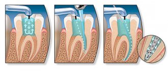
- Sampling of a section of the oral mucosa followed by histological examination. A biopsy is the most accurate method for diagnosing cancer and identifying the type of malignant tumor. Also, the results of the method may indicate precancerous changes in the epithelium, which increase the risk of cancer.
- Endoscopic examination. During the procedure, a small flexible tube equipped with a camera and a light source is placed down the patient's throat. The nasal cavity is examined in the same way. Endoscopy is necessary to detect the primary area of tumor spread if oral cancer is a secondary formation.
- Visualization. To obtain high-precision images of certain tissues and organs, doctors prescribe radiography, as well as computer or magnetic resonance imaging. These methods are useful for assessing tumor size and finding the primary site of malignant tissue spread.
Screening diagnostic methods aimed at detecting the early stages of oncology are also important. If a patient has certain risk factors, regular examinations are necessary.
What symptoms to look out for
To recognize a tumor at an early stage, dentists advise being guided by your own feelings. The first symptoms that raise suspicion may be:
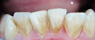
- slight bleeding of the gums that occurs as a result of gentle pressure on the gums;
- change in the color of the gum tissue and the appearance of swelling, which is often perceived as gingivitis;
- the appearance of neoplasms on the gums that have a pronounced red color, as well as light-colored ulcers and inclusions.
There is no need to panic if your gums are bleeding. This can be the cause of a number of diseases that are not related to cancer (periodontal disease, gingivitis, periodontitis). But plaques and ulcers should alert you and be the reason for an immediate trip to the dental clinic. At this stage, the disease can still be defeated and the likelihood of causing harm to the body can be reduced.
- Admin
- Oncology,
- Dentistry
Forecast
The survival period directly depends on the type of tumor and the stage of spread. During the first stage, when malignant cells remain on the surface of the mucous membrane, surgery helps to completely get rid of the problem.
A poor prognosis is typical for late stages, when tumor cells have spread to the deep cervical lymph nodes. In this case, survival rarely reaches two years.
Thus, oral cancer, whose symptoms may be nonspecific, is best treated in its early stages of growth. Screening diagnostics help detect precancerous changes and prevent tumor growth.
Informative video about oral cancer - in the video:
Metastases - what is it?
This term is quite scary for any person. Indeed, metastases are secondary, often distant, malignant lesions of any tissue in the body. The cancerous tumor itself can be localized in any part of the body, sometimes located very far from the area affected by metastases.
Metastases - what is it?
The presence of metastases significantly complicates the treatment of the underlying cancer, which often turns out to be completely powerless. These abnormal cells quickly and easily spread throughout the body without control. True, this happens only at a certain stage in the course of the underlying pathology.
Metastases to the spine
Metastases can also appear in the spine. Here they often affect the vertebrae themselves and structures close to them. They are usually detected in such parts of the spine as the lumbar, thoracic, and rarely in the cervical. Regarding the human skeleton, this is the most common malignant pathology.
Cancer metastasis
On a note! Most often, this type of secondary formations in the spine is found in lung cancer, malignant neoplasms of the prostate or mammary glands. Their “donors” are the kidneys, lungs, digestive organs, thyroid gland, etc. Metastasis of the spinal region often accompanies myelomas, sarcomas and lymphomas.
Table. Types of metastases in the spine.
| Type | Description |
| Osteoclastic or osteolytic | In this case, the so-called osteoclasts are activated, causing the destruction of individual bone elements. The vertebrae decrease in height, which can be clearly seen on x-rays. |
| Osteoblastic or osteosclerotic | In this case, the cells of the spinal tissue begin to grow uncontrollably, and the density of the bone substance increases. The shape and size of the affected area changes, which is clearly visible in the photographs. Almost all elements of the vertebra are affected. |
Osteoblastic metastases
Oncology as a contraindication to dental implantation
Oncology requires priority serious treatment; any concomitant treatment during this period is impossible. Implantation is no exception. The question of implantation of dental implants can be raised after achieving stable remission.
Received radiation and chemotherapy threatens to develop consequences that will make the implantation procedure impossible.
Irradiation causes the following complications:
- local reaction;
- radiation osteopenia;
- necrosis of bone structures;
- secondary osteoporosis;
- reduced ability of bone to recover (regenerate);
- bone demineralization;
- suppression of bone marrow functions;
- general intoxication.
Significant toxic effects and metabolic disorders after irradiation can be stopped no earlier than six months after treatment.
Is it possible to get implants after cancer treatment?
In addition to disrupting the aesthetics of a smile, the absence of teeth entails chewing dysfunction. After all, complete chewing of food is necessary for recovery. Previously, the installation of implants was not available to patients with severe bone atrophy due to cancer. After all, radiation and chemotherapy can make the bone brittle.
After oncology, it is impossible to carry out bone augmentation, so titanium implants cannot be used. The solution to this problem was a new material that replaces titanium and surpasses it in osseointegration properties.
Biopolymer Polyether ether ketone (PEEK, Polyether ether ketone) is a semi-crystalline thermoplastic. This is a dense material that is resistant to any environment and is actively used in the space and aviation industries. Based on it, the Perso-B and Perso-C implants were created, which can be installed when the bone is thinned. They are recommended by European and American clinics.
Polyetheretherketone has the following properties:
- Isoelasticity. The elasticity of PEEK-OPTIMA implants is almost identical to the elasticity of bone. It is possible to reduce stress concentrations that lead to bone resorption, change or displacement.
- Reinforcement or replacement of bone to support soft tissue. The polymer can serve as a basis for installing implants into bone. Its elasticity prevents fractures of the implant.
- The polymer provokes the formation of bone tissue. Implants based on PEEK-OPTIMA do not inhibit, but stimulate regenerative properties.
- The material is biocompatible and has good osseointegration.
- The biopolymer is biologically and chemically inert, so allergies and inflammation cannot develop, which is important for patients with allergies to titanium (4% of the total).
- The implants have good adaptability.
- Light weight and small thickness.
The polymer can be used after previous or current oncological diseases (Perso-B implantation system). In case of severe bone atrophy, implants do not increase the risk of bone damage; on the contrary, they stimulate growth. Implants are installed in one stage. Immediate loading of the prosthesis is allowed. Implantation of polymer rods does not lead to side effects or contraindications.
Polymer-based implants can be placed:
- smoking patients;
- patients with gum disease;
- with significant bone atrophy;
- to restore the alveolar ridge;
- to restore deficiencies of the facial skeleton;
- to avoid bone augmentation surgery.
There are opinions that polymer material can replace zirconium dioxide, the most advanced material in dentistry.
- Complete restoration of the dentition in just 4 days!
more detailsRoott Pterygoid Implants Sinus lift is no longer needed!
more details
Once and for life! Express implantation in 4 days with a permanent ReSmile prosthesis
more details
All-on-4, All-on-6, ReSmile, Zygomatic implantation We use all modern methods of dentition restoration
more details
What is the danger of metastases?
The process of development of a secondary tumor focus is of great importance in the course of the disease: having spread throughout the body in large numbers, mutated cells lead to organ failure and death of the patient.
Clinical practice shows that the process of formation of a secondary tumor focus is detected in almost 70% of patients at the diagnostic stage. Even in the absence of metastases at the time of cancer diagnosis, it is necessary to immediately begin treatment in order to prevent progression.