Timely detection and treatment of dental diseases is considered a necessary and important condition for maintaining dental health. Even with careful daily oral hygiene, no one can be protected from the development of severe diseases of the teeth and gums, which a preventive examination at the dentist will help identify, because at the initial stage the disease can develop almost asymptomatically.
Experts strongly recommend undergoing a professional examination every six months, even without visible dental problems. Many people are interested in the question: Why exactly after six months? Based on numerous studies, scientists have concluded that this period is optimal for most oral diseases to begin their clinical manifestation. Therefore, they are easy to diagnose and treat because they are in the primary stage of development.
Of course, today the level of dental care is very high, which makes it possible to restore even severely damaged teeth and cope with any ailments of the oral cavity. However, it is better to prevent the disease than to treat it later in a long and tedious manner. Preventive dentistry is designed to recognize pathology in advance and eliminate it as soon as possible.
What is a dental examination?
During the examination, an experienced dentist-therapist, having the appropriate knowledge and the necessary tools, will pay attention to even the slightest changes in the oral cavity, and if pathologies are detected, he will take measures to eliminate the disease.
External inspection includes:
- the condition of the gums is assessed to determine the prognosis of possible diseases;
- determining the correct bite;
- mobility and loosening of teeth is eliminated;
- the condition of previously installed fillings is assessed;
- the enamel of the teeth is examined for caries damage;
- the integrity of dentures and crowns is checked;
- An X-ray examination may be performed (when an image of the teeth is necessary).
After the examination, the doctor makes a conclusion about the presence or absence of dental diseases, and issues recommendations regarding preventive measures or treatment.
The oral cavity, its mucous membrane and lymphatic tissues of the maxillofacial area play a vital role in the interaction of the human body with the environment.
Microorganisms living in the oral cavity, on the one hand, contribute to the digestion of food and the synthesis of vitamins, on the other hand, they form products that damage dental tissue.
A practicing doctor must be fluent in the technique of examining dental patients and, first of all, the oral cavity and teeth. A complete examination of the patient makes it possible to establish not only the localization and nature of the pathological process, i.e., to substantiate the diagnosis, but also to foresee the further course of the disease and its probable outcome. Based on the results of the examination of the patient, the doctor chooses treatment tactics, establishes indications and contraindications for certain therapeutic measures.
Research methods in clinical dentistry include interviewing the patient, examination (both external and oral), examination of the oral cavity and teeth, as well as perimaxillary soft tissues, probing, percussion, and palpation. Methods based on the use of radiography, electroodontometry, laboratory and others are additional, since sometimes the diagnosis can be made without their use. For diseases of the mucous membrane and periodontium, special research methods are used.
SURVEY IN DENTISTRY
Questioning a patient in dentistry is one of the important stages of examining a patient. When interviewing the patient, passport data is clarified and the history of the present disease is clarified. The dentist must listen to the patient, guiding his story in such a way as to find out the most important of his complaints. By asking additional questions, the doctor finds out the nature of the pain, the cause of its occurrence, the duration of the pain, localization, prevalence, and so on.
It is also important to find out living conditions, dietary habits, profession, concomitant diseases, for what reason the missing tooth was removed, and how often you went to the dentist. When communicating, the doctor must carefully monitor the patient’s facial expressions, pay attention to the configuration of the face, how the patient’s mouth opens, whether there is bad breath or taste disturbances. For example, a taste disturbance may occur due to pathology of the central nervous system. All examination data are entered into the dental patient’s medical record.
INSPECTION
After a thorough questioning, the doctor begins the examination, observing the rules of deontology. The examination begins with an external examination of the face and surrounding areas. Facial deformation often indicates the presence of a pathological process not only on the face, but also in the oral cavity. During the examination, the general condition of the patient, the position of the head, skin color, and sclera are also determined. Thus, a yellowish coloration of the sclera can occur with periostitis, acute periodontitis, and necrotizing stomatitis. When conducting an examination, the doctor must pay attention to the patient’s manner of speaking, since a change in speech may occur due to some pathological processes. After a general examination, the dentist examines the oral cavity, carefully examining the contours and corners of the lips (with jaws and lips closed). Then the vestibule of the oral cavity is examined using a spatula. The oral cavity itself is examined using a dental mirror. At the same time, attention is paid to the condition of the salivary glands, hard palate, tongue, and floor of the mouth. After this, they begin to examine the teeth. This study uses a dental mirror, a probe (usually angular), and tweezers. When examining the teeth, the state of the bite, the number of teeth, their color, condition, their shape, size, the presence of periodontal pockets and the condition of the periodontal tissues are determined.
Thus, multiple caries can be a symptom of general disorders in the body, vitamin deficiency, and mineral metabolism disorders. Identification of dental plaque allows us to judge the function of chewing and oral hygiene. Tartar deposits indicate a violation of mineral metabolism.
Examination of the oral mucosa is carried out in natural light. The condition of the oral mucosa is affected by many pathological processes of the oral cavity and internal organs. Based on certain elements of the lesion, the first symptoms of diseases of the gastrointestinal tract, blood diseases, and malignant diseases can be established. Healthy oral mucosa is pale pink. Congestion and cyanosis indicate a chronic inflammatory process; sharp hyperemia of the mucous membrane is observed in many infectious diseases - measles, scarlet fever, diphtheria, trauma, inflammation, and general diseases. Blue coloration of the lips, mucous membrane of the cheeks, and sublingual area indicates a lack of cardiac function. vascular system. Swelling of the oral mucosa is observed in diseases of the heart, kidneys, and in the presence of scars. Enlarged or atrophied papillae on the tongue indicate the possibility of pathological changes in the gastrointestinal tract. These are important diagnostic signs. During the examination, attention is paid to the presence of bleeding of the oral mucosa, which can occur with periodontal diseases, stomatitis, blood diseases, vitamin deficiencies, endocrine disorders, hemorrhagic diathesis, as well as with chronic trauma with a crown, dental plaque, filling, and so on. The examination data is entered into the dental patient’s medical record or special records. When filling out the documentation, anomalies, deformations, pathological changes, elements of the lesion and their location are carefully described. The normal state is not described. Filling out a dental formula is mandatory.
PALPATION
After the examination, palpation and palpation of the maxillofacial area (superficial, deep, extraoral and intraoral) are performed. This study determines: tissue consistency, turgor, mobility, relief, pain. Palpation is carried out with both hands, starting from the healthy side. Apply superficial and then deep palpation. Palpation of the lymph nodes is also carried out with both hands, giving the head the appropriate position. At the same time, their soreness, adhesion to soft tissues or bone tissue, and mobility are determined.
The teeth can be palpated with fingers or tweezers.
When palpating the tissues of the oral cavity, they fix the tissues and organs with one hand, and with the other they feel every part of the palate, tongue, cheeks, and sublingual area. Palpation determines the condition of the salivary glands and ducts.
PERCUSSION
Percussion is tapping on the tooth with the handle of a probe or tweezers. Percussion can be vertical or horizontal. Vertical percussion is painful if there is an inflammatory process in the area of the apex of the tooth. Horizontal percussion determines the condition of the marginal periodontium. Percussion is carried out carefully. Percussion begins with a healthy tooth, moving to the diseased one. Percussion can be weakly positive, positive and sharply positive. Movements should be light and uniform. During this examination, the patient himself determines which tooth hurts by comparing his sensations.
PROBING
This study is performed using a probe (usually angular), which is held in the right hand, and a dental mirror is held in the left hand. Probing determines the presence of a carious cavity, softening in it, the depth of the carious cavity, the pain of the bottom of the carious cavity, as well as the sensitivity of the enamel - hyperesthesia. Probing is performed very carefully.
ADDITIONAL INVESTIGATION METHODS
- Thermometric research methods
- Electrometric examination (electroodontodiagnosis)
- Histological and histochemical research methods
- Cytological examination
- Bacteriological research
- Blood and hematopoiesis research
- Urine examination
- X-ray examination
- Serological study
- Specific studies
What problems can be identified promptly during a dental examination?
If a visit to the dentist is postponed for an indefinite period, then existing problems may begin to progress, as a result of which more time, effort and money will be spent on eliminating them.
The main dental diseases that are identified during examination in the dental chair include:
- Caries. If caries forms at an early stage, it is possible to cure it in one visit to the dentist. If treatment is not carried out in a timely manner, common complications such as pulpitis and periodontitis may occur, which complicates the preservation of the tooth.
- Gingivitis. Due to the formation of tartar and plaque, this disease begins to progress, which in turn can cause periodontitis, in which inflammation of the soft tissues around the tooth occurs, the ligament of the root and bone is disrupted, and the patient may lose the tooth.
- Periodontal disease. This disease manifests itself in the gradual destruction of periodontal tissue, which can lead to the loss of healthy teeth.
As we can see, the importance of the examination cannot be underestimated; it serves as a kind of starting point for drawing up a further action plan to maintain oral health.
By signing up for a preventive examination, you will receive competent advice and find out all the answers to questions that are of concern to most patients about proper oral care:
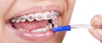
- which brush and toothpaste is right for you;
- Are you brushing your teeth correctly?
- what additional hygiene products should be used for prevention purposes;
- how to properly use an irrigator, floss, etc.
Also, if necessary, the dentist can prescribe a number of preventive measures, such as:
- Tartar/plaque removal procedure;
- whitening;
- fluoridation;
- fissure sealing, etc.
Don’t wait until dental problems make themselves known and begin to bother you, because it is much easier to set aside time for a professional examination than to spend days, and sometimes even whole weeks, on grueling treatment.
Prevention of oral diseases
- visit the dental office at least once a year;
- organize a special diet, in which lean meats, fish, fresh vegetables and fruits should predominate;
- rinse your mouth with special compositions after each meal;
- Strengthen your immune system and avoid colds.
To avoid any problems with teeth and oral cavity, you need to regularly visit the dentist and carry out preventive maintenance.
How does plaque affect the health of the oral cavity?
The development of many dental diseases is associated with the appearance of plaque. Harmful bacteria and their decay products have a detrimental effect on the enamel. Over time, plaque hardens and leads to the formation of tartar, which not only causes aesthetic problems, but also destroys the enamel structure and affects the health of the gums. And constantly multiplying microbes and bacteria provoke inflammatory processes and contribute to the progression of the focus of caries development. By causing bleeding and inflammation of the gums, pathogenic bacteria can cause the loosening and subsequently even loss of completely healthy teeth.
During an examination, an experienced dentist may recommend that the patient undergo a procedure for removing tartar and plaque to improve the health of the oral cavity and improve its general condition.
Effective measures to prevent the formation of plaque and tartar include:
- thorough oral hygiene;
- regular dental examinations;
- teeth cleaning at the dentist.
Diseases characteristic of the oral cavity
- A viral infection that includes herpes. This disease affects up to 90% of the world's population. The herpes virus can be present in the human body in a latent form for a long time and only appear if the person is under stress or has a cold.
- An infectious and inflammatory disease, which includes pharyngitis - inflammation of the pharyngeal mucosa. The disease can be triggered by inhaling tobacco smoke or any chemicals. Stomatitis. This disease can be caused by mechanical damage to the mucous membrane, after which ulcers appear on it.
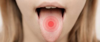
- Fungal diseases. The causative agent is a fungus that can be present in the body for a long time and become active only when the immune system is weakened. Fungal diseases include thrush, which manifests itself as whitish spots on the mucous membranes and lips. Atrophic candidiasis is the most common type of fungal disease. The patient feels dry, and redness appears in the oral cavity and a burning sensation may begin.
Other diseases include disc bacteriosis of the oral cavity. It can occur after long-term use of antibiotics.
Where can you find qualified dental specialists in Ivanteevka?
You can get professional dental care and undergo a preventive examination of the oral cavity at Sanident Dentistry, which provides a full range of services in Shchelkovo and in the urban district of Ivanteevka. We offer affordable prices and high-quality dental treatment. We have the most modern diagnostic equipment, proven painkillers, modern filling materials and instruments.
In our clinic you can undergo such hygiene procedures as: fluoridation, removal of dental plaque, AIR FLOW teeth cleaning, teeth whitening using professional preparations, complete professional oral hygiene and much more.
Entrust your health to professionals, do not allow the disease to enter the active stage of development, prevent the disease in a timely manner. Our experienced professionals are ready to help you with this.
The comprehensive dental clinic "Sanident" is located at the following addresses:
- Ivanteevka, st. Novoselki, 4 (Ivanteevka railway station);
- Shchelkovo, st. Central, 80 (railway station Voronok).
Types of pathologies of the oral mucosa
- Mechanical damage that can occur when the mucous membrane is exposed to poorly installed fillings, dentures and other solid objects, including solid food. The damage appears as scratches and may bleed.
- Thermal injuries occur when eating very hot food or drinks. A whitish spot appears at the burn site, from which the skin begins to peel off.
- Chemical injuries occur when eating hot, spicy foods, or when taking certain medications.
Palpation examination
Using this research method, inflammatory processes are identified, as well as the nature and stage of development of periodontal destruction. To more accurately understand the condition of the tooth, they begin to move it in different directions using tweezers, this makes it possible to understand how it moves in the alveolus:
- In three directions
- In five together with vertical
- In one
- In two.
If a tooth gives in very easily with a little force, this means that there are deviations from the norm, which may be accompanied by tissue swelling and an inflammatory process. These symptoms may indicate the presence of traumatic tooth lesions, such as periodontal disease, periodontitis and periodontitis. It is by palpating the cheeks and gums that the “One to One” doctor receives information about the presence of purulent or bloody discharge, pain, compaction and swelling. A big plus that can be given to the method is that it allows you to instantly obtain the research result upon examination. The only negative that unfortunately exists is the inability to make a definitive diagnosis.
Percussion (tapping)
Percussion with tweezers or a probe handle along the crown of the tooth allows you to determine the location of the area affected by inflammation. Any examination begins with healthy teeth; for this purpose, the edges responsible for cutting and chewing reflexes are tapped on the top and sides of the crown.
The process of tapping teeth (percussion)
A good idea of where the source of inflammation is located is given by the relationship between the nature of the pain and the direction of the blow: periodontitis is detected with a horizontal blow, damage to the root nerve can be determined with a vertical blow. Thanks to this technique, the doctor has the opportunity to identify the place where there is pain and prescribe the necessary treatment.
References
- Pomazanov, V.V. Microecological status of a person and its definition. — Prospects for the implementation of innovative technologies in medicine and pharmacy, 2022. — pp. 167-184.
- Gostry, A.V., Simonova, A.V., Mikhailova, A.N. and others. Chronic pharyngitis: etiology, pathogenesis, treatment. New approaches to assessing etiopathogenesis. - Archives of Internal Medicine, 2022. - No. 1 (45). — P.32-43.
- Man, W., Clerc, M., Pieters, W. et al. Loss of microbial topography between oral and nasopharyngeal microbiota and development of respiratory infections early in life. – American journal of respiratory and critical care medicine, 2022. – Vol. 200(6). — P. 760-770.