General information
Of all patient visits to a surgeon, 70% are for purulent diseases of soft tissues. Patients present with a mild to moderate infection— boil , hidradenitis , abscess , or carbuncle —referring to these conditions as “abscesses.”
In most cases, these are advanced processes due to “wait-and-see” tactics or unsuccessful self-medication. Most patients turn to the surgeon on the 4-6th day after the first symptoms of inflammation appear, which indicates a frivolous attitude towards their health. Wikipedia gives the following definition: “an abscess (abscess) is a limited accumulation of pus in organs or tissues that occurs during inflammation caused by pyogenic microbes.” Purulent inflammation is most often caused by streptococci and staphylococci, affecting not only the skin and subcutaneous tissue, but also the organs of the abdominal cavity and mediastinum. The high incidence is associated with the widespread distribution of staphylococci and streptococci . Microbes penetrate through the skin and mucous membranes, which are damaged, multiply and cause inflammation with the formation of a purulent focus. Inflammation develops when the body's defenses and sensitivity to pathogens decrease.
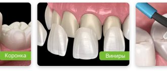
Types of dental abscess
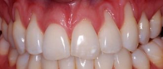
- Gingival Lump with a purulent cavity is formed on the soft tissue near the gum papillae between the interdental segments. The cause of inflammation is damage to the gum tissue, accumulation of food, plaque between the teeth, or injury to the gums by a toothbrush or floss. Gingival abscess (flux) affects only the gum tissue; the tooth and ligamentous apparatus with the root are not affected.
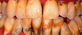
- Periodontal Exacerbation of periodontitis. The infection affects the tissue between the alveolar process and the gum, and hypertrophy of the periodontal pocket begins. Inflammation is provoked by subgingival deposits (plaque + tartar). Purulent exudate accumulates inside the gum pocket and forms a lump. The infection will penetrate deeper into the bone tissue and tooth root.
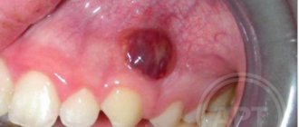
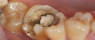
- Periapical (root) Most often occurs in units with non-living pulp. The cause of inflammation is poor treatment of caries, chronic periodontitis of root canals. Pus accumulates to a limited extent in the periodontal tissues at the root of the tooth; a granuloma with exudate is formed at its apex. Inflammation spreads to the supporting apparatus of the tooth and periodontal tissue. As it matures, a fistulous tract forms and pus comes out to the surface of the gum in the form of a ball.
- Pericoronal (pericoronitis) The soft tissue of the gums becomes inflamed during tooth eruption. Pericoronitis often occurs in partially erupted wisdom teeth. If bacteria gets into the “hood” above the tooth or plaque accumulates, a lump with pus forms. Surgical excision of the gum or tooth extraction is recommended to avoid swelling of the face, neck and the development of angina (in the lower jaw).
- Endo-periodontal (combined) Abscess is formed in the complex of endodontic tissues (pulp, dentin, root canals). The inflammatory process in periodontal tissues is supported by infection from the root canals through the apical foramen. Bacteria spread along the dentin canals, which can cause periodontitis in adjacent teeth.
International classification ICD-10
- K-04.6 Periapical with fistula. The cavity with purulent contents communicates with the maxillary sinus, nasal or oral cavity, and facial skin.
- K-04.7 Periapical without fistula. The abscess is associated with a complicated form of periodontitis.
- Abscess in acute form of periodontitis (K-05.20 periodontal or K-05.21 periodontal). The source of infection is hidden in the periodontal tissues, periodontal pockets swell and teeth become mobile.
Classification of abscess according to severity:
- mild - a purulent sac in one anatomical area;
- moderate severity - the pathological process affected two anatomical areas (for example, an abscess on the gum and face);
- severe - abscesses of the gums, sublingual space, neck and face.
☝️ The abscess affects the surface of the gums or deep layers of the periodontium. Tooth abscesses appear in the lower jaw in 72%, and in the upper jaw in 28% of cases. Most often, the causative teeth are molars, followed by premolars; most rarely, an abscess occurs on the incisors or canines.
Stages of abscess development
- Initial. Inflammation of the gums near the tooth or implant worsens. With strong immunity, the symptoms are almost invisible, and the pathological process can go away on its own. With a lack of immunity, necrosis of soft and bone tissue, hyperemia of blood vessels, and pain syndrome begin.
- Subacute. The inflammatory process intensifies. An infiltrate (tissue compaction) forms in the area of the gingival papillae, in the periodontal pocket, and at the tooth root. A throbbing pain begins when pressing on the unit.
- Spicy. Abscess formation begins - pus accumulates in the carious cavity. Exudate, swelling of the gums, sharp constant pain. The submandibular lymph nodes become enlarged and painful.
- Chronic. Resolution of the abscess - a fistula tract is formed, through which the purulent contents of the lump come out. The pain subsides.
Pathogenesis
In the pathogenesis of purulent inflammation, a decrease in antibacterial resistance, both general and local, is important. The integrity of the skin and the bactericidal properties of the secretion of the sweat and sebaceous glands prevent the introduction of pyogenic flora. If these conditions are violated, purulent-infectious inflammation develops. It consists of successive phases. histamine , prostaglandin , serotonin , etc.) in response In the exudative phase, under the influence of histamine, the liquid part of the plasma and proteins enter the intercellular spaces. During the infiltrative phase, tissues are saturated with lymphocytes and macrophages. As a result of exudation and infiltration, interstitial pressure increases, which significantly disrupts the condition of tissues, metabolic processes in them and reduces resistance to the effects of microorganisms. Then follows the immunological phase - the cellular and humoral response.
Inflammatory infiltrates have different outcomes, depending on the cellular composition of the infiltrate. Sometimes infiltrates resolve, but in most cases, with leukocyte infiltrates, lysosomal enzymes are released, which melt the tissue with the formation of pus - a purulent focus appears. Over time, areas of softening appear in the purulent focus and the abscess opens on its own or is opened surgically. The process ends with the reparative phase - the formation and growth of granulation tissue, which fills the resulting tissue defect as a result of purulent melting. With shallow lesions, no trace remains on the skin, but with deep lesions, a scar is formed.
Classification
According to localization, purulent infection can be:
- Skin and subcutaneous tissue of the arms, torso, legs.
- Skull skin.
- Pleura and lungs.
- Mediastinum.
- Abdominal organs.
- Pelvic organs.
- Bones and joints.
The most common purulent infection of the skin and its appendages (hair follicles, sebaceous glands, nails), as well as subcutaneous tissue (the lesion is located under the skin). The localization of purulent foci can vary - on the head in the hair, on the ear or on the lip.
Pustular skin diseases are the most common dermatoses. Purulent sores on the body ( pyodermitis ) are caused by staphylococci and streptococci. The greatest contamination with staphylococci is in the folds of the skin and subungual spaces.
There are:
- Superficial forms of purulent skin diseases ( ostiofolliculitis , folliculitis ).
- Deep ( furuncle , carbuncle , hidradenitis ).
Staphylococcal lesions are always associated with the sebaceous gland or sweat gland, hair follicle. The lesion spreads deeper, local and general temperature rises and creamy yellow-green pus forms. Streptococcal lesions are superficial purulent lesions of smooth skin that enlarge through peripheral growth.
Ostiofolliculitis is the mildest form of staphyloderma affecting the tissue near the hair shaft. It occurs in men in the mustache and beard area and is single or multiple in nature. The process begins with the appearance of a small red papule, in its place a yellow abscess forms. After 3 days it becomes covered with a dirty yellow crust, which falls off with the formation of a residual pink spot.
Folliculitis is an inflammation of the hair follicle with the formation of pus. Unlike ostiofolliculitis, the sores in this case are surrounded by a red, inflamed cushion and are painful. Over time, a pustule forms, which opens and pus is released, forming erosion and a crust. The infiltrate resolves or a scar forms.
A furuncle is an inflammation of the hair follicle, sebaceous gland and subcutaneous tissue. The cause of the disease is a staphylococcal infection. Most often, boils form in areas that sweat, chafe, or are covered in vellus hair (legs, neck, face, and underarms). A boil often develops from folliculitis.
Furunculosis is a recurrent form of boil. There are local furunculosis (rashes in a limited area) and disseminated (elements scattered throughout the body). Chronic recurrent furunculosis has a sluggish course, periodically worsens and is difficult to treat with antibiotics.
Carbuncle is a purulent-necrotic inflammation of many hair follicles. The infiltrate is large due to the involvement of new follicles and the spread of inflammation deeper. The formation is sharply painful, and deep necrosis develops around the hair follicles in the central part.
Hidradenitis is an inflammation of the sweat glands that occurs with the formation of pus. The disease occurs at a young age, when the apocrine glands begin to function. Most often, the lesion forms in the armpit, sometimes on the nipple, around the genitals, navel or anus.
The most severe forms of purulent foci are:
- An abscess is an accumulation of pus in tissues and organs, delimited by a pyogenic capsule (membrane). Abscesses can form due to deep infection, hematogenous introduction of microbes, as well as as a result of medical manipulations. Many people are familiar with post-injection abscess. Often, an abscess of the buttock occurs after the administration of analgin , baralgin , magnesia , oil solutions and hormonal drugs. In the development of a purulent focus, the stage of infiltration and suppuration (or abscess formation) is distinguished. In the first stage, the skin turns red, swells, and deep in the tissues a dense, painful formation is detected. With suppuration (abscessation), the skin becomes bluish and areas of softening (fluctuation) appear. The abscess code according to ICD 10 is L02.3. An abdominal abscess can develop in any part of the abdominal cavity. Most often it appears after injuries, surgical operations, peritonitis and perforation of a hollow organ and intestinal tumor. Abscesses develop when infection spreads due to appendicitis , pancreatitis , Crohn's disease , diverticulitis . Patients have a high fever and severe abdominal pain. Computed tomography is used for diagnosis. Treatment is surgical with antibiotics.
- Cellulitis is a diffuse inflammation of the tissue without demarcation (there is no pyogenic membrane), and pus spreads throughout the tissue space.
- Empyema is an accumulation of pus in hollow organs.
- Mediastinitis is an inflammation of the mediastinal tissue, often purulent in nature. This disease is life-threatening. Mediastinitis often develops after surgery on the heart, lungs, aorta, vena cava, esophagus, thoracic lymphatic duct, nerves (vagus and phrenic) and mediastinal lymph nodes. Prevention of surgical infection is preoperative and postoperative antibiotic therapy. All patients are prescribed beta-lactam antibiotics ( penicillins , cephalosporins ) or vancomycin . In addition to surgical trauma, the development of mediastinitis is caused by an infection that spreads from adjacent organs through the cellular spaces: the esophagus, lungs, descending infection of the neck, oropharyngeal and dental infections.