Perforation of the maxillary sinus (or, as it is also called, the maxillary cavity) is an iatrogenic complication that occurs during dental procedures on the upper jaw. It consists of perforating the bottom of the sinus, which causes an abnormal communication to appear between it and the oral cavity. Air begins to flow into the mouth through the hole, bleeding from the hole and a feeling of compression in the projection of the cavity are possible. If infectious agents get into the wound, sinusitis develops.
The CELT Dentistry Department invites you to undergo a course of treatment for perforation of the maxillary sinus in Moscow. Our multidisciplinary clinic welcomes leading domestic dentists with decades of scientific and practical experience behind them. They have a modern equipment base for accurate diagnosis and treatment in accordance with international standards. Our dental department has all certificates and licenses, treatment is carried out on the basis of an official contract with guarantees. We use the most effective methods for closing maxillary sinus perforations that provide the best results.
Consultation with a dental surgeon - 1,000 rubles.
Plastic perforation of the maxillary sinus - 7,500 rubles.
At CELT you can get advice from a dental specialist.
- The cost of a consultation with a dental surgeon is 1,000
Make an appointment
Structural features
The maxillary (maxillary, main) sinus is located inside the bone of the upper jaw. It is delimited from the oral cavity by the alveolar process. It forms the bottom of the cavity. The volume of the cavity of the upper jaw can be up to ten cubic centimeters. She has a communication with the nasal cavity. The inside of the cavity is lined with mucous tissue.
The structure has features that make it easy to damage:
- Sometimes the thickness of the bone plate between the bottom of the cavity and the roots of the teeth does not exceed one millimeter.
- There is a variant of the location of the roots of the second and first molars, when they penetrate into the cavity and are delimited from it only by the mucous membrane that lines the sinuses.
- The bone plate quickly thins out during inflammatory processes.
- Small thickness of trabeculae of the upper jaw bone.
Such structural features cause slight damage to its wall, even when the doctor did not violate any rules and did not apply significant force.
What is perforation of the maxillary sinus?
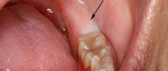
The formation of a defect in the maxillary sinus is a complication during manipulations on the upper jaw. A hole is formed between the oral cavity and the main sinus. This can happen when removing the molars of the upper jaw (molars and premolars) or during prosthetics. And also for complex endodontic treatment of tooth roots and removal of cystic formations. The defect is formed at the site of the tooth socket.
What factors are taken into account by the dentist when performing an intervention?
The likelihood of sinus perforation increases when the tooth root is located in close proximity to it at the time of extraction. Therefore, surgical treatment should be carried out with extreme care and subsequent X-ray monitoring of the tissue condition after surgery. The dentist also monitors the condition of tissues when using pins for implantation or when treating root canals for filling purposes, since there is a high risk of filling material and root fragments penetrating into the maxillary sinus.
If the process occurs at the time of implantation into bone tissue or when filling canals, then this is a direct mistake that is made by the doctor during his therapeutic tactics. The only way to control manipulations is X-ray control and the experience of the dentist, who, when working, must take into account the anatomical structure of the patient’s upper jaw. In this case, damage to the bottom of the sinus is fraught with serious complications that are difficult to eliminate. Especially if this happens during the implantation of artificial roots. The bone tissue in the upper jaw quickly undergoes degenerative changes, and this leads to a decrease in the height of the alveolar process.
When performing root resection to eliminate cystic formations, as in the above cases, the dentist must fully examine the patient. If the history is insufficient, perforation may occur in a situation where the doctor does not know the exact size of the bone plate that separates the bottom of the sinus from the wall of the cyst itself. In this case, the prognosis worsens if it is necessary to remove a large volume of bone tissue.
Causes of perforation.
The dentist is not always to blame for this complication. It can often appear due to the individual anatomical characteristics of the patient. It may also be due to the course of the inflammatory process in the tissue surrounding the root.
The causes of perforation may be the following:
- Perforation of the maxillary sinus during tooth extraction occurs most often. Its bottom is perforated when a tooth is suddenly removed with great force.
- In some patients, the roots of the upper teeth penetrate into the sinus cavity. When teeth are removed, the integrity of the bone plate is automatically violated. During treatment, filling material may enter the cavity.
- For technically complex endodontic treatment. With this type of treatment, the inflammatory focus is located deep in the gums or under the root of the tooth. Instead of removing the diseased tooth, the dentist tries to save it. During this type of treatment, the bone plate is easily damaged.
- In the process of installing an implant in the upper jaw bone with subsequent dental prosthetics, the maxillary cavity can be easily damaged. This is because the implant is similar to a screw and must be screwed into the bone. If there are defects in this manipulation or anatomical and topographical features, the patient’s bone plate of the upper jaw may be damaged (the size of the implant was incorrectly selected, there were defects in preparation for implantation). Before placing an implant, the doctor does not take into account the fact that when a tooth is removed, the thickness of the bone plate quickly decreases.
- Perforation can occur due to chronic inflammation of the tissues surrounding the tooth (periodontitis). With this pathology, the bone plate delaminates and becomes thin. If a tooth has to be removed in such a situation, perforation almost always occurs.
- Carrying out manipulation to remove an impacted tooth from the maxillary cavity.
- Perforation often occurs during tooth root resection procedures. The need for this manipulation arises when extracting a root with a festering cyst.
Prevention
Perforation of the maxillary sinus is a problem that is easier to avoid than to fix it later. Since perforation is caused by dental intervention, preventing the problem falls on the shoulders of the dentist. He is obliged:
- responsibly examine the patient before performing procedures;
- clearly understand the anatomical features of the client before major intervention;
- strictly adhere to the intervention technology.
The dentist is also obliged to respond adequately to any signs of perforation that has just occurred due to his fault. If for some reason the doctor has not fulfilled his own duties, then it is up to the patient - he must refuse self-medication and put aside the fear of dentists, in no case try to “endure” the discomfort, but immediately seek help.
Symptoms of perforation of the maxillary sinus
How does perforation of the maxillary sinus manifest itself? There are specific symptoms when this happens.
Signs of perforation:
- Bleeding from the tooth socket with the inclusion of air bubbles. When you exhale through the nose, the number of bubbles will increase.
- With perforation, bleeding occurs not only from the tooth socket. It may come from the nasal passage, which is close to the sinus.
- The patient speaks “in his nose” or nasally.
- Then there is a feeling of free passage of air through the tooth socket.
- The patient sometimes notes distension and a feeling of heaviness in the middle third of the face on the affected side.
If the perforation was not recognized immediately and treatment was not carried out, then the symptoms of sinusitis are added to the previous clinic.
Sinusitis manifests itself:
- Body temperature rises.
- The feeling of fullness in the projection of the maxillary sinus intensifies.
- Nasal breathing is difficult.
- The nasal mucosa on the affected side is swollen.
- General weakness increases.
- Aching pain in the nasal region.
- Purulent discharge from the nasal passage on the side where the manipulation took place.
Methods for determining perforation
This pathology can be identified only on the basis of a characteristic clinic. If there is any doubt, a full range of diagnostic procedures is performed, including instrumental methods.
To diagnose a bone plate defect and subsequently eliminate it, it is necessary to perform the following manipulations when examining the patient:
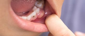
- Carefully examine the tooth socket after the tooth root has been removed.
- Perform sounding of its bottom.
- Ask the patient to pinch their nose and exhale through the nose. The air will escape into the mouth through the tooth socket.
- If the patient puffs out his cheeks, air passes into the nasal cavity. But this technique can provoke sinus inflammation and should not be used frequently.
Instrumental diagnostic methods include:
- probing the dental and perforation canal using a thin probe;
- CT scan;
- radiography, in the pictures you can see the defect and foreign bodies;
- general blood analysis.
Treatment of sinus perforation during tooth extraction
The tactics for managing a patient with perforation depends primarily on the condition of the sinus itself and the time of detection of this defect. This defect should only be treated by a qualified specialist.
Treatment of perforation of the main sinus of the upper jaw has the following objectives:
- Closing the defect.
- Prevent the process of inflammation in the sinus.
- Prescribe treatment if there is inflammation.
- If there are foreign particles, they must be removed.
If the perforation was immediately noticed and there are no signs of infection, then the treatment measures are as follows:
- Preservation of a blood clot in a tooth socket.
- Take measures to prevent its infection (application of a tampon with iodine solution).
- Apply stitches to the gums, if necessary.
- Treatment is carried out until the granulations grow and the defect is closed.
- The tampon is not removed from the hole.
- If the defect does not close on its own, it is covered with a plastic plate. It is fixed to the teeth.
- Prescribing a course of drug therapy aimed at counteracting inflammation.
If the perforation is complicated by gum rupture and penetration of foreign particles into the soft tissues surrounding the tooth socket, plastic closure of the defect is performed on the same day. Or after some time, when you are confident that the fabric will hold the seams. Before this, all foreign bodies are removed and areas that have undergone necrosis are excised. The manipulation is performed under x-ray control to make sure that there is no foreign body. If penetration of a foreign body into the cavity occurs, then it is necessary to perform surgery in a hospital setting.
Operation stages:
- Opening of the main sinus of the upper jaw.
- Removal of a tooth from the maxillary sinus (its fragments) and other foreign bodies.
- Excision of necrotic areas.
- Closing the defect.
How to treat?
The tactics for eliminating the problem depend on exactly what causes led to the perforation and what the general clinical picture is. Almost always, repairing a perforation requires surgery, the only exception to this rule is when a hole was created during a tooth extraction, provided that the dentist found it immediately and no foreign objects or infection got inside. If the problem is identified immediately, it is important to keep the blood clot in the place where it formed and prevent it from becoming infected - it will become a natural barrier to the infection entering the sinus. Protecting a blood clot from infection is carried out using a tampon with an iodine solution, which will have to be kept for at least a week.
In some cases, the doctor chooses the tactic of suturing the gum tissue. Another alternative is to install a compact plastic plate that is attached to adjacent teeth and acts as a barrier between the oral cavity and the sinus until the septum is restored.
For obvious reasons, physical barriers cannot provide a 100% guarantee that infection will not penetrate the hole, so the doctor additionally prescribes vasoconstrictors and anti-inflammatory drugs. They can be taken at home, but sometimes require the patient’s outpatient presence.
Penetration of any foreign objects into the maxillary sinus is regarded as a serious complication, which automatically means the need for surgical intervention and hospital treatment. In such a situation, the sinus is opened, removing foreign objects and tissues that cannot be restored, after which the fistula is covered with the patient’s tissues.
Consequences of perforation of the maxillary sinuses
If the presence of a defect is not noticed and the patient, despite the symptoms, does not consult a doctor, this threatens the onset of serious and dangerous consequences for health.
Possible consequences:
- Severe inflammatory reaction.
- Formation of osteomyelitis.
- Generalization of infection.
- Development of abscess and phlegmon.
- Loss of healthy teeth in the fistula area.
- Chronic sinusitis.
- Meningitis.
- Encephalitis.
- Cavernous sinus thrombosis.
To avoid the formation of complications, the patient should visit the dentist in case of any problems after tooth extraction.
Hospitalization
The day hospital at our Center is a post-operative support service for patients. Necessary if the patient has concomitant cardiac issues, such as hypertension, arrhythmia, AOS, etc.
Gentle ultrasound surgical protocols for low-impact PiezoSurgery operations in combination with microscopic surgery performed by operating teams consisting of pairs of the most experienced surgeons at our Center allow you to carry out treatment so delicately that you will not need mandatory hospitalization, which is so popular in Moscow hospitals.
All operations are performed only in medicated sleep, without pain or nervous overload.
The treatment will be carried out with respect for your personal time, in a short time and at a comfortable time, the operating teams and anesthesiology department work seven days a week and holidays.
Old sinus perforation
If the defect is not detected and eliminated in a timely manner, acute inflammation will subside. Within a month, the patient develops a fistula. It connects the gum surface and the sinus cavity. Signs of a chronic inflammatory process appear. This will be a serious complication.
The patient has complaints:
- The presence of dull pain in the upper part of the cheek, of a constant nature. They radiate to the eye area and temporal region.
- Feeling of nasal congestion on one side.
- Separation of pus from the nose and from the fistula on the upper jaw.
- Swelling of the middle third of the face on the affected side.
- Air movement through the defect.
- Difficulty speaking.
- Getting fluid from the mouth into the nose.
Therapy of old processes is associated with significant difficulties. Patients are indicated for surgical treatment in a hospital.
Operation stages:
- Opening of the main sinus of the upper jaw.
- Removing foreign bodies.
- Excision of necrotic areas and granulations.
- Excision of tissues forming the fistula.
- Closing the defect.
After the operation, drug therapy with the use of antibiotics, anti-inflammatory and decongestant drugs is mandatory for a course of two weeks.
Preventive measures before and after sinus lift to prevent the development of a fistula
Fistula after sinus lifting and bone grafting is quite easy to prevent by taking certain groups of medications before and after surgery, as well as observing the rules of oral hygiene.
Rules of conduct after surgery
The protective regimen should last from 5 to 7 days after the sinus lift. It includes a number of behavioral features:
- When brushing your teeth, special dental pastes with antibacterial and anti-inflammatory properties are used: Solcoseryl, Lakalut, Sensodyne. Oral hygiene procedures should be carried out twice a day (in the morning and before bedtime). It is necessary to brush your teeth on the operated side with great care so as not to disturb the condition of the tissues and implant after surgery.
- When eating, chewing movements should be carried out exclusively on the healthy side. Drinking all kinds of liquids through a straw is strictly contraindicated. You should also give up sweet foods for a week.
- During the first two weeks, you must stop playing any sports. This is especially true for athletics, gymnastics, cycling, swimming and martial arts. You should avoid the latter for a month.
- Try, if possible, not to sneeze or cough, avoid head injuries and bruises, and shaking.
- When sleeping, do not lie on your operated side.
Advantages of treatment in our clinic
Summing up, we can understand that it is very easy to get a septal defect in the cavity of the upper jaw. This depends both on the actions of the dentist and on the individual anatomical characteristics of the patient.
When seeking treatment at our clinic in Moscow, you can be sure that you are guaranteed not to have such complications. We prevent complications.
To do this, we prescribe a mandatory set of preventive measures to prevent them:
- We conduct a complete examination of patients before performing any dental procedures.
- We perform a comprehensive assessment of the patient’s anatomical and topographical features.
- We follow all stages of dental procedures.
You will be satisfied with the quality of dental services if you contact us at the clinic. Our clinic has the most modern equipment at the level of the standards of the best European and American dental clinics. We can offer you a full range of all diagnostic procedures in one place. Our doctors have been trained in the world's leading dental centers and are proficient in the latest treatment and prosthetic techniques. We will help even in the most difficult cases. We develop an individual course of treatment taking into account all the characteristics of the patient. We use only proven treatment methods and materials of the highest quality. Our clinic has made the highest quality dental services available. You can view the full range of our treatment and diagnostic capabilities on our website.
Why you should entrust your treatment to the ENT Department of Dentistry
ENT dentistry is a comprehensive approach to the treatment of complications in the maxillary sinuses after dental treatment
The symbiosis of two areas - dentistry and otolaryngology - makes it possible to identify the cause, assess the situation, and select competent treatment tactics.
The ENT department of the Doctor Levin Center has been providing assistance for many years when problems arise after treatment and removal of teeth located on the border with the maxillary sinus. Surgical treatment is carried out by candidates of medical sciences, maxillofacial surgeons with otolaryngological training .
Patients come to the Center after a painful search for a solution to the problem. Repeated treatment by an ENT doctor in the hospital does not bring results. But a thinking patient should understand that if there is concern on the side of the sinuses where endodontic treatment once took place, you need to contact an oral and maxillofacial surgeon with ENT training. Only in this case can a comprehensive assessment of the situation be made and an adequate treatment plan drawn up.