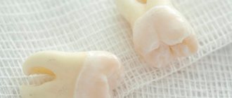
Impacted teeth are abnormally located teeth that cannot erupt into the oral cavity on their own and remain “sit” in the jawbone for years. To designate such an anomaly of the dental system, dentists use the term “retention” (translated from Latin as “retention”, “restraint”). Many dental patients have had to deal with problems caused by impacted wisdom teeth. Unfortunately, not only the “eights” are susceptible to retention, but also the canines, as well as the lateral and central incisors. Impacted teeth can be called a time bomb, as they can create a lot of trouble for their owner.
What is an impacted tooth?
Retention is an abnormality in tooth eruption, in which the tooth erupts partially or remains completely in the tissues of the jaw.
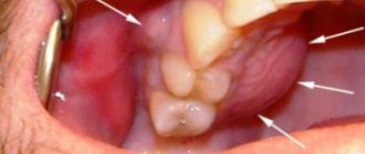
An impacted tooth is a tooth that has failed to erupt and “sleeps” in the gum or even in the jaw bone. There are cases when only part of the tooth crown erupts and is visible, and the rest of the tooth is covered by the gum - such teeth are called semi-impacted.
Most often, impacted canines and wisdom teeth are found - the notorious eights, but there are cases of impaction of the central and lateral incisors, and the first - a little more often.
Reasons for delayed eruption
Tooth eruption may be delayed for various reasons:
- underdevelopment of jaw bones;
- abnormal position of the tooth germ;
- fusion of the root of an impacted tooth with the root of an adjacent tooth;
- delayed resorption of the root of a baby tooth;
- pathological neoplasms near the impacted tooth;
- premature loss of a baby tooth and displacement of the neighboring one in its place.
Risk factors that increase the likelihood of retention include:
- genetic predisposition;
- weakness of the body due to frequent infectious diseases;
- unbalanced nutrition in childhood;
- disorders of embryonic development;
- supernumerary teeth;
- rickets.
What are the types of tooth impaction?
There are two types of retention - partial, in which part of the tooth is visible from the gums, and complete - in which the tooth is completely located under the gum tissue and/or jaw bone.
Such teeth can also be positioned differently, both being in “their” place in the dentition and occupying an incorrect position outside the dentition.
- Vertical
- Horizontally.
- At an angle to the jaw
If a tooth does not erupt in the right place beyond the border of the dentition, they speak of tooth dystopia.
There are even reverse retentions, when the tooth lies “upside down,” that is, upside down with its roots.
There may be only one impacted tooth in the dentition, or two impacted teeth may be located symmetrically. This problem concerns both milk and permanent teeth.
Symptoms
Retention of baby teeth is rare; retention of permanent teeth is said to occur if the timing of eruption is delayed by more than 10 months. Most often, the lower and upper wisdom teeth (third molars) are delayed in their appearance; in second place in terms of delay are the upper canines, incisors, and second premolars. The impaction of the upper canines is usually symmetrical.
A delay in the eruption of wisdom teeth until the age of 25-27 is considered normal; after this age, third molars should appear in most people. But it happens that wisdom teeth do not erupt at all: 10% of the population lack their rudiments.
An impacted tooth may not cause complaints from the patient for a long time; it is often discovered by chance during an X-ray examination during the treatment of neighboring teeth. A missing tooth can cause:
- trigeminal neuralgia;
- headaches;
- destruction of the roots of adjacent teeth;
- odontogenic cysts, complications of which can be purulent periostitis, odontogenic sinusitis, abscess.
A semi-retained wisdom tooth causes pericoronitis - inflammation of the gums (inflammation of the hood): the gum hangs over the tooth, and uncleaned food debris rots underneath it. In this case, pain appears when chewing, pus is released from under the gums, and there is an unpleasant odor from the mouth.
When an impacted wisdom tooth erupts, patients feel aching pain in the jaw, which is caused by:
- displacement of adjacent teeth: in a horizontal position, the impacted tooth rests against the wall of the adjacent tooth;
- obstruction of the jaw bone from tooth eruption.
Symptoms such as increased body temperature, enlarged lymph nodes, swelling of the tongue and gums are also added.
What complications do impacted teeth cause and why do they need to be “extracted”?
It is difficult for the patient to immediately understand what danger “hidden” teeth can pose, but it is really serious.
Semi-retinated teeth cause inflammation of the “hood” with which they are covered and adjacent tissues, and this provokes purulent inflammation.
Fully impacted teeth that lie in the jaw can behave like a foreign body; they put pressure on neighboring teeth, causing their displacement and a number of other problems.
In this case, the patient experiences pain and fever in case of inflammation, a feeling of pressure and discomfort in the jaw, neuralgia or numbness of part of the face.
If we list the diseases that can result from impacted teeth, it will become clear that this is a real “time bomb” that can detonate at any moment.
What diseases can be caused by an impacted tooth:
- periodontal cyst,
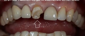
- caries of neighboring teeth and their roots,
- pulpitis,
- pericoronitis,
- periostitis,
- periodontitis,
- purulent lymphadenitis,
- inflammation of the trigeminal nerve,
- odontogenic sinusitis,
- abscess,
- phlegmon,
- resorption (resorption) of the roots of adjacent teeth,
- bad habits: mouth breathing, infantile swallowing, inserting the tongue into a dental defect,
- problems with biting food,
- malocclusion, displacement of interdental contacts, crowding of teeth.
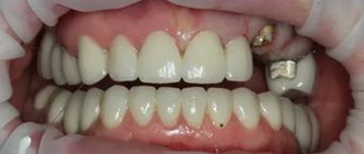
Can the root “come out” on its own?
Some patients, due to fear of pain and other discomfort, wrongly believe that if no action is taken, the base will fall out on its own over time. They walk around for many years with rotting bone remains in their mouths, creating a favorable environment for the development and spread of pathogenic microorganisms. An unpleasant odor appears in the mouth. Sometimes the root can actually come out without outside intervention, but this happens extremely rarely.
How events usually develop if you don’t visit the dentist on time:
- Hard deposits and stones form on the surface. The mucous membranes surrounding the base are irritated.
- The gums become overgrown and a severe inflammatory process develops. Subsequently, soft tissue dissection will be required.
How are impacted teeth diagnosed?
Impacted teeth are often “found” by accident during an X-ray diagnosis for another dental reason. If a patient asks about orthodontic treatment and is missing some teeth, or has symptoms that may be caused precisely by the presence of hidden impacted teeth, then a special diagnosis is carried out.
The problem of impacted teeth is solved by two specialized dentists - an orthodontist and a surgeon, so the patient will have to undergo diagnostics by an orthodontist and diagnostics by a surgeon. Most of the diagnostic procedures will be general, and the key is x-ray diagnostics, and it is preferable to do CT - computed tomography (3D diagnostics). Unlike an orthopantomogram (OPTG), it shows the spatial location of the impacted tooth in the jaw and relative to other teeth, which is very important for the surgical stage of traction - exposing its crown, because allows the surgeon to choose the optimal “approach”.
Features of the technique
A technique based on teeth traction allows you to restore the natural anatomical position of problem units. The correction involves the use of controlled orthodontic force, leading to a mechanically mediated change in the primary state of the element. When performing the operation, the features of the anatomical structure of the root part are taken into account, the proper use of which makes it possible to create pressure sufficient for histological changes.
It is important to note that traction is not a one-step procedure and is carried out in several sessions. The duration of restoration of the natural anatomical position reaches several months, but in rare cases it can be significantly reduced. A factor influencing the rate of correction is the patient’s age.
What to do if an impacted tooth is found?
There are 3 solutions to the problem of an impacted tooth:
- Orthodontic traction and placement in the dentition. This is often done with impacted teeth that are located in the smile zone - canines and incisors, provided that there is a place for them in the dentition and the tooth itself is healthy.
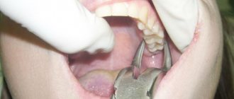
- Removal is a common fate for impacted eights, especially if the patient is undergoing orthodontic treatment. Very often, wisdom teeth erupt partially – this provokes inflammation and complications. Even a completely impacted (“hidden”) tooth serves as a potential source of caries, tooth displacement, and instability of orthodontic treatment. There are many reasons - there is only one verdict: it is better to delete it. Other indications for the removal of different types of impacted teeth are cysts, signs of inflammation, lack of space in the dentition, destruction of the neck of the tooth, caries.
- Preservation and surveillance. It is justified when the patient has no problems with the place in the dentition or bite, orthodontic treatment is not required, the tooth does not provoke complications and its absence does not spoil the aesthetics of the smile. Such teeth are observed; in case of the slightest problem, they are recommended to be removed.
Diagnostics
A semi-retained tooth is easily detected: its tip is visible above the gum, and its contours are determined by palpation.
A completely impacted tooth cannot always be detected during routine examination; impaction can be assumed based on the long-term failure of the permanent tooth to erupt. Definitive diagnosis is possible only after an X-ray examination. Carry out:
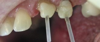
- targeted radiography - the image is taken on a limited area of the jaw, allows you to assess the condition of 3-4 teeth;
- orthopantomography - a survey x-ray, on it you can see the condition of the teeth and tissues of both jaws.
To make the right decision on further treatment, a computed tomography scan is often required - using a special device, the doctor receives a layer-by-layer image of the jaw. The resulting images can be combined into a 3D image, which makes it possible to make an accurate diagnosis and determine the position of the impacted tooth relative to other teeth. Computed tomography is indispensable in the diagnosis of supernumerary impacted teeth, the detection of which using conventional x-ray examination may be difficult due to the overlay of their image on the contours of permanent teeth.
In children, a sign of retention is the prolonged absence of replacement of a baby tooth with a permanent one.
How to pull out and place an impacted tooth in the dentition?
In some cases, if the root of an impacted tooth has not yet formed and there is an obstacle to its eruption, then removing such an obstacle is enough for the tooth to come out of the gums and take its place in the dentition. If the tooth root is already formed, this method will not help.
To “get” and move a fully formed impacted tooth into the dentition, treatment is carried out in 3 stages. Here we present the “classical” scheme, because in individual cases there may be deviations from this scheme.
- Preparatory orthodontic stage - you need to prepare a place in the dentition to which the orthodontist will move the extracted tooth. To do this, a braces system is installed, which aligns the teeth in the dentition and frees up the necessary space.
- The surgical stage is the image of the crown of an impacted tooth, on which a bracket or button is installed to transmit force from an arch or elastic element.
- The main orthodontic stage, during which the impacted tooth is pulled out and placed in its “rightful” place in the dentition.
How is surgery performed to expose the crown of an impacted tooth?
- As a preliminary stage, preparation for the operation is carried out. The patient undergoes professional hygiene and sanitation to reduce the amount of infection in the oral cavity and speed up postoperative healing.
- The operation to expose an impacted tooth is performed under local infiltration anesthesia and is considered a fairly serious surgical intervention.
The operation to expose the crown of an impacted tooth and install an orthodontic element on it can be carried out according to 2 schemes:
I Delayed bracket installation.
- The mucous membrane in the projection of the crown of the impacted tooth is excised, the entire crown of the tooth is exposed, and a special tampon is installed in the wound.
- After 2-3 days, a button or bracket is installed on the crown of the exposed tooth, which is tied to the orthodontic arch and traction begins.
II Bracket installation during surgery
- The dentist-surgeon peels off a small mucoperiosteal flap and exposes part of the crown of the impacted tooth, onto which the locking element is immediately fixed.
- The bracket is tied to an orthodontic arch or additional devices.
- After installing the orthodontic element, the flap of tissue is placed in place and the wound is sutured.
The disadvantage of this method is that in this case, repeated surgical intervention is possible if the bracket on the tooth comes off.
After surgery, the patient is prescribed antibiotics and antiseptic rinses, if necessary, to speed up healing. Light, non-traumatic food is recommended.