- Published by: Laima Jansons
The appearance of any pigmentation on the lips should not be ignored . If a blue dot appears on the lip, even more so. Because this always indicates the presence of malfunctions in the body.
Interesting! Doctors distinguish between two main types of spots - youthful and senile. Both cases have their own characteristics and causes. Before starting treatment for a defect, you should understand what caused it to appear.
Causes of dots on lips
Spots and dots on the lips can vary in size, shape and color. However, special attention should be paid to blue, intensely pigmented dots, since most often they appear as a consequence of serious diseases :
- Disorders of the liver and adrenal glands . Spots and dots on the lips in this case are similar in appearance to chloasma that appears on the face during pregnancy;
- Worm infestation . Pigmentation defects on the lips can be caused by the presence of parasites in the body;
- Peutz-Jeghers syndrome . This is a serious disease, the main symptom of which is the appearance of dots on the lips that look like freckles. The patient develops polyps up to 5 cm in size in the intestines. The syndrome is inherited;
- Melanomas on the lips appear after prolonged exposure to the sun and too frequent visits to the solarium . Such a defect can cause malignant degeneration of cells and cause the development of oncology ;
- Hormonal disorders . Blue spots and dots on the lips may appear due to long-term use of hormonal contraceptives;
- Venous lake . This defect appears on sun-damaged lips and is more common in older people. Sometimes these formations can be quite painful;
- Wart (papilloma) . This unpleasant defect can appear not only on the body, but also on the lip. There are many types of warts, and each case requires special treatment, including surgery.
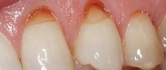
Photo 1: Brown and blue spots often appear on the lips of smokers, usually on the lower lip. The spot has a pronounced color, its diameter does not exceed 1 cm. Sometimes the spot may look like a small tubercle or nodule under the skin. Source: flickr (Adrian Trenholm).
results
Detailed clinical examination results were available for 38 of the 41 study participants. In 17 cases, the genital lesions were irregular in shape and color. In 22 cases, the lesions had at least two colors, including several shades of brown and black, zones of white or gray regression, and mixed zones of hyper- and hypopigmentation.
The size of 12 lesions exceeded 1.5 cm. 22 patients had multiple lesions, and 19 patients had only one.
The researchers found no significant correlations between clinical manifestations and histological results.
The researchers also checked for a history of human papillomavirus (HPV) infections and inflammatory dermatoses. HPV and inflammatory dermatoses are considered risk factors for the development of genital melanosis. During the analysis, no connection was found between HPV infection and inflammatory dermatoses with clinical or histological manifestations of genital melanosis.
The history of melanoma in close relatives and the person himself was analyzed in 32 and 34 patients, respectively. 7 out of 32 had melanoma in relatives, and 5 out of 34 had melanoma in their personal medical history. Only one patient was diagnosed with genital melanosis and genital melanoma simultaneously. Those. on the genitals of one person in different places (!) both genital melanosis and melanoma were present (approx. D.S.)
No correlation was found between family history of melanoma and clinical manifestations of genital melanosis.
In patients with melanoma, a tendency towards an increase in the number of suprabasally located melanocytes was revealed. The average number of melanocytes per square millimeter was 53.6. In patients without melanoma, this figure was 35.5.
Of the three patients with nuclear atypia in melanocytes, two had a history of melanoma.
Follow-up history is known for 30 of the 41 patients. None of the 30 participants developed genital melanoma. In one patient, genital melanosis developed 10 years after identification and successful removal of genital melanoma.
Detailed descriptions of clinical examination findings are available for 23 patients. Nine of the 23 study participants had unchanged genital lesions over 33 months. Ten of the 23 patients had no evidence of residual lesions 56 months after diagnosis of genital melanosis. Only 2 out of 23 people had relapses after excision of the formations. No correlation was found between the recurrence rate and any histological or clinical features.
Treatment of blue spots and dots on lips
Only a doctor can make a correct diagnosis and prescribe treatment in each specific case.
Spots that appear due to the harmful effects of ultraviolet rays can be successfully eliminated with the help of bleaching agents and appropriate vitamin complexes.
If the spots are intensely colored , injections of ascorbic acid help well. The appearance of a large number of blue dots on the lips (hyperpigmentation) requires oral administration of folic acid, aevit and riboflavin.
If any other diseases are discovered during research, the intervention of specialists in the required field is required - oncologists, gastroenterologists, dermatologists. Treatment in this case will be comprehensive, and as your overall health improves, the blue spots will disappear on their own.
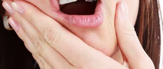
Photo 2: Treatment of any pigmentation on the lips depends on the causes of the defect, so first of all you will need to undergo an examination and a blood test. Source: flickr (Dave Black)
Treatment
Age spots are not an independent disease, but a symptom. Therefore, before treating pigment spots that have arisen, it is necessary to find out the cause of their formation, and for this you will have to undergo examination by a gynecologist, endocrinologist, gastroenterologist and neurologist. Treatment begins with treating the underlying disease, and only then does it make sense to begin to discolor spots and eliminate skin defects.
Among the methods for eliminating age spots, the following are currently widely used:
- laser removal. This is one of the most effective and harmless ways to get rid of age spots.
- chemical peeling, which is effective only on superficial stains;
- photoremoval (photorejuvenation);
The treatment option must be determined by the doctor, since only a specialist can, taking into account the characteristics of the skin and the condition of the patient’s body, identify all possible contraindications.
Find out the cost
Homeopathic medicines
The success of homeopathic treatment, first of all, lies in an individual approach to each patient. The homeopathic doctor prescribes a drug suitable for the constitutional type of the patient . In this case, the chosen remedy will affect the health of the body as a whole .
Depending on the type of stain on the lip and the patient’s psychotype, the following drugs may be prescribed:
- Arnica (Arnica montana). The drug promotes the resorption of compactions and is used to treat warts and venous nodules on the lips. The constitutional type of Arnica is full-blooded, good-natured people. Most often they are friendly, but during illness they become moody and irritable.
- Calcarea fluorica. Effectively fights vascular tumors, increases the tone of capillaries and blood vessels, and helps in case of helminthic infestation. Prescribed to patients with malocclusion and severe asymmetry of the bone skeleton.
- Silicea . The product effectively fights papillomas and helps eliminate hyperpigmentation on the lips. Psychotype - thin, sickly people who tend to get nervous over trifles. They often freeze and do not tolerate mental stress well.
- Phosphorus (Phosphorus). The drug is prescribed if the appearance of defects on the lips is caused by dysfunction of the liver and adrenal glands. The constitutional type of the drug is tall, stooped people with soft blond hair. The character is sensitive, touchy and vulnerable.
- Bellis perennis. The drug fights the manifestations of excessive pigmentation on the lips and has whitening properties. Most often prescribed to older people who complain of constant fatigue and memory problems.
The popularity of homeopathic treatment is primarily due to the proven effectiveness of the drugs . In addition, the absence of side effects that often occur when using traditional medications can be considered a big plus. Any drug is prescribed individually by a homeopathic doctor , therefore, when a blue dot appears on the lip, consultation with a specialist will be the first step towards a successful cure.
What groups of ingredients are used to lighten skin?
Representatives of these groups should be sought in the compositions of combination drugs.
- Tyrosinase inhibitors – azelaic acid, arbutin, flavonoids, resveratrol .
- Destruction and reduction of melanosomes: niacinamide, soy extracts .
- Inhibition of DOPA and dopaquinone synthesis: kojic acid.
- Exfoliation of the stratum corneum: AHAs and retinoids.
- Stimulation of the conversion of melanin to leukomelanin: vitamin C, E and glabridin.
Summary : to effectively lighten pigmentation and reduce the likelihood of side effects, it is more advisable to use combined preparations containing ingredients from different groups.
Laser therapy
A modern, highly effective method of removing pigmentation is laser technology. The method of laser therapy for HSV is based on the phenomenon of photothermolysis - the ability of pigment cells to absorb the energy of a laser beam, which subsequently leads to their destruction. The laser targets the chromophore-melanin found in melanocytes, keratinocytes or macrophages.
According to scientific studies, in more than 45% of patients, the manifestations of IGP disappeared after laser therapy, and in 35% of cases, lightening of the spots was noted.
Although in most cases the effectiveness of laser therapy for VGP is quite high and 1–3 sessions are enough to completely remove spots, constant exposure to UV rays on the facial skin leads to recurrence of VGP 3–4 years after successful treatment. Therefore, the therapeutic complex includes:
- daily use of medications or medicinal cosmeceuticals with a whitening effect for a long time;
- regular use of photoprotective products, even on a cloudy city day;
- professional manipulations aimed at enhancing desquamation (exfoliation) of the epidermis and destruction of melanin-containing cells.
A universal method for treating skin hyperpigmentation has not yet been developed, but over the long history of research and treatment of pigmentation disorders, experience has been accumulated in the use of a variety of correction methods, including a range of remedies from external applications and ointments to high-tech invasive procedures using surgical methods of dermabrasion and high-intensity laser radiation.
Our clinic’s specialists are proficient in various methods for removing skin hyperpigmentation and will help you choose a set of drugs and procedures that are most suitable for the skin type and individual characteristics of each patient.
Diagnosis of melanoma of the mucous membranes
When diagnosing mucosal melanoma, mistakes often occur. Due to its hidden position and lack of noticeable early signs, detection of mucosal melanoma is usually delayed.
When diagnosing a primary melanoma, especially a rare one, it is important to exclude the possibility of metastatic lesions from the primary cutaneous or ocular melanoma.
If melanoma of the mucous membranes is suspected, endoscopic examinations are performed:
- tracheobronchial tree;
- upper respiratory tract;
- esophagus and stomach;
- large intestine;
- rectal segment.
During the diagnostic procedure, the doctor takes fragments of the changed mucosa for analysis. A biopsy of a sample of suspicious tissue and subsequent pathological examination are the main points in the diagnosis of mucinous melanomas.
Amelanotic forms of tumors, which are often found among mucosal lesions, further complicate diagnosis. Immunohistochemical staining of the material to detect tumor protein (S-100, HMB-45, Melan-A, Mart-1) and the tyrosinase enzyme helps in the diagnosis of non-pigmented forms of the tumor.
If the prevalence and metastasis of mucous melanomas is suspected, a body scan with visualization is performed: CT, PET CT, MRI.
Exfoliating treatments
One of the methods to combat hyperpigmentation is a variety of peelings:
- phytic;
- mandelic acid;
- alpha hydroxy acids;
- retinol, etc.
For photoaging, medium peeling with TCA (trichloroacetic acid) will be most effective, for post-acne - salicylic, and for melasma - pyruvic.
One of the popular methods of injection cosmetology is biorevitalization using components that lighten melanin.
The influence of hormones on skin pigmentation
According to the free radical theory of melanogenesis, due to a lack of antioxidants, mitochondrial DNA in some areas is damaged. The skin is hormone-dependent, that is, even minor hormonal imbalances affect its condition. Many functions of the skin - mitotic activity of the epidermis, activity of pilosebaceous follicles, hair growth, etc. – are directly influenced by hormones, in particular sex hormones. Therefore, about 30% of women using combined oral contraceptives are affected by melanosis. However, stopping medication does not always lead to the disappearance of hyperpigmentation.
Hormones such as adrenocorticotropic, somatotropic and thyroid-stimulating hormones also affect melanogenesis. Scientists note that hormonally caused dyschromia is less susceptible to therapeutic effects than pigmentation resulting from exposure to sunlight or inflammatory processes.