• CRT (color reaction time) – color reaction time (Walter, 1958; Mayvold, Jager, 1978)
• TER (enamel resistance test) (Okushko V.R., 1984)
• Laser reflexometry (Grisimov V.P., 1991)
• Electrometry (Ivanov G.G., 1984; Zhorova I.A., 1989)
• Determination of the amount of calcium and phosphorus in enamel ash
• Enamel biopsy (determination of intravital enamel solubility); (Leontyev V.K., Distel V.A., 1974)
• Spectrometry
CRT (color reaction time)
• Purpose: to study the rate of dissolution of enamel in acid
• Method: study the time required to neutralize a standard amount of acid with ions more or less actively emerging from enamel apatites dissolved by this acid. The transition from an acidic to a neutral environment is determined using an acid-base indicator
• Materials and equipment: 1N hydrochloric acid solution, micropipette, filter paper disk with a diameter of 3 mm, soaked for 30 seconds in a 0.02% aqueous solution of crystal violette, which is yellow in acidic pH and violet in neutral pH, stopwatch.
• Methodology: tooth 12 is isolated from saliva, cleaned of plaque with a brush and dried. A paper disk is placed on the vestibular surface, and 1.5 μl of 1N HCl is applied to it using a micropipette (after the test, remineralizing agents must be applied)
• Interpretation of results: CRT˃60 s – solubility is low, caries resistance is high; CRT˂60 s – high solubility, low caries resistance
TER (enamel resistance test)
• Purpose: determining the degree of destruction of the surface layers of enamel under the influence of acid
• Method: visual assessment of an enamel defect resulting from the use of a standard acid solution under standard conditions, using a dye that is fixed in larger or smaller quantities in the irregularities of the damaged enamel and therefore gives a more or less intense color.
• Material and equipment: 1N hydrochloric acid solution, micropipette or glass rod, 1% methylene blue solution, 10-point blue scale (standard or prepared using serial dilution of the original solution 1:2 - from 100 to 0.18%)
• Methodology: tooth 12 is isolated from saliva, cleaned of plaque with a brush and dried. A drop of acid with a diameter of 1.5-2 mm is applied to the vestibular surface. After 5 seconds, remove the drop with a dry cotton swab in one movement. A drop of dye is applied to the damaged and adjacent intact enamel for 5 s, after which the dye is wiped off with a dry swab until the intact enamel returns to its original color (the pellicle acquires a barely noticeable blue tint)
• Interpretation of results: pale color 1-3 points – high caries resistance; 4-5 points – moderate caries resistance; 6-7 points – low resistance; 8 points or more – very low caries resistance
Enamel biopsy (determination of intravital solubility)
• Purpose: quantitative analysis of the mineral composition (Ca, P) of enamel, or rather that part of its apatites that react with acid.
• The method is based on the theory that calcium-saturated enamel can, in relatively larger quantities than caries-weakening enamel, release ions of this element to neutralize acid, while maintaining the apatite structure. Enamel is studied in vivo.
• Material and equipment: hydrochloric acid buffer solution (97 ml of 1N HCl and 50 ml of KCl) is mixed and added to 200 ml with distilled water; For viscosity, add glycerin 1:1. Microsyringe for application of buffer solution and aspiration of acid enamel biopsy.
• Methodology: The tooth is isolated from saliva, cleaned, and dried. A drop of demineralizing buffer solution with a volume of 3 μl is applied to the enamel surface. After 1 minute, the drop volume (biopsy) is taken with a microsyringe, the biopsy is transferred into a test tube with 1 ml of distilled water and used for quantitative chemical analysis.
• Interpretation of results: The technique allows you to assess the condition of the enamel in a comparative aspect, therefore it is actively used to study the degree of risk of developing caries in some people compared to others, to study changes in enamel that occur under the influence of mineralizing prophylaxis, etc. (it is important to remember that with an increase in enamel caries resistance due to the formation of fluorapatites in it, the solubility of the enamel decreases, and the amount of calcium in the biopsy drops).
Laser reflectometry. (Grisimov V.P., 1991)
• Purpose: to determine the density of the crystal lattice of the enamel surface. The method is based on the differences in the optical properties of resistant and labile enamel: well-mineralized, dense enamel reflects light more and absorbs it less (i.e. diffusely scatters) than loose caries-labile enamel.
• Material and equipment: helium-neon laser LGN-105 with a wavelength of 0.63 microns. A device for photographing laser light reflected by enamel. Meter of reflected light characteristics.
• Method: the tooth is cleaned, dried, and a beam of laser light is directed at it. The beam of light reflected by the enamel is photographed.
• Interpretation of the results: the diffuse component is less than 0.24 – the enamel is caries-resistant; more than 0.30 – caries-weakening.
According to the degree of resistance of teeth to caries, P. A. Leus identifies 5 groups, which, in descending terms of resistance, are distributed as follows:
1. First and second molars of the lower jaw.
2. First and second molars of the upper jaw.
3. The second premolar of the lower jaw and the first, second premolars of the upper jaw.
4. Maxillary central and lateral incisors, maxillary canine and mandibular first premolar.
5. Central and lateral incisors of the lower jaw, canines of the lower jaw.
If all teeth are healthy, then they should be considered a highly resistant type.
When molars and premolars are affected (the first three groups of tooth resistance according to the classification of P. A. Leus), the average level of tooth resistance in the examined individual is determined.
Involvement of central and lateral incisors in the carious process in addition to molars and premolars (the fourth group of teeth resistance to caries according to the classification of P. A. Leus) indicates a low level of resistance.
Caries damage to all functionally oriented groups of teeth should be considered as a consequence of a very low level of tooth resistance.
Date added: 2015-02-22; | Copyright infringement
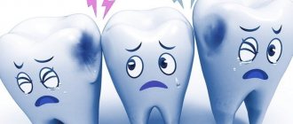
Caries is the most common lesion. This disease appears in almost everyone, and is accompanied by unpleasant sensations and sometimes serious complications. However, in dentistry there is such a thing as caries resistance. In everyday life, few people have heard this word and therefore it is unlikely that anyone will be able to give an exact definition of this concept.
What is caries resistance?
In dentistry, a condition called caries resistance is often encountered, but what does this mean? How is this condition characterized?
Caries is a disease of our time; it occurs in almost every person in one form or another. But there are those who have innate immunity, this phenomenon is called caries resistance.
Focal demineralization of tooth enamel is a widespread disease that occurs in more than 97% of the population of our country [14]. The development of new methods for diagnosing focal demineralization of tooth enamel is determined by the requirements of clinical practice related to the early and timely diagnosis of this pathology in order to use the least invasive treatment methods. The question of assessing the relationship between objective diagnosis of demineralization and adequate treatment in each specific case, taking into account the characteristics of the course of the disease, is also relevant. Most existing methods for diagnosing focal demineralization of tooth enamel are relatively imperfect, since they are based on subjective perception. In this regard, it is relevant not only to introduce objective diagnostics of enamel demineralization into practice, but also to create a clear scale for assessing its degree, which would reflect the depth and area of the lesion.
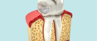
Focal demineralization of enamel is a systemic lesion of the cervical areas of teeth in the form of spots as a result of underdevelopment of tooth tissue and a decrease in the amount of calcium in it. This condition is formed during the period of maturation of tooth tissue after its eruption and corresponds to the ICD-10 code K02.0 - “Enamel caries. Stage of “white (matte) spot” [initial caries].” Caries in the spot stage is characterized by changes in color (matte surface) and then texture (roughness) of the enamel resulting from demineralization, which have not spread beyond the enamel-dentin border, in the absence of a carious cavity [2].
A feature of focal demineralization is that almost all teeth are affected. This process is the only reversible one. Enamel is a mesoporous substance; it does not contain cells and is not capable of regeneration when damaged, but there is a constant exchange of mineral ions in it, which come from saliva through the pores and are adsorbed on its surface, which makes it possible to carry out remineralization therapy [2]. The fundamental local factor in the development of this pathology is a local change in pH on the tooth surface due to the accumulation of dental plaque on it containing acid-forming streptococci ( Streptococcus mutans
,
S. sanguis
,
St.
mitis ,
St.
salivarius ), which are characterized by anaerobic fermentation, and some lactobacilli [21]. Caries develops cyclically, the rate of its development depends, firstly, on the frequency of action of lactic acid produced by microorganisms on the tooth, and, secondly, on the degree of demineralization, i.e., on the penetration of calcium and phosphorus ions into the hard tissues of the tooth (the process catalyzed by fluorine). In addition to fluoride deficiency, for demineralization of tooth tissue to occur, the pH of the oral environment must be below a critical point. On average, the critical pH point is estimated at 5.5 (Stefan curve) [1, 3].
Pathomorphological changes are characterized by varying degrees of enamel demineralization. On thin sections of the enamel, the lesion looks like a triangle with the base facing its surface. When studying enamel in polarized light, G. Gustafsonson (1975) identified 5 zones, depending on the intensity of demineralization. The zone of hypermineralization with the disappearance of structural details of the enamel is located deepest in the thickness of the enamel. In the 2nd zone, a decrease in its hardness was noted due to the partial dissolution of minerals, in the 3rd - an increase in mineralization. In the subsurface, 4th demineralization zone, minerals are washed out almost completely. In the superficial, 5th zone, complete disintegration is possible, but the enamel remains fairly mineralized and intact for a long time, even if the carious process extends to more than half the thickness of the enamel. A. Darling (1958) indicates that if in normal enamel microspaces constitute 0.5% of its volume, then in the body of the lesion they reach 25% of the volume, in dark enamel - 4%, and in transparent enamel - 2% [1].
With a white spot area of up to 3 mm2, changes reach 1⁄2 the thickness of the enamel layer. With more extensive lesions, changes in the enamel are observed down to the enamel-dentin junction. The formation of brown pigment in the spot is associated with the accumulation of the amino acid tyrosine with its further conversion into the pigment melanin. Under a white carious spot, changes in the enamel-dentin junction have not yet been detected; under a brown one, its damage may already be observed [1, 3, 21].
Focal demineralization of enamel is a borderline pathology for which both conservative and invasive treatment is possible. At the same time, there are no clear algorithms for determining a particular treatment plan, despite the wide variety of definitions of the degree of demineralization in enamel caries.
Existing methods for determining demineralization are divided into qualitative and quantitative. Qualitative assessments include clinical assessment. The main quantitative methods for determining enamel demineralization include the following:
1. TER-test (enamel resistance test; V.R. Okushko, L.I. Kosareva, I.K. Lutskaya, 1983). This test reflects the predisposition to caries (based on the functional resistance of enamel to acid). The TER test method boils down to isolating the tooth from saliva, cleaning it from plaque, and drying it. Then 1 drop with a diameter of 1-2 mm of 1% HCl is applied to the prepared tooth. After 5 seconds, the drop is washed off with distilled water, dried, and 1 drop of a 1% solution of methylene blue is applied. The dye is removed with a dry cotton swab in one erasing motion. The etched area is painted blue, the intensity of staining is assessed on a 10-point scale (1-10 points; the maximum score corresponds to the least caries resistance of the enamel)[6].
2. KOSRE-test (clinical assessment of the rate of enamel remineralization according to the method of T.L. Redinova et al.). This test is based on assessing both the condition of tooth enamel and the remineralizing properties of saliva. The enamel surface of the tooth being examined is thoroughly cleaned of plaque with a dental spatula and a 3% hydrogen peroxide solution and dried with compressed air. Then a drop of hydrochloric acid buffer pH 0.3-0.6 is applied to it, always in a constant volume. After 1 minute, the demineralizing solution is removed with a cotton swab. The etched area of tooth enamel is also exposed for 1 minute to a cotton ball soaked in a 2% solution of methylene blue. The compliance of enamel to the action of acid is assessed by the intensity of staining of the etched area of tooth enamel. The degree of staining is judged by the blue topographic tint scale (10-point scale: the least stained part is 10%, the most saturated is 100%). After 1 day, the etched area of tooth enamel is re-stained without repeated exposure to the demineralizing solution. If the etched area of tooth enamel becomes stained, this procedure is repeated again after 1 day. The loss of the etched area's ability to stain is regarded as a complete restoration of its mineral composition. The degree of resistance of tooth enamel to the action of acid is taken into account as a percentage, and the remineralizing ability of saliva is calculated in days. Resistance to caries is characterized by low compliance of tooth enamel to acid action (<40%) and high remineralizing ability of saliva (from 24 hours to 3 days), and caries susceptibility is characterized by high resistance of tooth enamel to acid action (≥40%) and low remineralizing ability. ability of saliva (>3 days) [5].
3. Determination of the initial level of mineralization of erupting teeth using the electrometric method (L.P. Kiselnikova, 1990). The degree of mineralization was assessed using the Dentest electrometry apparatus (ZAO Geosoft). The principle of the electrometric method is that in the hard tissues of fully mineralized teeth and without signs of demineralization, the electrical conductivity is 0. In teeth with reduced mineralization processes, the electrical conductivity values increase. The lower the degree of mineralization, the higher the electrical conductivity. An electric current of 1.8 to 4 μA passes through areas of focal demineralization, localized on the visible surfaces of permanent teeth with enamel mineralization ending. Permeability impairment from 1 to 8 points on the methylene blue staining intensity scale corresponds to an increase in the current value from 1.68 to 5.17 μA. With an increase in the size of the spots, the magnitude of the current passing through the tooth tissue significantly increases from 2.55 to 3.31 μA [7].
The electrometric method makes it possible to determine the general mineralization of the tooth crown, but can only be used to a limited extent for local changes. The absolute values of electrical conductivity vary significantly depending on the tooth, the local thickness of the enamel layer, the ability to carry out insulation and other parameters. The electrometric method is applicable for the relative assessment of changes in the mineralization of a spot on a specific tooth in a specific patient with complete repetition of measurement conditions, which during a clinical appointment can lead to quite serious errors in the results obtained.
4. Determination of focal demineralization of tooth enamel using infrared fluorescence using the DIAGNOdent apparatus.
The DIAGNOdent device (KaVo, Biberach, Germany) was introduced in 1998 by Hebst and Gall as an aid to the detection of caries and focal demineralization of tooth enamel on occlusal surfaces in addition to inspection and radiographic examination. This device works using light emanating from a diode laser; wavelength 655 nm, peak power 1 mW. Light travels through the fiber assembly to the tip of the handpiece. The tip of the handpiece is placed on the surface of the tooth and the laser beam passes through the tooth. Both organic and inorganic molecules in the tooth absorb light. Infrared fluorescence is used. Fluorescence, as well as return light, passes through the tip and enters through the ascending tissues to the photodiode sensor. The fluorescent light is measured and its intensity indicates the size and depth of the tooth's decay or focal demineralization. Fluorescence intensity is presented in the range from 0 to 99. According to prof. Reich (Univ. Hamburg), digital values from 5 to 25 correspond to carious damage to the enamel, 25-35 to damage to half the thickness of dentin, ≥35 to deeper damage to dentin. According to O.A. Krasnoslobodtseva and L.Yu. Orekhova (2000), the indicators for caries in the spot stage corresponded to 9.0±2.0, for superficial caries - 15.0±3.0; average caries - 50.0±30.0. Differences in digital indicators characterizing the condition of dental tissues are explained by different degrees of initial mineral maturity of the diagnosed teeth (EA Kidd, D. Beighton, L. Zoitopoulos, 2001). The reason for the fluorescent glow in the tooth is the presence of protoporphyrins and mesoporphyrins, waste products of bacteria (Hibst and Paulus, 1999, 2000). DIAGNOdent is widely used to detect caries on occlusal surfaces and focal demineralization on smooth tooth surfaces (Aljehani et al., 2006, 2007; Antonnen et al., 2003; Bamzahim et al., 2004, 2005; Lussi et al., 2003, 2006 ). Studies have shown that the sensitivity of the DIAGNOdent device for detecting caries and focal demineralization of enamel ranges from 0.17 to 0.78, and in some cases from 0.72 to 0.98 (Lussi et al., 1999; Shi et al., 2000) [8, 16].
Most studies highlight that DIAGNOdent is much more sensitive to pathology detection than traditional methods. But the increasing number of false-positive diagnoses makes it impossible to use this device as the only reliable one (Bader et al., 2004). Therefore, a definitive diagnosis cannot be made based on DIAGNOdent data alone.
5. Transillumination of focal demineralization of enamel - bright illumination of the coronal part of the tooth to study its structural components; carious lesions stand out as a dark shadow against the background of tooth enamel. The transillumination method also makes it possible to identify enamel cracks and assess the condition of tooth tissue around previously applied fillings [4, 12—14].
This method can only identify pathology, but does not evaluate it qualitatively and quantitatively, which deprives it of objectivity.
Treatment of focal demineralization of tooth enamel has been studied for decades and has a serious scientific basis. Theoretically, remineralization therapy for focal demineralization is justified by the preservation of the protein matrix in the tooth enamel in the early stages of caries (white carious spot), as well as the possibility of remineralization. Many studies have noted that after a course of restorative therapy, the enamel surface becomes more uniform and microporosity decreases. Thanks to the remineralization of enamel, it is possible to stabilize the initial process of its demineralization and even eliminate the chalk stain. The pigment spot remains preserved when the process stabilizes [9].
There are a huge number of remineralizing products - remineralizing pastes, gels, mousses. Let's look at some of them.
Profilak JSC "StomaDent" is created on the basis of domestically produced cedar balsam using a special technology that allows you to maintain the active component - fluorine in a dispersed state, preventing its precipitation.
Significant factors that unite fluoride-containing varnishes are the transformation of hydroxyapatite into fluorapatite and the formation of calcium fluoride in the outer layer of the tooth. After applying fluoride-containing varnishes, the tooth surface is protected by an acid-resistant layer of calcium fluoride.
Clinical studies of the use of fluoride-containing varnishes have proven their high effectiveness in the progression of proximal caries. They should be used immediately after the eruption of permanent teeth, since there are no contraindications for their use.
The use of Profilak is also indicated:
— for the prevention of tooth root caries (2-3 times a year);
— for the treatment of hypersensitivity of teeth (course of 1-3 procedures over 7-10 days);
— on low-calcified teeth (4–8 times a year) [15–17].
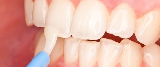
Remineralizing gel ROCS Medical Minerals. It is a source of bioavailable calcium, phosphorus and magnesium compounds that strengthen enamel. Thanks to special additives, it forms a stable invisible film on the teeth, ensures gradual penetration of minerals into the tooth tissue, and allows prolongation of the exposure of active components. After brushing your teeth, apply it to your teeth with a brush and refrain from eating or drinking for 30 minutes. Recommended course duration is 2 weeks. From 1 to 3 courses are conducted per year. It is possible to use the gel on an ongoing basis, as it is harmless and does not cause side effects [20].
GC Tooth Mousse is an application cream containing calcium and phosphorus, used by dentists in accordance with the recommended indications. GC Tooth Mousse is a water-soluble cream containing Recaldent™* CPP-ACP (casein phosphopeptide - amorphous calcium phosphate). In the oral environment, CPP-ACP binds tightly to biofilm, plaque, bacteria, hydroxyapatite and soft tissue, delivering calcium and phosphorus. Saliva increases the activity of CPP-ACP, and the pleasant taste of the mousse increases salivation. The longer CPP-ACP and saliva are in the oral cavity, the more effective the result. Recaident™ CPP-ACP is derived from cow's milk casein and should not be used in patients with milk protein allergies. There are known cases of recovery from enamel hypersensitivity after taking GC Tooth Mousse [18, 19].
One of the innovative areas for the treatment of focal demineralization is the method of caries infiltration of an area of pathological enamel with a viscous-flowing composite material Icon, developed by the German company DMG in 2009. The method is based on layer-by-layer “impregnation” (infiltration) of the focus of enamel demineralization with a light-curing resin, which is carried out after preliminary etching of the surface, relatively highly mineralized “pseudo-intact” layer of enamel. According to foreign authors [22], the infiltrate penetrates into the intercrystalline pores of the enamel throughout the entire volume of the affected area and, after hardening, strengthens (“reinforces”) the demineralized area of the enamel, generally preventing further progression of the disease [10]. Currently, laboratory and clinical studies are being actively conducted all over the world, which confirm the prospects of using the infiltration method for the treatment of caries in the spot stage [11].
Analysis of publications devoted to methods of diagnosis and treatment of focal demineralization of tooth enamel allows us to conclude that, despite numerous studies in this direction, many unclear questions remain. There are many recommendations and subjective methods for diagnosing this pathology of tooth enamel, but there are no objective standards and a method that gives an accurate quantitative assessment of the lesion. The question of optimizing the indications for the use of certain methods and means of remineralizing tooth enamel, depending on the degree of demineralization of its structure, remains open.
The authors declare no conflict of interest.
Features of caries resistance
Caries resistance includes several main properties of enamel:
- Degree of acid resistance;
- Microhardness;
- Permeability.
The development of caries depends on the following factors:
- On the degree of aggressiveness of various factors;
- From the condition of the enamel.
It follows from this that during the pathogenesis of the development of dental caries, the main role is played by the decalcifying effect of organic acids of microbial origin. It is believed that the degree of resistance of the enamel structure to acid demineralization directly reflects all the properties of resistance to carious lesions.
Caries resistance – resistance of dental tissues to carious effects. Caries resistance includes not only the condition of the tooth tissues, but also the condition of the oral cavity, oral fluid, and the condition of the body as a whole.
Therefore, resistance to dissolution is considered the most important quality of enamel, which allows maintaining the integrity of the structural and functional appearance in the presence of acid-forming bacteria inside the oral cavity and plaque. As a result of numerous studies, it was revealed that the degree of solubility of enamel tissue on different surfaces differs. The gingival areas of all teeth have the highest degree of solubility. This is due to the fact that it is in these places that there is a reduced level of mineralization. According to V.K. Leontiev, which was put forward in 1977, various factors may underlie the heterogeneous structure of enamel:
- Differences in mineralization of different parts of the tooth;
- The presence of various defects in the structure of the crystalline lattice;
- Individual qualities of interactions between the protein matrix and the mineral phase and other reasons.
Professor Okushko V.R. in 1984 determined that the acid resistance of enamel is associated with the properties of centrifugal permeability, due to which cerebrospinal fluid is released on the surface of the enamel. As a result of a series of experiments, it was found that by regulating the speed of movement of the cerebrospinal fluid, it is possible to change the acid resistance, and therefore the resistance of teeth to caries. In general, acid resistance and caries resistance depend on the properties of dental tissue and enamel. These factors are provided by the functional and structural features of the tooth structure.
Material and methods
To achieve the stated goal of the study at the Department of Pediatric Dentistry with Orthodontics at VSMU named after. N.N. Burdenko, as well as on the basis of the state educational institution of the Voronezh region “Boarding School No. 1 for orphans and children left without parental care,” a prospective cohort examination of the oral cavity of 138 children was conducted, of which 27 (19.6%) were aged 7-10 years old, 39 (28.3%) children aged 10 to 14 years and 72 (52.2%) adolescents aged 14-16 years old, living in the same type of environmental conditions of Voronezh, receiving the same nutrition, having intact dental row, foci of initial demineralization of enamel, as well as carious cavities of teeth.
In accordance with the purpose of assessing the effect of a fluoride-containing coating with tricalcium phosphate, a study was carried out on 45 patients with high caries resistance, without carious cavities and foci of initial demineralization of teeth, covered with a small amount of soft plaque, who made up the 1st group, 62 patients with sufficient average caries resistance, having foci of initial demineralization and primary foci of destruction of enamel and cement, as well as carious lesions of only chewing teeth, the absence of pulpless teeth, the presence of a small amount of soft plaque, according to the surveyed - new carious cavities did not appear every year - group 2 and 31 patients with reduced average caries resistance, having carious cavities in the chewing frontal teeth, the presence of several cavities in one tooth, the presence of pulpless teeth, a significant amount of plaque on the teeth, according to the surveyed, the annual appearance of new carious cavities, the rapid “loss out” of fillings within 1-2 years — 3rd group [4].
All groups were divided into 2 subgroups to evaluate the effectiveness of treatment with the use of therapeutic and prophylactic fluoride-containing coating with tricalcium phosphate and without the use of this product, but using individual oral hygiene products.
In laboratory conditions, 10 extracted teeth of patients aged 7 to 16 years were examined for orthodontic indications. Intact teeth were subjected to special research methods: scanning electron microscopy, X-ray spectral microchemical and energy dispersive analysis.
The objectification of clinical data was facilitated by the use of laboratory research methods, the results of which were recorded at certain time points - a clinical examination after 6 months. When carrying out the study, ethical principles were observed, written consent was obtained from the administration of boarding school No. 1, as well as the Department of Education, Science and Youth Policy of the Voronezh Region for the examination and implementation of preventive measures.
Dental fluoride material Clinpro White Vamish is a complex remineralizing fluoride coating with tricalcium phosphate, containing fluoride in the form of 5% sodium fluoride and the innovative ingredient tricalcium phosphate with fumaric acid, which does not allow it to react with the fluoride inside the package until the product is applied on the teeth. The coating is an alcohol solution of a modified resin. Clinpro White Vamish is sweetened with xylitol and has a pleasant minty flavor. The product is supplied in packs containing a single dose of 0.5 ml. Each 0.5 ml dose contains 25 mg of sodium fluoride, which is equivalent to 11.3 mg of fluoride ion (22,600 ppm fluoride).
Visible and accessible tooth surfaces were assessed by visual inspection in good light using a dental mirror and probe, and vital staining of the teeth with a 2% aqueous solution of methylene blue. To assess the hygienic state of the oral cavity, the expanded hygienic index according to Fedorov-Volodkina was determined; the plaque index was assessed using a probe from four surfaces without staining with preliminary drying with an air jet.
To assess the rate of reduction in caries growth, we proposed an index of clinical and laboratory assessment of the resistance of hard tissues of teeth (ICLORZ) as a more sensitive assessment result to initial changes in enamel, determined in combination with X-ray diagnostics, electrometric diagnostics and light-induced fluorescence, in contrast to the CPZ index (Table 14].
Table 1. A set of methods for clinical assessment of the condition of hard dental tissues Note. * — R0 — no signs of destruction; R1 - demineralization of the outer half of the enamel in the affected area; R2 - demineralization of the entire enamel layer in the affected area; R3 - demineralization of enamel and the outer half of dentin in the affected area; R4 - demineralization of enamel and deep layers of dentin in the affected area.
If you have filled teeth, the score is given depending on the electrometric resistance at the border of the filling and the hard tissues of the tooth.
The index was calculated as the ratio of the sum of points obtained to the total number of examined teeth in the oral cavity.
Intraoral X-ray diagnostics were carried out using a dental apparatus de Götzen srl (Italy) sistema radiografico per diagnosi intraorale - x-ray system, electrometric diagnostics of hard dental tissues were carried out using a DentEst apparatus, Russia. The measurements were carried out at a constant voltage of 4.26 V, and the results obtained in microamperes were recalculated to the resistance value of the hard dental tissues under study, taking into account the factor of the full maturation of the enamel. The assessment of light-induced fluorescence of hard dental tissues was carried out using an LED activator LED active 05, Russia, at a wavelength of 530 nm, illumination of 100,000 lux, and also a wavelength of 625 nm at a radiation power density of 140 mW/cm².
To determine the microcrystallization index of oral fluid on a drop of this liquid, the ratio of the number of ocular grid points projected on the crystals to the total number of ocular grid points projected throughout the entire drop of oral fluid was calculated.
The buffer capacity of saliva was determined using the CRT buffer kit, and the acidity of the oral fluid was determined using the I-500 basic ion meter.
Scanning electron microscopy and X-ray spectral microchemical analysis (XMA) of 10 extracted teeth were assessed using a low-vacuum scanning electron microscope model JEOL JSM-6380LV (Japan). The distribution of chemical elements in the area of the interface of the fluorine-containing coating and tricalcium phosphate with tooth tissues was studied by micro-X-ray spectral mapping of transverse chips of teeth using an INCA-250 energy dispersive analysis (EDA) system. The planar distribution of chemical elements was assessed by coloring the X-ray image with different colors specified by the operator.
Statistical analysis of the materials obtained as a result of the study was carried out using the mathematical software package Statistica 6. X, Biostat, which is an integrated environment for statistical analysis and data processing.
Insufficient mineralization of enamel as a factor predisposing to the development of dental caries
Tooth enamel
- highly mineralized tissue of a living organism: the content of mineral salts in it is 95%, organic substances - only 1.2%, water - 3.8%.
The morphological structure and mineral composition of enamel are not constant and can change under the influence of various factors: age, characteristics of mineral metabolism in the body, composition and properties of saliva, diet, etc.
There are two phases in enamel mineralization: primary mineralization, which occurs during the intramaxillary period of tooth development, and secondary mineralization, or “maturation” of the enamel, which continues for 3-5 years after teeth eruption.
By “maturation” we mean an increase in the content of calcium, fluorine, phosphorus and other mineral components and an improvement in the structure of the enamel. The processes of “maturation” of enamel occur especially intensively in the first 12 months after the eruption of a tooth in the oral cavity.
Before tooth eruption, the developing enamel is in close contact with blood serum and tissue fluid and is mineralized by the substances contained in them. The enamel matrix of an unerupted tooth is similar in structure to a mature one. However, it differs from mature ones in its higher content of organic substances and water and a smaller amount of mineral components - about 25 - 30%.
The combination of these formations forms the microporosity of the enamel.
The total volume of pores in newly erupted enamel ranges from 3 to 6%. Features of the chemical composition and structure of immature enamel, combined with microporosity, determine its low caries resistance, high solubility and permeability.
Numerous clinical observations indicate that caries develops most intensively in the first years after tooth eruption.
, which coincides with the period of immature enamel.
Complete mineralization of enamel
after tooth eruption occurs due to the supply of minerals from saliva.
Mineral components can be introduced into the enamel purposefully
in the form of remineralizing solutions, fluoride-containing gels, varnishes and other means of local prevention. Mineralization is ensured by a high degree of permeability of the enamel of immature teeth, which has important physiological significance during this period.
As the enamel matures, the homogeneity of its structure increases, the surface relief smoothes, and the volume of microspaces decreases to 0.1-0.2%, which leads to an increase in the density of the enamel. The amount of water in the enamel decreases. Due to the entry of fluoride ions into the enamel, its resistance to caries increases.
Fluoride plays an important role in the maturation of enamel.
the amount of which gradually increases after tooth eruption. Its inclusion from saliva into enamel has been proven. Fluoride regulates the absorption of calcium by the hard tissues of the tooth. The rate of mineralization increases significantly in the presence of fluoride. Even with such a low fluorine concentration as 1:1000, the rate of mineralization increases by 3-5 times.
Fluoride has the most pronounced anti-caries effect when it is supplied during the period of mineralization and maturation of enamel. Additional introduction of fluoride reduces the solubility of enamel and increases its microhardness. Thus, information about the structure and physiological properties of the enamel of immature teeth allows us to formulate the task of local prevention of dental caries - this is to ensure the physiological process of maturation of hard tooth tissues and stimulate it, if necessary, in order to form caries-resistant enamel.
Caries
Classification of caries Dental caries is the most common human disease. Anatomical and physiological characteristics, reactive properties and general resistance of the body in childhood leave their mark on the course of caries. Caries of primary teeth under the age of 2 years is localized mainly on those tooth surfaces that formed in the antenatal period (smooth surfaces of the incisors of the upper and lower jaw), especially if it was unfavorable for the development of the fetus (hypoxia of various etiologies, malnutrition, chronic extragenital diseases of the mother, anemia, toxicosis of pregnancy, etc.). After 3 years, caries affects the chewing surfaces of molars, and after 4 years, the contact surfaces of temporary molars. It should be noted the high incidence of caries on the chewing surface (80.8%) of the first permanent molars. A feature of the carious process is its occurrence during the period of teething (6-7 years - first permanent molars, 11-13 - second permanent molars) and rapid progression due to incomplete mineralization. The largest percentage of the occurrence of initial forms of caries occurs precisely during the period of tooth eruption. An increase in intensity is observed at a later age and is due to the progression of existing foci of initial caries. In accordance with the International Classification of Diseases (ICD-10), the following are distinguished: - By 02.0. Enamel caries. — By 02.1. Dentin caries. — By 02.2. Cement caries. — By 02.3. Suspended dental caries. — By 02.4. Odontoclasia. — By 02.8. Other dental caries. — By 02.9. Dental caries, unspecified. Stain stage (macula cariosa) Focal demineralization of enamel, depending on the nature of the course, is divided into slow and fast-flowing. A differential diagnosis between these forms can be made on the basis of anamnesis, clinical picture (color, size, shape of the lesion), and data from staining of teeth with a methylene blue solution. The clinical picture indicates that demineralization of tooth enamel goes through at least three stages. The early stage is a white spot measuring 1-3 mm. In the 2nd, developed stage, distinctive signs of slow and rapid demineralization of the enamel appear. Slow-flowing demineralization is characterized by uniform changes in the enamel surface: on several teeth one of the stages of development of focal demineralization of the enamel predominates, which suggests the possibility of the simultaneous occurrence of foci of demineralization. The rapid demineralization of enamel in the 2nd stage is characterized by the activity of the process. Foci of demineralization lose clear boundaries, their edges become blurred. The enamel surface is rough, matte. The probe easily gets stuck in the demineralization area. The enamel loses its density and is easily scraped off with an excavator. The intensity of staining is on average 60 points. Increased staining is associated with an increase in enamel porosity. Rapid demineralization moves into the 3rd stage - the defect stage. At this stage, characteristic signs for both forms of damage are also noted. Summarizing the above, G. N. Pakhomov et al. propose the following classification of dental lesions with focal demineralization. Focal demineralization of tooth enamel: 1. Slow: - initial stage; - developed stage; — stage of the defect. 2. Fast-flowing: - initial stage; - developed stage; — stage of the defect. In children who frequently consumed sweets, the slow-moving form of enamel demineralization was 1.7 times more common and the fast-moving form was 3.5 times more common than in children who consumed sweets in moderation. After removing plaque from the entire surface of the tooth, an area of dull white or pigmented (from gray to black) enamel is discovered; the surface is smooth, sometimes rough, but painless and dense. Lesion in the stain stage on the vestibular and cervical surfaces of the tooth most often appears in children with the third degree of caries activity on a large group of teeth, up to the defeat of all teeth. It can occur in children of any age. The slow-onset form of enamel demineralization most often affected the incisors of the upper jaw (54.9%). In 2nd place in terms of the frequency of detection of a cervical slow process was the group of lower incisors (17.9%), followed by the group of small molars of the lower (8.7%) and upper (6.7%) jaws. Foci of rapid demineralization of enamel were more common on the upper incisors (45.8%) than on the lower jaw (21.5%). The canines of the upper (7.2%) and lower (7.4%) jaws, as well as the small molars of the upper (9.1%) and lower (9%) jaws were affected equally. Superficial caries (caries superficialis) . It is characterized by softening of the affected enamel, which is removed with a little effort by an excavator. Most children at this stage do not complain. Some of them indicate pain from sweets and sours, and children 1-3 years old refuse sour fruits. Upon examination, an enamel defect is detected, usually round in shape. When the process is chronic, its edges are flat, and when it is acute, they are steep. Exposure to cold and chemical irritants is often painful. Average caries (caries media) . Depending on the activity of the process and age, average caries has some clinical differences. In children 1-3 years old: in most cases it is very active, but they still cannot localize their sensations and express them, so average caries is detected during preventive examinations of children by a dentist, less often by parents. A feature of the clinical course of caries in children is the so-called planar form of caries, when the process of tissue demineralization spreads faster over the surface of the tooth than in depth. Occasionally, planar, slow-moving caries does not progress without treatment, but “stabilizes.” The affected enamel is erased when chewing, the exposed dentin has a color from light yellow to dark brown, dense, shiny, painless when probing. This form is called arrested caries. It is most often found on the chewing surfaces of the first temporary molars in children 4-7 years old. The slow progression of caries in children is observed relatively rarely: carious dentin is brown, dry, and difficult to remove with an excavator in the form of scales; at the bottom of the cavity the dentin is dense and often pigmented. In preschoolers and schoolchildren, there is an intermediate course, when both decalcification and pigmentation of carious tissues are moderately expressed. Deep caries (caries profunda) In temporary and permanent teeth with incomplete root formation, this form of pathology practically does not occur. Due to the morphofunctional characteristics of dentin and pulp, deep destruction of dentin is always accompanied by pronounced reactive and dystrophic changes in the pulp. These changes, under the influence of irritations caused by the treatment of the carious cavity with a drill and medications, easily turn into inflammation and necrosis. Each case of deep destruction of tooth dentin should be thoroughly examined using clinical (response to temperature stimuli, probing, percussion, etc.), electrodiagnostic and radiological research methods. During the active course of caries in children aged 1.5-3 years, replacement dentin is practically not formed, the dentin of the bottom of the carious cavity is deeply infected, there are changes in the pulp characteristic of developing forms of chronic pulpitis or pulp necrosis, even in the absence of complaints from the child on the day of the visit doctor In the differential diagnosis of deep caries and complicated caries, it is necessary to take into account the anatomical features of the teeth. The process is most active in children aged 1-3 years, when the activity of the pulp is reduced, there is little functional ability to produce replacement dentin, and the protective properties of the pulp are minimal. The process progresses rapidly, causing in most cases complicated forms of caries. In relation to permanent teeth, the diagnosis of deep caries is justified for any activity of the process. With regard to primary teeth, this diagnosis is made with great caution, mainly in older preschool children and in cases of compensated caries. Temporary teeth are smaller than permanent teeth and their enamel layer is thinner. When determining a carious cavity, i.e. proximity of the pulp, it is necessary to take into account the group affiliation of the teeth, their size, the age of the child, and the location of the cavity. For example, on the contact surface of the lower incisors in children 2-3 years old, a cavity with a depth of 1 mm is considered deep, and in schoolchildren 12-15 years old on the chewing surface of the molars, a cavity with a depth of 3-3.5 mm can be considered medium. With active caries, not only is there no or almost no replacement dentin, but also the protective sclerosis in the dentin of the cavity bottom is weakly expressed. Dentinal tubules remain wide, the cytoplasmic processes of odontoblasts are destroyed, the tubules are filled with mixed bacterial flora, therefore, irreversible changes in the pulp of temporary teeth often occur in shallow carious cavities. Despite the fact that caries in temporary teeth develops in accordance with the same patterns as in permanent teeth, the clinic identifies a number of features in the manifestation of the main symptoms of the pathology. These features, in turn, are determined by the degree of maturity of the tooth in which caries develops, as well as risk factors predisposing to a certain localization of the carious cavity, the intensity of destruction of tooth tissue, pulp reaction, etc. As already noted, the main feature of the carious process in children is the rapid course of the pathological process. Children more often than adults experience acute, or “blooming” caries (CC), which in a short time (from several weeks to 3 months) can completely destroy the tooth crown. “Blooming” caries is an acute carious process that affects many or all erupted teeth, quickly destroys coronal tissues, often localized on surfaces that are usually not susceptible to caries, with early involvement of the dental pulp in the process. In one recent study, individuals with active CD were defined as having 5 or more new carious lesions per year. "Blooming" caries usually affects primary teeth in the order of their eruption, with the exception of temporary incisors on the lower jaw. These teeth are likely to be resistant to caries because they are in close proximity to the submandibular salivary glands, which secrete secretions, and because of the cleansing action of the tongue during pacifier sucking. The first carious lesions are usually found on the labial surface of the maxillary incisors near the gingival margin as an area of whitish decalcification or softening on the enamel surface shortly after their eruption. These lesions quickly become pigmented and acquire a light yellow color; within a short time they reach the proximal surfaces and the cutting edge of the incisors. Less commonly, areas of decalcification may initially be localized on the palatal surfaces, and in some extreme cases even near the incisal edges of the incisors. As the process progresses, it often spreads around the circumference of the tooth, leading to pathological fracture of the crown with minimal trauma. Gradually, other teeth are involved, namely the first and second primary molars and, ultimately, the canines. Blooming caries can also affect permanent teeth due to children's tendency to frequently consume cariogenic breakfasts and sugary drinks between meals. Typical “blooming” caries in adolescents is characterized by carious lesions of the buccal and lingual surfaces of molars (buccal and lingual caries), the proximal and labial surfaces of the mandibular incisors (proximal and labial caries). A specific form of “blooming” caries can develop in children and adolescents whose saliva production has significantly decreased under the influence of radiotherapy undertaken to treat cancer in the head and neck area, or due to surgical excision of tumors in the oral cavity. In such patients, fillings fall out soon after treatment, teeth are often covered with plaque, saliva is viscous and scant. Children suffering from acute dental caries have a history of acute infectious diseases, chronic concomitant diseases - rheumatism, chronic tonsillitis, etc., and a tendency to colds. A dental examination reveals caries damage to a large number of teeth, often exceeding 12-14. All groups of teeth are affected, including the canines and lower incisors, which are usually resistant to caries. As with acute caries, symmetrical teeth are often affected. Acute (“blooming”) caries is characterized by a multiplicity of carious defects (up to 3-4 defects on the crown of each affected tooth). So-called “bottle” caries develops quickly in a group of children when they are bottle-fed at night. Usually occurs in 2.5-15% of children. It is characterized by rapid damage to the anterior teeth of the upper jaw, which later spreads to the chewing teeth of both jaws. Due to late eruption, canines are less affected than first molars. “Bottle” dental caries is typical for all socio-economic groups of the population and is often an indicator of the social level of the family. The same system for the occurrence of caries with indiscriminate and prolonged breastfeeding. The cause of this pathology is prolonged exposure to a cariogenic substrate, which comes into contact with the vestibular surface of the upper anterior teeth for 8 hours. In addition, at night there is a low level of salivation and a reduced buffer capacity of saliva. The peculiarity of the course of acute caries manifests itself at different depths of the defect. Thus, initial caries in its most acute form is characterized by the formation of a dirty gray spot (spots) or area(s) of enamel clouding with unclear contours. Such lesions are usually detected by a painful reaction when exposed to mechanical, temperature or chemical stimuli. In case of superficial caries, when the defect is localized in the enamel or reaches the enamel-dentin junction, the enamel appears heterogeneous, fragile, brittle; such defects are usually extensive, with uneven edges, as the process quickly spreads in breadth, along the plane. This picture is especially often observed in temporary teeth, with a characteristic cervical localization, encircling the neck of the tooth. In such cases they talk about “circular” caries . In the most acute cases of superficial caries, there may be complaints of pain associated with eating sweet, sour, and salty foods. When such a defect is localized on the approximal surface, as with other forms of caries, complaints about food getting stuck come to the fore. In the most acute course of moderate caries, a cavity (cavities) with uneven contours, undermined edges, formed by brittle whitish enamel is discovered. Usually there are complaints of pain from chemical, temperature irritants, and when localized on the proximal surface - of food getting stuck. In the most acute cases of moderate caries, there may be complaints not only about the effects of cold. Sometimes pain occurs from hot foods, which may be due to the involvement of the pulp in the chronic inflammatory process. Deep damage to primary teeth in the most acute course of caries has to be diagnosed extremely rarely, since the progression of the process is complicated relatively early by inflammation of the pulp. A detailed diagnosis, reflecting both the depth of the lesion and the nature of the course of caries, is the basis for complex treatment and is of great importance for the practical use of preventive means. Complications of caries Without timely and proper treatment, caries can develop into more severe forms of tooth disease (pulpitis, periodontitis) and lead to tooth loss.