16.11.2019
Such a complication as stomatitis after tooth extraction is well known in dental practice. Moreover, the source of inflammation can appear not only on the gum, where the tooth used to be, but also on other places on the mucous surface of the oral cavity. The cause of the disease may be the unscrupulous work of the dentist. The patient himself can influence the formation of ulcerative defects. In any case, it is necessary to carry out full treatment of the disease.
Carrying out the procedure
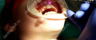
The tooth extraction procedure is carried out using effective, modern painkillers, so, as a rule, there is no pain during the operation itself.
The operation begins immediately after the anesthesia takes effect. A scalpel is used to loosen the ligament supporting the tooth.
If the procedure was traumatic, or the edges of the wound are too wide, the dental surgeon may use self-absorbing sutures. But most often, the wound is simply closed with a gauze pad with a special hemostatic agent. To stop bleeding, you need to lightly but firmly press the tampon onto the wound with closed jaws. After 20 minutes, the gauze can be spat out.
Diagnostics
The main symptom of the disease is the appearance of acute pain, which does not subside either 24 hours or 2–3 days after tooth extraction. Sometimes a dentist can identify chronic alveolitis during routine oral examinations. In this case, an empty socket without granulation tissue appears in place of the impacted tooth. The bone is already visible at the bottom of the hole.
The dentist will be able to determine the presence of tissue changes during the examination; radiography and radiovisiography of the affected area may also be prescribed.
Gum condition
Pain in the gums may occur after the anesthetic drug wears off, that is, after 3-4 hours. After the procedure, the patient is often worried about the return of painful sensations and the release of ichor (for 4–6 hours. After the operation, the wound looks quite scary, especially if a wisdom tooth was removed.
In the absence of pathology, the healing process occurs in several stages.
Catarrhal, ulcerative with the appearance of ulcers under the plaque, acute aphthous stomatitis
Chronic recurrent aphthous stomatitis CHRAS Symptoms of stomatitis, causes
Oncological diseases and the appearance of stomatitis on the background of their treatment
Sore throat
Stomatitis is the name unites a whole group of diseases of the oral mucosa in both children and adults. The diagnosis of stomatitis can be made by either a general practitioner or a doctor's tooth in the examination room during initial treatment. Later, during the examination by a narrower specialist and after additional examination methods, the diagnosis is refined and sounds more accurate. The main signs of stomatitis are the same, regardless of the cause of this disease. At the clinic Denterum in Samara, an accurate diagnosis will always be made.
This is redness and swelling of the oral mucosa, white or gray patina in the mouth. If the defeat goes to the tongue, the plaque can cover it with a thick layer and then the diagnosis expands, as an additional diagnosis is made “glossitis”. The tongue is an extremely mobile organ and lesions of its mucous membrane are accompanied by severe soreness and a host of accompanying problems—it is painful to eat, talk, or just hot tea gets on the tongue, the pain immediately makes itself felt.
Types of stomatitis stomatitis herpes vesicles
Manifestations and stages of stomatitis formed the basis of clinical classification. The teeth of dentists are differentiated depending on the visible manifestations.
Catarrhal stomatitis
The causes can be very diverse, but usually catarrhal stomatitis develops against a background of numerous carious lesions and poor oral hygiene. The superficial epithelial layer of the mucous membrane suffers. There is always a large amount of soft plaque, there may be a hard plaque. On the mucous membrane of the oral cavity, edema and redness, white and gray coating, the gum between the teeth is swollen, and when the instrument is touched, blood is extracted from the interdental papilla. If catarrhal stomatitis lasts several days, an unpleasant odor from the mouth is attached.
Tooth doctors treat catarrhal stomatitis based on an obvious underlying cause. Our specialists, after anesthetizing the oral mucosa and gums, first conduct a gentle professional hygiene, removing visible plaque from the teeth and partially from the mucous membrane. More thorough cleaning of the teeth is difficult for the doctor, bleeding appears because of the presence of gingival inflammation. For the patient, the first visit can be painful. Further, mouth baths are prescribed at home for 2-3 days with antiseptic solutions. If the main cause-the virus or bacterial infection is established, a general appropriate etiologic treatment is prescribed. At the second visit the patient's gums bleed much less, they can easily anesthetize. Purification by ultrasound and AER-FLOU is carried out. Further treatment is carried out based on the specified diagnosis.
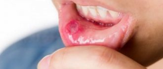
Ulcers in the mouth. Ulcerative stomatitis. Ulcers in the language of stomatitis Photo
The appearance of ulcers under the plaque in the mouth suggests that catarrhal stomatitis has passed into the stage of ulcerative stomatitis. The mucous membrane of the mouth becomes inflamed to the full depth. Necrotic ulcers are extremely painful, covered with putrefactive gray bloom. Remove the plaque for antiseptic treatment is obtained with great difficulty due to the fact that it is closely welded to the walls of the ulcer, and the procedure itself causes severe suffering. The saliva becomes thick, the patient can only take liquid food. The smell from the mouth becomes sharply putrefactive, felt by the surrounding at a considerable distance. The pain that appears in the tongue of the ulcer causes extremely strong, the person can not talk, sometimes the tongue swells and does not fit in the mouth. Becomes dense and enlarges the lymph node inflammation is indicated near the ear and under the jaw, the temperature may rise. It also hurts to swallow because of the spread of inflammation from the mucous membrane of the mouth to the tonsils. Often the throat ache starts first and the patient consults the otorhinolaryngologist. The prescribed general treatment in the case of stomatitis and tonsillitis is very close, but to conduct a full-fledged hygiene of the oral cavity the ENT doctor can not-he does not have special tools, so recovery can be delayed. How to treat stomatitis ulcerative? To carry out the treatment of the affected areas, the tooth doctor first needs to conduct anesthesia mucosa. The mucous membrane is covered two to three times with an anesthetic gel, sometimes an injection of small doses of anesthetic is done. Injection system ARTI-DZHET allows you to make a very accurate calibration of the injected drug, and the needle is extremely thin.
After anesthesia, treatment with hydrogen peroxide 1% on tampons and rollers, mouth baths. In a large number of cases of acute stomatitis in the microbial landscape there is an anaerobic flora, oxygen containing preparations.
Acute aphthous stomatitis.
Most often, this disease accompanies other chronic diseases of the gastrointestinal tract gastritis, colitis, cholecystitis amid provoking local factors in the mouth and signs of seasonality. On the mucous membrane of the oral cavity appear rounded aphtha areas of inflammation, covered with a yellowish coating. They are surrounded by a red border. Aphids are moderately painful. Their periodic appearance in combination with diseases of the stomach and intestines gives reason to recall the unity of the innervation and trophism of the whole gastrointestinal tract. Consequently, similar aphthae can be present on the mucous membrane of the stomach and intestines. But the oral cavity in this chain takes the first, starting place. It is in the mouth that the whole process begins. And if the dentist notices numerous carious lesions in the teeth, it should be reminded to the patient that the cause of many of his troubles in the stomach is a poorly sanitized oral cavity, where microorganisms rush along the entire length of the gastrointestinal tract, provoking his diseases.
Chronic recurrent aphthous stomatitis. Very painful and dangerous disease, when his attacks and the appearance of aft in the mouth are replaced by remissions, but complete recovery does not occur.
If, in all the above cases, the physician achieves complete recovery with vigorous treatment of the general and local character, then in case of chronic recurrent aphthous stomatitis the disease returns again and again, despite carefully treated teeth, rejection of all bad habits and high level of oral hygiene. The causes of the disease are likely to be polyethiologic, multiple. The main ones are nervous overstrain (stress), hypovitaminosis in ascorbic acid and routine, carriage of some streptococcal strains in the oral cavity, more often hyaluronidase-producing. People with a second blood group are predisposed. The hereditary connection is traced. Very close to the symptoms of chronic herpetic stomatitis (see below). But with well-designed treatment, the herpes virus can be suppressed for a long time and the disease is clinically cured. In the case of chronic aphthous stomatitis, antiviral drugs are ineffective. There are different areas of rashes, with herpes marked by seasonality and the connection with common colds.
It is especially necessary to note the high probability of the appearance of severe forms of stomatitis against the background of treatment of oncological diseases. If at the same time there are traumatic factors in the mouth, a low level of hygiene, inflammation develops, up to necrotic lesions of the alveolar process.
In general, we must admit that chronic recurrent aphthous stomatitis is not only a dental disease. In its treatment should be attended by doctors immunologists and gastroenterologists.
Day after the procedure
At the initial stage, the hole remaining in the place of the pulled out tooth is filled with a scarlet blood clot. It is not recommended to remove it, as it performs several functions:
- protects the wound from infections;
- eliminates bleeding from blood vessels;
- promotes the formation of new tissue that will fill the empty space.
To avoid breaking up the blood clot, it is recommended not to brush your teeth on the day of surgery. Smoking is accompanied by inhalation of smoke, which creates negative pressure in the oral cavity. This may help pull the clot out of the socket. It is not recommended to blow your nose or spit. Rinsing your mouth should also be avoided; you can simply put the solution in your mouth and hold it for a while without rinsing. Gentle rinsing can be indicated only in the presence of inflammatory and purulent processes.
What systemic diseases can manifest as aphthae?
- Reiter's syndrome
- Behçet's disease
- AIDS
- cyclic neutropenia
- PFAPA syndrome
The gastroenterologist is especially interested in the connection between recurrent ulcers and specialized systemic diseases.
Celiac disease is found in 5% , and for a long time canker sores may be the only manifestation of a reaction to gluten.
Let me remind you that the estimated prevalence of celiac disease in the general population is about 1%.
Enamel defects and aphthous stomatitis in celiac and healthy subjects: Systematic review and meta-analysis of controlled studies
Other important conditions that manifest as canker sores are inflammatory bowel diseases: Crohn's disease and ulcerative colitis.
Extraintestinal Manifestations of Pediatric Inflammatory Bowel Disease: Prevalence, Presentation, and Anti-TNF Treatment
After three days
The blood clot begins to change and thicken. Gray and white fibrin stripes appear on it, after which the formation of new gum tissue begins. Painful sensations may still occur. But they are much weaker and have a pulling character. The patient may experience bad breath. This is considered normal and is due to the formation of a blood clot. Failure to brush your teeth also leads to the formation of bacteria and an unpleasant odor.
To rinse the mouth, you must use special solutions recommended by your doctor. The appearance of pain from touching the gums, increased pain during eating, serves as a signal for the need to return to the dentist's office. If the tissue of the edges of the hole has acquired a red tint, you should also immediately consult a specialist.
Therapeutic treatment
Treatment is carried out according to a standard scheme and it is important to start it in a timely manner:
- Cleaning the hole from food debris, plaque, and dead tissue;
- A thorough inspection of the hole, checking for the absence of roots and foreign elements;
- Washing the wound with hydrogen peroxide;
- Drying the wound;
- Putting anti-inflammatory drugs into the hole.
After treatment, the doctor gives recommendations to the patient on oral care. A few days later, the patient comes back for an examination to make sure that the treatment has given positive results and the wound is healing. It is important to rinse your mouth with herbal decoctions at home. Do not eat hard foods. For pain relief, you can use Nimesil or Ketanov.
After 2–3 months and beyond
The gum gradually hardens, and the space remaining from the tooth is filled with maturing bone tissue. By the beginning of the 4th month, the gum bone tissue completes its formation. The gum can be called completely healed.
If the wound heals with suppuration, then healing of the wound can last up to six months.
On the top
The most common complication of extraction of figure eights in the upper jaw is a puncture of the base of the maxillary sinus (maxillary sinus).
Puncture of the bottom of the maxillary sinus
The dental roots of molars number three in the upper jaw are located too close to the base of the sinus, and sometimes even grow into it. Chronic inflammation of the periapical tissue provokes resorption of the axillary septum, due to which the roots of the molars fuse with the mucous membrane inside the sinus. It is this part that most often ruptures during the extraction of the right or left tooth. This complication can be noticed by the following symptoms:
- nasal sound, bleeding air bubbles;
- blood from the nose on the side of the figure eight.
In such a situation, urgent medical assistance is necessary.
Gum healing process
The speed of gum healing depends on several factors:
- individual characteristics of the patient’s immunity;
- the success of the operation;
- location of the extracted tooth.
If the tooth is in a hard-to-reach place, has crooked roots, or its crown has been significantly damaged, then the removal procedure becomes more complicated. During manipulation, the tooth may begin to crumble, leaving fragments in the gum. In this case, it is necessary to cut the gum tissue, detach it from the bone, remove the tooth in parts, and use a drill. These traumatic procedures prolong the period of gum healing after surgery.
Slight swelling of the gums is considered normal. The temperature may rise slightly (due to an immune reaction). The swelling usually subsides within three days.
Noticeable swelling of the gum is also observed after cutting it. This swelling goes away in about one week.
Vitamins are the key to health
At the same time as using disinfectants, it is necessary to strengthen the immune system. This can be done with a diet rich in vitamins, macro and microelements. On the recommendation of a specialist, you can take a vitamin and mineral complex: “Alphabet”, Vitrum, “Multi Tabs”, “Centrum”.
Decoctions or extracts of echinacea, eleutherococcus, ginseng, and Schisandra chinensis will help. If necessary, the doctor prescribes drugs to enhance immunity: “Ribomunil”, “Imudon”, “Irs-19”, “Likopid”, “Derinat”, “Timaktide”.
If stomatitis occurs due to problems with the gastrointestinal tract, complex treatment should include drugs containing beneficial microorganisms - probiotics. They will help restore the microflora of the digestive tract. These include “Bifidumbacterin”, “Lactobacterin”, “Acilact”, “Bifilong”, “Narine”.
To speed up the healing of damaged mucosal surfaces, sea buckthorn or rosehip oil, vinylin, and propolis preparations are prescribed.
Possible complications
Signs of alarm should be considered an enlargement of the cheek, further spread of swelling, a persistent increase in temperature, increased pain, nausea, and weakness. If the healing process is disrupted, the following complications may occur:
- Cyst formation. It is a fibrous neoplasm filled with fluid.
- Flux. Formed after infection penetrates into the socket and then into the periosteum. The resulting inflammation is characterized by severe swelling of the cheek on the side of the diseased gum. There is severe pain and redness of the gums. The formation of flux requires immediate medical attention. Therefore, it is necessary to carefully protect the site of the extracted tooth from possible infection.
- Alveolitis. This is a complication that occurs during the inflammatory process of the hole in the jaw bone. The infection occurs due to a violation of the integrity of the protective blood clot. The onset of the disease is characterized by inflammation of the outer layers of the socket, spreading into the deep layers of the bone. Alveolitis is accompanied by aching pain during eating, swelling and redness of the gums. There is a putrid odor from the mouth. The patient feels chills, headache, and fever. The occurrence of the disease most often occurs during the extraction of molars located on the lower jaw. It is necessary to obtain medical attention in a timely manner to avoid the spread of infection to other organs. One of the dangerous complications of the disease is osteomyelitis.
Removing wisdom teeth is a more complex procedure, so gum inflammation often occurs after surgery. At the same time, discoloration or swelling of the gums should not cause concern to the patient. Often after surgery there are difficulties opening and closing the mouth. This is a consequence of surgery. To get to a hard-to-reach place, the doctor asks the patient to open his mouth as wide as possible. The pressure exerted on the tissues leads to their swelling. On the 3rd day, the discomfort usually goes away completely. The appearance of purulent contents in the hole, increased temperature, acute pain, heavy bleeding - all these signs require immediate contact with the dentist.
The consequences are dangerous
Without timely treatment, seemingly harmless stomatitis can bring many negative consequences. Pathogenic microorganisms begin to secrete toxins - poison of biological origin.
Once in the blood or stomach, toxins poison the body. Intoxication leads to deterioration of health. Headaches and joint pains appear, appetite disappears, lethargy, nausea are felt, diarrhea begins, and the temperature may rise.
Other pathologies may develop: from inflammation of the throat or respiratory tract to destruction of the jaw bone or general infection of the circulatory system.
Comprehensive treatment of stomatitis will help to avoid complications. But before you begin self-therapy, you need to contact your dentist again. Or visit a therapist for adults, or a pediatrician for children. Only a doctor can determine the type of disease and prescribe appropriate medications.
Recommendations after tooth extraction
There are recommendations that, if followed, significantly speed up the gum healing process. Not only the speed of wound healing depends on them, but also the absence of possible complications. The doctor’s main recommendations in the postoperative period may be the following:
- you need to avoid foods that are too hot, spicy, or irritating to gum tissue;
- in the first days you need to carefully avoid damaging the clot; It is necessary to be extremely careful in maintaining oral hygiene;
- about 3 hours after the tooth extraction procedure you need to refrain from eating;
- in the next three days you need to eat only soft food, without consuming sweets, alcohol, or hot drinks;
- on the first day after the procedure, it is recommended to sleep on a high pillow;
- During the week, it is recommended to avoid visiting the sauna, solarium, sunbathing on the beach, and reduce physical activity;
- on the first day, it is forbidden to brush your teeth to avoid damaging the blood clot;
- Do not try to pick the clot with your finger, toothpick or tongue;
- a cold compress should be applied to the surgical area for 20 minutes, every 2 hours;
- It is recommended to use oral baths and then rinse with antiseptic agents.
The postoperative period requires careful attention to the patient's health status. A complication resulting from neglect of the rules will require much more time, money and effort to heal the wound.
Causes of stomatitis in adults
Pathogenic bacteria, viruses. Inflammation begins due to the action of pathogens of viral or infectious diseases. Normally, they are located in the oral cavity and do not cause irritation. The mucous membrane is protected from inflammation by the immune system. If it is weakened, viruses or bacteria can cause stomatitis.
Deficiency of vitamins and microelements. If the body receives little zinc, iron, folic acid, or B vitamins, the risk of stomatitis increases. Such a deficiency occurs due to irregular or unbalanced nutrition and consumption of foods with low nutritional value.
Injuries to the mucosal surface. The mucous membrane can be injured mechanically, by temperature or chemical irritants. Mechanical injuries occur when biting the cheek, damaging its inner surface with the edge of a crown, prosthesis, or hard food. When injured by a chemical irritant, contact occurs with acids, alkalis, and salt. Thermal injuries occur when consuming hot food or drinks. Minor and irregular injuries heal without stomatitis. It develops if injury to a certain area occurs regularly or when the immune system is weakened. In particular, when smoking, the surface of the mucous membrane is constantly injured both thermally and mechanically.
Insufficient hygiene. This reason is more typical for children. The risk also exists for adults. It is associated with insufficient personal hygiene. Inflammatory agents enter the oral cavity if you do not wash your hands before eating or eat unwashed foods.
Improper oral hygiene. Too frequent brushing or rinsing, constant brushing of teeth with a paste that contains sodium lauryl sulfate, reduce salivation, which causes the mucous membrane to become dehydrated and vulnerable to irritants.
Drug treatment. Taking diuretics and some other drugs can reduce salivation and provoke dehydration of the mucous membrane. If the mucous membrane is dry, the risk of mechanical injuries and accumulation of pathogenic microorganisms increases.
Accompanying illnesses. If the body is weakened, the risk of developing an inflammatory process increases. Stomatitis can be provoked by cancer, systemic diseases, HIV, gastrointestinal diseases, hormonal imbalances, diabetes mellitus, candidiasis, helminthic infestation, and bronchial asthma. Often, inflammation of the mucous membrane is provoked not by the disease itself, but by the use of medications that are used in its treatment.
Do you have questions about stomatitis in adults?
We will call you back within 30 seconds
+7