The maxillary (maxillary) sinus, despite its significant volume (up to 10 cubic centimeters), in a healthy person is hidden in the thickness of the bone of the upper jaw and does not betray its presence in any way. However, it is not so far from the mouth - the roots of an adult’s teeth sometimes reach it. In some cases, perforation of the bottom of the maxillary sinus is possible, meaning its direct connection with the oral cavity. This is possible due to some physiological characteristics of a particular patient:
- the close arrangement of the roots of the teeth can thin the bone border between the oral cavity and the maxillary sinus, from normal 1 centimeter to a very dangerous 1 millimeter;
- with low bone thickness, the roots of the molars can reach the cavity, separated from it only by the mucous membrane;
- a cyst, periodontitis or periodontitis can contribute to the thinning of the bony septum between the sinus and the oral cavity.
Perforation occurs against the background of the listed factors after dental intervention, even if the surgical technique was not violated.
What causes perforation?
The most common causes of perforation are:
- removal or implantation of teeth;
- root resection;
- endodontic treatment.
When the tooth roots are located directly in the maxillary sinus, the doctor’s skill level or his accuracy do not affect the situation in any way - perforation simply cannot be avoided. But it must be recognized that with low professionalism of a specialist, a problem can appear even in cases where its occurrence could have been prevented.
Endodontic treatment leads to perforation in cases where the insertion of a pin or compaction of filling cement was carried out too aggressively, as well as in pathological expansion of the root canals. In such a situation, perforation occurs directly through the thickness of the tooth, and foreign objects in the form of fragments of dental roots and cement can get inside the maxillary sinus.
When implanting teeth, filling root canals and installing pins, the occurrence of a fistula is always considered a consequence of a medical error.
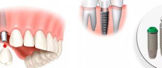
Installation of an artificial root requires maximum attention from the doctor, since it threatens to perforate the maxillary sinus. The patient’s bone tissue after the removal of a “native” tooth has a tendency to rapid degeneration - the body does not see the point in maintaining tissue that is not assigned any functions. If you delay the installation of the implant, choose its size incorrectly, or perform the operation without proper preparation, perforation will be a predictable result.
In the case of tooth root resection, there are no alternative treatment options - a cyst in the upper part of the root can only be removed this way. However, the doctor must first obtain a clear idea of the thickness of the alveolar process (the bony septum between the maxillary sinus and the oral cavity), and how far the root is from the maxillary sinus.
Features of the maxillary sinus
The maxillary sinus (its other name is the maxillary sinus) is located in the thickness of the bone tissue of the upper jaw. It is separated from the oral cavity by the alveolar process of the upper jaw, which forms its bottom. The volume of such a sinus is quite large, and in adults it can reach 10 cubic centimeters.
This sinus, or sinus, is not airtight. It communicates with the nasal cavity through a narrow slit.
Typically, perforation of the maxillary sinus occurs in the area of its bottom. Some of its features contribute to this:
- Close proximity of the roots of molars and premolars. In some cases, the thickness of the bone layer between the tooth roots and the bottom of the maxillary sinus can be relatively large - up to 1 cm, but in some people the bone border between these formations is very thin - no more than 1 mm.
- Sometimes the roots of the first and second molars are located in the sinus cavity itself, separated from it by just a layer of mucous membrane.
- Rapid thinning of the bone layer in the presence of acute or chronic inflammatory diseases: periodontitis, periodontitis, cysts.
- Relatively thin bony trabeculae in the tissue of the upper jaw.
All this predisposes to the occurrence of perforation during dental procedures, even if the treatment technique was not violated and the doctor did not apply significant traumatic force.
Symptoms
When a tooth is removed, sinus perforation can be diagnosed by characteristic and clearly visible signs:
- there are air bubbles in the blood released at the site of the extracted tooth, their number increases with targeted, accentuated exhalation through the nose;
- blood will also come from the nose - on the side where the tooth was removed and perforation occurred; it can be mixed into nasal secretions in small quantities, only slightly tinting them a light pink shade;
- The patient's voice changes, a previously uncharacteristic nasal tone appears in it.
A visitor to a dental office can sometimes feel that air is passing through the hole where the extracted tooth was located. This phenomenon is accompanied by unusual feelings of pressure or heaviness in the maxillary sinus.
Endodontic procedures and implantation, accompanied by perforation of the maxillary sinus, are distinguished by their own characteristics:
- the installed element or equipment seems to fall inward after applying additional force;
- there is a sharp change in the position of the tool in the hole made;
- the blood protruding from the hole is saturated with oxygen bubbles.
It is important to understand in time that there is a problem and promptly address it. If this is not done, an infection will get into the maxillary sinus. Symptoms will become more than obvious to the patient:
- the sinus area delivers extremely unpleasant sensations in the form of acute pain;
- the nasal mucosa on the corresponding side swells, breathing through the nostrils becomes difficult;
- Possible discharge of pus from the nose.
In addition to the specific symptoms, there are also general signs typical of intoxication: the temperature rises, weakness is felt, the patient has a headache, and the patient is shivering.
Old perforations that occur after tooth extraction or therapeutic treatment
If perforation was not detected after tooth extraction, the patient may attribute the discomfort to the consequences of surgery. It is worth saying that after a couple of weeks the stage of acute pain passes, and in the area of the resulting defect in the maxillary sinus, a so-called fistula appears, which connects the surface of the gum to the sinus.
This process is accompanied by symptoms characteristic of chronic sinusitis; among other things, patients complain of pain, purulent discharge from the nasal cavity, and swelling of the cheek. Treatment consists of the use of therapy aimed at stopping the inflammatory process and surgical methods that eliminate foreign bodies in the maxillary sinus.
If we summarize the above, we can say that the removal of teeth in the upper jaw, as well as their treatment, should be carried out as competently as possible. Perforation of the maxillary sinus can have quite serious consequences for the patient’s body. In some cases, long-term hospital treatment is required. The addition of an infectious process is especially dangerous.
Such a complication can be avoided if the dentist is sufficiently competent, who must take into account the anatomical position of the teeth and the maxillary sinus before performing any complex dental procedures related to the upper jaw. Based on this, the dentist is obliged to highlight all the anatomical and topographical features of the patient and conduct qualified treatment.
Before the procedure for removing, implanting or treating upper teeth, the doctor must take a targeted photo, which will help visualize the area with which he will be working. If these conditions are met, such a serious complication as perforation of the maxillary sinus can be avoided.
Diagnosis
At the time of a dental operation that led to perforation of the maxillary sinus, the doctor draws a conclusion about a new problem based on the signs described above. If perforation is questionable, the dentist's findings are rechecked using the following methods:
- probing with a thin probe;
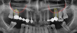
- radiography - shows dark accumulations of blood, tooth root fragments and filling cement located inside the maxillary sinus, can be done with the preliminary administration of a contrast agent;
- CT scan;
- general clinical blood test - necessary when identifying an old problem to make sure there is an infection in the body.
Causes of perforation
The physiological location of the upper teeth and associated pathologies are a risk factor that can cause perforation of the maxillary sinus. Complications can be characterized by dental operations. In particular, this is the sanitation of dental tissues in case of deep caries, the removal of part of the tooth or its root. It is important for the dentist to carry out surgical intervention extremely carefully without fragmenting or causing fractures of the tooth roots, which is why mandatory x-ray monitoring is required.
When carrying out dental treatment associated with the removal of an upper wisdom tooth, perforation can occur when manipulating the root of the tooth, which is often removed in fragments. Pathology also occurs during manipulations in the root of the tooth during the treatment of caries and when intensive canal filling is done.
Perforation may occur during root resection and installation of implants in the upper jaw. The dentist must act with extreme caution when removing the upper tooth. First, an image of the upper jaw is taken, which allows one to assess the location of the roots relative to the maxillary sinus, and then the doctor determines the subsequent tactics of therapeutic or surgical treatment.
Article on the topic: Open and closed sinus lift surgery
How to treat?
The tactics for eliminating the problem depend on exactly what causes led to the perforation and what the general clinical picture is. Almost always, repairing a perforation requires surgery, the only exception to this rule is when a hole was created during a tooth extraction, provided that the dentist found it immediately and no foreign objects or infection got inside. If the problem is identified immediately, it is important to keep the blood clot in the place where it formed and prevent it from becoming infected - it will become a natural barrier to the infection entering the sinus. Protecting a blood clot from infection is carried out using a tampon with an iodine solution, which will have to be kept for at least a week.
In some cases, the doctor chooses the tactic of suturing the gum tissue. Another alternative is to install a compact plastic plate that is attached to adjacent teeth and acts as a barrier between the oral cavity and the sinus until the septum is restored.
For obvious reasons, physical barriers cannot provide a 100% guarantee that infection will not penetrate the hole, so the doctor additionally prescribes vasoconstrictors and anti-inflammatory drugs. They can be taken at home, but sometimes require the patient’s outpatient presence.
Penetration of any foreign objects into the maxillary sinus is regarded as a serious complication, which automatically means the need for surgical intervention and hospital treatment. In such a situation, the sinus is opened, removing foreign objects and tissues that cannot be restored, after which the fistula is covered with the patient’s tissues.
Causes and mechanisms of fistula development
The oral cavity, due to the peculiarities of its anatomical structure and physiological load (food intake and salivation), is aseptic. The presence of microorganisms creates conditions in which the penetration of infection into all kinds of wounds and surgical incisions is almost inevitable. Despite the fact that during bone grafting surgery the most sterile conditions are created to prevent bacteria from entering the wound, their infection cannot be ruled out both during the intervention and in the postoperative period. Most inflammatory processes after sinus lifting develop due to the patient’s violation of the dentist’s recommendations, namely: ignoring disinfectant rinses, antibacterial ointments and oral baths, eating on the side of the bone graft.
Once in tissues that have recently undergone surgery, bacteria find themselves in ideal conditions for reproduction. The presence of minor blood clots, tissue detritus, pieces of food, if hygiene requirements are not followed and the optimal temperature is present, create a breeding ground for them. In the process of life, microorganisms destroy the structures surrounding them, and the release of leukocytes from the bloodstream to destroy hostile microflora leads to the release of pus and inhibition of healing processes.
The fistula after sinus lifting and bone grafting is not able to heal on its own due to the presence of a defect in the bone tissue. The development of such a pathological course leads to the appearance of a focus of chronic infection, the penetration of bacteria into the maxillary sinus with the formation of sinusitis and other complications. In this case, the fistula after an open sinus lift can be located not only at the implant site, but also on the lateral surface of the gum. The appearance of a fistula tract more often occurs after closed bone grafting due to worse fixation of the denture.
Among other things, the formation of a fistula can occur when the tooth root is not completely removed during a sinus lift.
Perforation discovered after the fact
If the patient suffered discomfort in the first 2-4 weeks and did not contact the dentist, who, in turn, did not identify the problem at the time of its occurrence, the perforated wound turns into a permanent fistula that is not prone to healing.
Typical signs of chronic sinusitis appear:
- the nose on the side of the fistula is constantly stuffy;
- the parasinus area gives off a dull pain, its waves can roll to the nearest eye and temple;
- pus is discharged from the nostrils and in the mouth (from the fistula);
- possible swelling of the cheek on the side of the fistula with visible deformation of the face.
In most cases, a fistula is felt as air passing between the nose and mouth while talking or sneezing. Pronunciation of a number of sounds becomes more difficult. Liquid food may enter the nose from the mouth. Therapy for an old fistula shows rather weak results, and relapse with such a problem is not uncommon. There is no alternative to surgical intervention - it is necessary to open the maxillary sinus, remove foreign objects and non-viable tissue. The fistula requires excision throughout its entire thickness, the defect is closed with healthy tissues of the patient. After the operation, an antibiotic course lasting one and a half to two weeks is prescribed; in parallel, antihistamines and anti-inflammatory drugs must be used.
Why shouldn’t you treat perforation of the maxillary sinus yourself?
To date, there are no effective treatments for perforation other than surgery. An attempt to cure yourself at home using “traditional medicine” means that time will be lost and the situation will become neglected. You can start the problem before complications arise:
- the sinus cavity becomes inflamed, the infection spreads to the bone tissue, the patient begins to suffer from osteomyelitis of the upper jaw;
- inflammation penetrates into other intracranial sinuses, and there are more foci of infection;
- next to the untreated perforation, the alveolar process is weakened, as a result of which healthy teeth may fall out;
- foci of suppuration develop.
The maxillary sinus is also located in close proximity to the brain. The lack of a timely response and the development of suppuration inside the sinus is fraught with meningitis and meningoencephalitis - these are diagnoses that directly threaten life.
Classification and complications of perforations of the maxillary cavities
Depending on the diameter of the hole, it is customary to distinguish:
- Small perforations - from five millimeters;
- Medium perforations - from six to seven millimeters;
- Large perforations - more than eight millimeters.
Depending on the period of development, the process proceeds:
- In acute form - it is detected either during the procedure or immediately after it;
- In its chronic form, it is detected only a few days after dental treatment.
Localization determines the presence of the following types of perforations:
- Palato-sinus - located on the palatal side;
- Alveolar sinus - located on the alveolar process;
- Vestibulosinus - located in the vestibule of the oral cavity.
Perforations that were not diagnosed on time always provoke infection and the development of inflammatory processes in the form of perforated sinusitis. The patient suffers from a feeling of nasal congestion and intense purulent discharge mixed with blood. Due to the entry of liquid food from the mouth into the sinus, infectious and inflammatory processes of the periosteum of the alveolar process and purulent lesions of the jaw bone develop.
Prevention
Perforation of the maxillary sinus is a problem that is easier to avoid than to fix it later. Since perforation is caused by dental intervention, preventing the problem falls on the shoulders of the dentist. He is obliged:
- responsibly examine the patient before performing procedures;
- clearly understand the anatomical features of the client before major intervention;
- strictly adhere to the intervention technology.
The dentist is also obliged to respond adequately to any signs of perforation that has just occurred due to his fault. If for some reason the doctor has not fulfilled his own duties, then it is up to the patient - he must refuse self-medication and put aside the fear of dentists, in no case try to “endure” the discomfort, but immediately seek help.
A joint operation performed by an implant surgeon and an ENT specialist at Dial-Dent
According to the conclusion of the consultation of doctors, it was decided to remove the teeth on the upper jaw on the right, remove the mycetoma from the maxillary sinus through the sockets of the removed teeth, and suture the fistulous tract. It was decided to carry out the operation under sedation – a state of medical sleep. Before the operation, the patient passed the necessary tests and had a consultation with an anesthesiologist.
A family dentist is licensed to provide treatment under anesthesia. Before the operation, the patient takes the necessary tests and undergoes examinations. When using any form of anesthesia (full general anesthesia or sedation), the patient is under the supervision of an anesthesiologist throughout the operation. There is a recovery room where you can rest after treatment.
In this case, the implant surgeon removed the problem teeth. Then the ENT specialist, under the control of an endoscope, removed the mycetoma from the maxillary sinus through the sockets of the extracted teeth. After removing the mycetoma, the surgeon sutured the fistulous tract.