VITAL COLORING
Volume 5. Moscow, 2006, p. 390
Copy bibliographic link:
VITAL STAINING (vital staining), a special method of staining living cells. dyes used in non-toxic. concentrations. Such dyes can be basic (for example, methylene blue) and acidic (for example, phenol red). Penetrating into animal cells, some dyes diffusely stain the cytoplasm, while other dyes are deposited in the form of granules in the area of the Golgi complex, leaving the nucleus and cytoplasm unstained. When cells are damaged, staining with diffuse dyes increases, while granular dyes lose the ability to form granules and stain the cytoplasm and nucleus diffusely. In living plant cells, dyes are concentrated in vacuoles; in dead ones, they stain the entire protoplast. These features make it possible to distinguish dead and damaged cells from living ones. Quantitative accounting of the dye bound by cells makes it possible to judge more subtle changes in their functional state. Acid granular dyes are used to identify elements of the reticuloendothelial system and study their condition; methylene blue – for selective coloring of departments. neurons. The distribution of fluorescent dyes in cells is used to assess cell viability and in some cytochemical studies.
Source
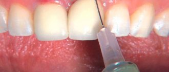
The main examination method for identifying caries: visual inspection with a mirror
This is the very first and main method used in clinical practice. Conclusions about the condition of a patient's teeth were previously based solely on visual examination data. But this method of diagnosing caries has significant drawbacks: low insufficient information content, as a rule, almost complete impossibility of identifying fissure or proximal caries.
The quality of the visual inspection depends on subjective factors. Mandatory testing conditions include preliminary cleaning and drying of the teeth being examined, and good lighting. This is a suitable method for diagnosing superficial caries, as well as medium or deep caries - at the initial stage of the disease, a visual examination does not reveal defects. Approximal (contact) caries can sometimes be identified by changes in the color of the enamel (it darkens, becomes grayish or chalky), floss gets stuck between the teeth, and the interdental papilla is inflamed.
VITAL COLOR
VITAL COLOR
(lat. vitalis vital, living) - intravital coloring of animal or plant cells and tissues. For this purpose, almost harmless or low-toxic dyes are used. For the first time, vital coloring was performed by the Austrian scientist Unger (F. Unger, 1848), who introduced the juice of red Phytolacca fruits into the white petals of hyacinth. In the histophysiology of animals V. o. with the help of ammonia carmine and sodium indigo sulfate was first used by the Russian scientist N.A. Khrzhonshchevsky (1864, 1866) when studying the function of the kidneys and liver.
V. o. is one of the important experimental methods of cyto- and histophysiology. Sometimes V. o. used in the clinic as a functional test to study the physiological usefulness of the kidneys (see Chromocystoscopy).
With the help of V. o. it is possible: 1) studying the patterns of intake, accumulation and release of substances involved in the metabolism of the body; 2) visual examination of some fiziol, processes (secretion, excretion); 3) marking of certain parts of the body (organs, embryonic rudiments of animals and plants, cells or their organelles); 4) study of physiol, the state of the cell normally and under various irritating and damaging influences; 5) detection of certain chemicals in the cell. substances (nucleic acids, proteins). The most convenient object for carrying out V. o. is cell and tissue culture, however V. o. It is also successfully used for pieces of organs and even small animals and plants, completely immersed in the dye. V. o. It is also carried out by injecting a dye into animal or plant tissue. The distribution of the dye in cells and tissues is usually observed under a light-optical microscope, and in those cases when for V. o. fluorescent dyes (fluorochromes) are used using a fluorescent microscope, which significantly increases the sensitivity of the study.
For V. o. use ch. arr. various aniline dyes used in histol, research methods: acidic (anionic), basic (cationic) and neutral (tsvetn. fig. 1-3). Aqueous solutions of acid dyes do not penetrate normal cells well. Since permeability increases dramatically with damaging stimuli, these dyes (eosin) are used to detect cellular damage. Colloidal solutions of acidic dyes (trypan blue, lithium carmine, etc.) penetrate the cell by phagocytosis and are used by Ch. arr. to study the reticuloendothelial system.
Basic dyes, as a rule, penetrate well into living cells. Their further distribution in the cell depends on its condition. In an undamaged or slightly damaged cell, the main dyes accumulate in lysosomes in the form of clearly defined cytoplasmic granules, therefore acridine orange fluorochrome is widely used for intravital detection of lysosomes. With significant damage to the cell, the accumulation of dyes in the lysosomes stops and diffuse staining of the nucleus and cytoplasm appears. The color change can be reversible - the so-called. paranecrosis (see).
You can see the spores
Ziehl fucci staining allows bacterial spores to be seen.
Having a pink color after staining, they are clearly visible against the background of blue bacteria. This method is also a tool of bacteriology and is of great practical importance.
Comments
Similar materials
Education Tinctorial properties - what are they?
Before examining microorganisms under a microscope, the sample being studied is subjected to special preparation.
In their natural form, almost all bacteria are transparent, so they are stained with dyes. Coloring is allowed...
Business Dural is a high-strength aluminum-based alloy with additions of copper, magnesium and manganese: properties, production and application
Dural is a multicomponent alloy made from aluminum, magnesium, zinc and manganese.
During the production process, other components are added to the mixture.
Health Linden: beneficial properties and contraindications to the use of products based on it
Linden is a wonderful cure for many ailments, sent by nature itself. Its yellow flowers with a pleasant strong aroma are a real storehouse of health. However, not everyone knows how best to collect these gifts...
Health Bacteria and viruses are the basis of the microcosm
According to K. Wese, all living beings are divided into several domains. There are three of them: bacteria, archaea and eukaryotes. Viruses are treated as a rankless category. The fact is that not all scientists classify this group...
Health Wheat porridge. Useful properties of a product based on different cereals
The first of the porridges that humanity tried in ancient times was undoubtedly wheat.
Even in biblical sources this product is mentioned repeatedly
Taste qualities make wheat an important “per... News and Society Wormwood
Useful properties and recipes based on it
News and Society Wormwood. Useful properties and recipes based on it
Every summer in the fields and city limits you can find a tall plant with an unforgettable bitter smell. Common wormwood is familiar to many - in the vastness of our Motherland there are more than a hundred varieties of it, distinguished...
Education Cultural properties of bacteria: definition, description, features and functions
Microbiology is a broad modern science that studies the biochemical and physical properties, morphology and systematics of bacteria.
The world of prokaryotes is rich in a huge number of different species. To explore everything in pairs…
Education Riddles based on descriptions of properties and characteristics. Descriptions-riddles for children
Everyone is familiar with riddles. This small form of folk art provides the necessary…
Education Basics of chemistry: properties, applications and production of nitrogen
Nitrogen is one of the most common elements on Earth - its content in the atmosphere exceeds 78%.
The existence of such a large amount of nitrogen in a free state indicates its inertness and it is difficult...
Business Zirconium: alloys based on it. Properties, application
A rare, but at the same time very important metal in many industries, zirconium was first isolated only in 1824. However, it still contained a certain percentage of other elements. Only in the 20th century was it possible to semi...
Vital coloring
Vital coloring
in microscopy - staining organisms or living preparations of their tissues to increase contrast when observed under a microscope. Intravital staining allows one to simultaneously observe the structure and functioning of organisms, cells and tissues. [1]
Sometimes vital staining is also called the introduction of a dye into a living organism, followed by its killing and preparation of a microslide. [2]
A number of methods for vital staining of microorganisms have been developed and used in microbiology.
Granulocytes
Granulocytes differ in two main characteristics of cell structures: the presence of specific granules (granules) and a segmented nucleus in the thickness of the cytoplasm. Each type of granulocyte contains two types of granules: specific and nonspecific.
Under the guise of nonspecific granules, special forms of lysosomes appear - structures that contain hydrolytic enzyme substances.
Depending on the properties of specific granules in each cell, basophil, neutrophil and eosinophil cells are distinguished. The main difference between neutrophils is the smallest granule size, amounting to only 0.1-0.3 microns. The composition of these granules differs in the content of phagocytins, lysozyme, and alkaline phosphatase. The main function of neutrophils as human cells is protective.
Bacteria and microorganisms
Literature
Useful
See what “Vital coloring” is in other dictionaries:
VITAL STAINING - staining of living organisms, cells or their parts for the purpose of studying ... Dictionary of botanical terms
Vital staining - (from Latin vitalis vital, living) a histophysiological method of staining living plant or animal cells with special dyes, in which the cell does not die; the same as intravital staining ... Great Soviet Encyclopedia
VITAL Staining, INTRAVITAL Staining - (infravital staining) the process of staining living tissue by introducing a dye into the body. For comparison: Supravital staining ... Explanatory dictionary of medicine
vital staining - intravital staining of microbial cells (usually with aniline dyes) to study morphology and motility. (Source: “Microbiology: a dictionary of terms”, Firsov N.N., M: Drofa, 2006) ... Dictionary of Microbiology
Vital Staining, Infravital Staining is the process of staining living tissue by introducing a dye into the body. For comparison: Staining is supravital. Source: Medical Dictionary ... Medical Terms
Intravital staining is vital staining, a method of staining living cells with special dyes used in non-toxic concentrations. Such dyes can be basic, for example neutral red and methylene blue (chromophoric group... ... Great Soviet Encyclopedia
LITERAL COLORING - see vital staining ... Dictionary of botanical terms
VITAL STAIN - VITAL STAIN, or intravital coloring, is the phenomenon of staining tissues during the life of an organism by introducing various dyes into it. The definition of V. coloring as the coloring of living tissues is not entirely correct, since it is extremely difficult in a number of ... ... Big Medical Encyclopedia
Khrzhonshchevsky, Nikanor Adamovich - (1836 1906) Russian. physiologist and histologist. In 1864 he graduated from Kazan. University of T. Prof. Kharkiv. (since 1867) and Kyiv. (since 1869) un comrade The works are devoted to the study of the structure and physiology, function of the lungs, kidneys, liver, circulatory and lymphatic systems. blood vessels, skin functions, ... ... Large biographical encyclopedia
Most typical dyes
To obtain a colored sample, aniline dyes with different pH values are used.
- Basic (emphasis on the second O) dyes - the coloring agent is a cation. The use of certain reagents allows microbes to be stained in different colors:
- red – neutralrot, basic fuchsin, safranin, pyronin;
- violet – gentian-, methyl- and crystal violet, thionin;
- blue – Victoria and methylene blue;
- green – methylene and malachite greens;
- brown – chrysoidin and vesuvin;
- black – indulin.
- Acidic (pH less than 7) dyes – the anion has coloring properties. The most common microbial dyes are:
- red – eosin and acid fuchsin;
- yellow – picric acid;
- black – nigrosin.
- Neutral paints - color both cations and anions; an example is rhodamine B.
Vital staining Intravital staining
| Practical advice Vital staining Intravital staining Home laboratory Entertaining microscopy Making microslides Goryaev's camera Classification and marking of microscope lenses Combinations of colored glasses for highlighting the spectrum Microscopy methods Methods for studying protozoa Methods and techniques of biological experiment Microscopy for beginners Microscopic measurements Modifications of Gram contrast staining Necessary equipment General methods of conclusion preparations Organization and equipment of a histological laboratory Keller lighting Preparation of slides Polarizing microscopy Rules for working with a microscope Rules for keeping a laboratory journal Purchasing a microscope Preparation of microslides of arthropods Technique for preparing histological preparations Phototube for digital cameras Formidron instructions for use |
| When studying living objects, especially those of small size, staining with vital dyes is often used, i.e., those that in known concentrations color the object without killing it. There are basic and acidic vital dyes, and in zoological work the former are more often used. It should be noted that under normal conditions, vital dyes do not stain the nuclei of a living cell or stain the basic substance of protoplasm to a very small extent. Paints are adsorbed mainly by various cellular inclusions. With prolonged exposure to cells, paints are deposited in the protoplasm in the form of granules. We cannot consider the theory of vital staining here and will limit ourselves to indicating some of the most common basic dyes. Neutral red is especially often used for vital staining of protozoa, when studying their processes of intracellular digestion, etc. It is used in very weak concentrations from 0.1 to 0.001%. In stronger concentrations it kills the cell. Neutral red is an indicator. In an acidic environment it has a bright red tint, in an alkaline environment it is yellowish. Congo red produces colloidal solutions with a low degree of dispersion, so it only penetrates into the protoplasm with difficulty. It is an indicator; it is blue in an acidic environment and red in an alkaline environment. It is used mainly in the study of intracellular digestion. Methylene blue is one of the common vital dyes. It is used, like neutral red, in very weak concentrations. Sometimes it is used to stain certain organ systems - the nervous system, excretory system (for example, in Dinophilus). Janus green occupies a special position among intravital dyes. It stains selectively mitochondria. Very weak aqueous solutions (0.01-0.05%) are used. It is better to color the object without covering it with a coverslip. This is due to the fact that the presence of free oxygen is necessary for the coloring of mitochondria. Source |
Working with fixed drugs
Based on the complexity of the work, methods for staining fixed (non-living) cells are divided into:
- Simple methods. In this case, only one paint is used, usually red (magenta) or blue (methylene blue). The difference between these dyes is the speed of exposure. Fuchsin gives results within 1-2 minutes, while the result of blue paint needs to wait 3-5 minutes. It is also convenient to use a solution of fuchsin in carbolic acid (the so-called Ziehl fuchsin) because the prepared preparation does not lose its coloring properties for several months. Methylene blue dye can also be prepared in advance in a strong alcohol solution.
- Complex (differential) methods. Here you will need several dyes (at least two) of different colors. This will allow you not only to see the bacteria, but also to study their internal structure in more detail, since different parts of the cell can perceive dyes differently. Complex methods include Gram, Ziehl-Neelsen (for acid-fast bacteria), Benignetti (staining of bacterial flagella), Gins (detection of capsules), Romanovsky-Giemsa differentiating method (staining of spores) and some others.
The Romanovsky-Giemsa differentiation method is based on the use of a special ready-made powder, on the basis of which laboratories prepare a dye solution of the required concentration. The advantage of this method is that the cytoplasm and nucleus of the cell receive different colors, which facilitates the identification and study of microorganisms.
The Neisser method is used in medicine to detect volutin grains (granules containing food for many prokaryotic cells) in diphtheria pathogens. As a result of staining, the bacterium becomes yellow, and the volutin granules become blue.
Transillumination
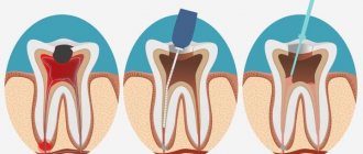
The method is intended to identify various types of caries in the initial stage. Based on the heterogeneous light-absorbing ability of dental structures. Transillumination is carried out by shining a light from inside the mouth. Hidden carious cavities are revealed in the rays of light passing through the hard tissues of teeth.
The doctor makes conclusions about the degree and type of the disease based on the visible results. Fissure caries is characterized by a dark, fuzzy shadow, the intensity of which depends on the degree of damage to the fissures: the deeper they are, the darker the shadow. If the caries is approximal, then the carious lesions look like brown hemispheres, delimited from healthy tissue. Transillumination can also detect mineral deposits (tartar).
Luminescent diagnostics
A research method for identifying superficial caries, based on the ability of enamel to change its color under the influence of UV rays. A beam of UV rays is directed at the teeth from a distance of 20-30 cm. Their hard tissues begin to glow. Normally, dentin and enamel emit a bright blue glow. With carious lesions of internal structures or enamel, the intensity of luminescence changes significantly: for example, in areas with chalk spots it is noticeably extinguished. This method of diagnosing dental caries allows you to detect changes inside them at the earliest stages.
Electroodontodiagnosis (EDD)
This clinical method is used in dentistry most often for the diagnosis of deep caries. Its essence is as follows: the tooth pulp is exposed to an electric current of minimal voltage until slight pain appears. For a healthy pulp, the values are in the range of 2-6 µA, for a pulp affected by caries - no more than 8 µA. This method also has disadvantages: it does not detect initial caries and does not allow one to determine the depth and extent of the process. The EDI method is not prescribed for children.
Probing
The doctor uses a periodontal or button probe to determine the condition of the teeth. By examining different areas of the tooth with a probe, he finds out how sensitive the reaction of the dental tissues is.
Probing also makes it possible to identify an inflamed pulp - in this case, the patient feels a sharp pain from the touch of the probe. At an advanced stage of the disease or an acute course, the enamel may be loose and even break off when touched by a probe.
Silk thread method
Despite the fact that modern dentistry has high-precision computer diagnostic methods in its arsenal, the silk thread method is still used by doctors during the initial examination, mainly to identify approximal caries. To do this, floss is passed between the teeth using sawing movements (it used to be a silk thread, hence the name).
If there are carious lesions between the teeth, their edges are uneven, and the thread begins to cling. This method is used after cleaning the enamel and between the teeth that do not border the filled ones. The thread may touch tartar or filling, which reduces the reliability of the method.