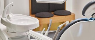
When a tooth is destroyed, caries forms, or a filling is lost, the question of filling arises. Any defect in dentistry can be replaced using various types of fillings . Conservative therapeutic intervention allows you to stop the spread of caries and maintain an aesthetic appearance.
Dental clinics today use various types of materials : cement, plastic, ceramics, photopolymer components. One filling material is suitable for the front teeth, the other for the lateral and chewing elements of the dental system. In each case, the choice takes into account the clinical picture, the degree of pathology and possible contraindications.
Classification of filling materials
Depending on its purpose, the filling composition can be temporary or permanent. For the first, a less durable material is used, since the dental filling is designed for a short period. It is usually installed in cases of pulpitis to isolate the diseased unit. Permanent ones are made from more durable materials; they are usually designed to last for several decades. The process of setting them up is longer and more scrupulous. Let's take a closer look at each of the options.
Temporary filling
A filling composition that is diagnostic. It is made from artificial dentin using special medicinal additives. The main purpose is to isolate the tooth cavity. It performs an important protective function, preventing the spread of infection and the accumulation of food debris. It is usually installed before nerve extraction. The doctor removes damaged enamel and dentin, cleans the canals, and then installs a special dental filling made of a certain material.
The temporary filling material must meet the following criteria.
- Effective sealing.
- Resistance to the influence of medicinal drugs.
- Easy to remove.
- No allergic reactions or toxic effects.
- Excellent ductility.
- Fast hardening properties.
A temporary filling composition is installed for a certain period of time, from a couple of days to several weeks. What material are these fillings made of? most often used in clinics are Vinoxol, Sympath and special cement-containing compounds. Their advantages include the fact that they do not have a negative effect on the tissue of the dentition element, they have good adhesion and sealing properties.
Permanent filling
The filling material is characterized by increased strength and wear resistance, and has the required coefficient of thermal expansion. Various types of permanent dental fillings are installed for a long time. Their main task is to hermetically seal the cavity of an element of the dental system, protect against the spread of a pathogenic environment and give the dental unit an impeccable appearance. In each case, a suitable option is selected. For example, for chewing dental units, various types of cement fillings are used, and for the anterior ones, compositions based on photopolymers are used.
Modern filling materials for permanent fillings must meet certain requirements.
- Good adhesion. Excellent connection with the tissues of the dental unit.
- No toxic effects on the body.
- Increased resistance to any chemical attack.
- Mechanical strength. The light-polymer filling must withstand significant loads.
- Good marginal seal. The composition should adhere tightly to the walls of the arious cavity and adjacent tissues.
- Increased wear resistance. Composite filling materials should not lose their properties when in contact with abrasive substances.
- High aesthetic performance.
When using various dental compounds, it is important to follow standards, regulations and relevant protocols. This will ensure a long service life of the filling component.
We looked at what fillings are and what important characteristics they must meet. In each case, you should consult a specialist. The doctor will be able to explain the difference between a gutta-percha filling and a light-polymer filling. In each case, functional load, possible diseases and contraindications are taken into account.
Basic requirements for fillings
The purpose of filling is to seal the cavity formed as a result of the pathological process and prevent bacteria from entering the tooth tissue. In order for this problem to be solved without complications, fillings must meet certain criteria [1]:
- safety for the patient - non-toxic, minimal risk of developing an allergic reaction;
- tissue compatibility - for example, amalgam in dental fillings can cause increased sensitivity to food temperature due to irritation of the pulp;
- stability - a high-quality filling does not expand or contract, but fills the cavity evenly and hermetically when working with it;
- strength, ability to withstand loads - when chewing or holding food, the filling should not crumble or crack;
- good adhesion is attachment to the canal walls, adhesion to living tooth tissues;
- aesthetics - even unnoticeable, “distant” fillings should not be noticeable and look contrasting;
- acceptable cost is one of the important criteria for patients, since many, due to the high cost, may refuse treatment for years, worsening the condition of the dental system and health in general.
Types of materials for dental fillings
Dental clinics today use various filling compounds. More information about the names of fillings and materials used can be obtained directly from your doctor. Filling materials are made based on the following components:
- Amalgam. A metal filling compound that is strong and durable. Quite rarely used in practice. The advantages include strength and abrasion resistance. The disadvantages are unaesthetic and the risk of developing allergies. There are contraindications.
- Cement. The main material for chemical fillings. The most commonly used powder is zinc oxide with the addition of silicon and magnesium. The advantages are affordable cost, ease of installation, and reduced risk of developing recurrent caries. The downside is quick erasing and poor aesthetics.
- Plastic. Composite product for durability and reliability. The best option when you don’t know what filling to put on a tooth and have a limited budget. Compositions based on acrylic acids are especially popular. They are distinguished by good durability and the ability to select colors. The downside is the high risk of microscopic pores.
- Photopolymers. Light-curing filling materials are the most popular today. The paste mixture hardens under the influence of ultraviolet light. The advantages are natural shine, strength, aesthetics, minimal toxicity. A light seal made of photopolymer material is practically free of disadvantages.
- Composites. Plastic products with special inorganic fillers. Among them is a gutta-percha filling. It is distinguished by strength, indelibility, and resistance to external factors. Used as a filling material for root canals.
- GIC (glass ionomer cements). High-strength filling material used for fixing crowns. Has excellent adhesion to metal and ceramic products.
More information about glass ionomer and light-based fillings can be obtained directly from your dentist. The choice of filling composition must be approached responsibly and trusted by your attending physician, since the performance of the chewing function will depend on this. At our AlphaDent dental clinic you can get detailed advice on selecting the necessary filling to suit your individual characteristics.
How does the procedure work?
Sequence of actions when filling teeth:
- An insulating gasket is applied (if necessary);
- The tooth surface is treated with adhesive;
- The doctor carefully introduces the material into the cavity, after which he compacts it well;
- The dentist models the filling;
- For photopolymer fillings - exposure to a special lamp is carried out for rapid hardening;
- Grinding and polishing is carried out.
The filling should be comfortable and not interfere with biting. If you experience the slightest inconvenience, you should inform your doctor; adjusting the filling is a matter of a few seconds.
Filling of anterior teeth
In the case of front teeth, the aesthetic aspect comes to the fore, so the filling material must have the appropriate properties. Although the front teeth receive better hygiene due to their accessibility, they are also susceptible to caries. Installing a filling on the front teeth is considered a more complex procedure: it is necessary not only to give the filling the desired shape, but also to choose the right color. In most cases, filling of anterior teeth is carried out using compomers, as well as light composite fillings, which harden under the influence of ultraviolet radiation. Be that as it may, in case of serious damage, it is impossible to achieve good aesthetics with the help of filling, so you often have to turn to orthopedic solutions.
Teeth filling process
- Initial consultation, taking x-rays.
- Coordination of treatment methods and selection of the type of fillings to be installed.
- Anesthesia before therapeutic intervention (local anesthesia, sedation, in rare cases - general anesthesia).
- Preparation of damaged or caries-affected tooth tissues.
- Grinding and processing the surface of the filling for proper closure.
Filling temporary teeth (baby teeth) follows the same plan, however, in pediatric dentistry, more compatible and safe materials are used.
Restoration in children's practice - to be or not to be?
The particular relevance of the use of composite materials in pediatric dentistry is due to the following factors: in childhood, the percentage of traumatic lesions of the frontal group of teeth is very high, because they are one of the first to erupt and protrude from the occlusal plane of the primary teeth that have not yet been replaced. In addition, some dental pathologies can be observed mainly in children. For example, with destructive forms of hypoplasia or fluorosis, teeth are destroyed so quickly that, due to the desire of doctors to follow the old treatment method (namely, wait until the root tips are closed), the only method of treating such teeth is often orthopedic restoration of the crown part.
For a long time, pediatric dentists were afraid to use composite materials in their practice, citing the following reasons:
* inappropriateness of restoring a tooth that may still be erupting;
* the inability to bring teeth into bite after various types of injuries, because microfractures appear at the root, which, if the load is not applied in a timely manner, can increase and lead to the death of the pulp and resorption of the tooth root.
* unsafe use of composite materials, because they are highly toxic and in teeth with uncovered apices and still wide dentinal tubules can lead to pulp death.
* inappropriateness of using composite materials in the treatment of destructive forms of hypoplasia and fluorosis at an early age, because their abrasion coefficient is lower than that of natural enamel. And in this regard, restorations made from composites require repair or complete replacement after some time.
In addition, often doctors and relatives of patients do not consider aesthetic restoration of teeth at a young age important and limit themselves to temporary structures, forgetting about the psychological aspects. But the trend of today is such that it is fashionable to be healthy and beautiful.
The achievements of modern dentistry dispel fears of using composites in pediatric practice. For example, with regard to toxicity, it is now known that the bonding system has a direct effect on the tooth. The latest generation adhesive systems are not only non-toxic, but may also contain fluorine compounds. The toxic monomer contained in chemically cured composites has practically sunk into oblivion along with the use of chemical composites themselves.
Of course, before starting restoration, it is necessary to carry out all examination methods (x-ray, EDI). However, we must not forget that the protective forces of the child’s body are very great, and in each case we try to individualize the algorithm of actions.
Despite the fact that the evolution of composite materials is advancing by leaps and bounds, pediatric dentistry places increased demands on restorative materials:
— Low toxicity.
— High degree of adhesion of the material to tooth tissues.
— Abrasion coefficient as close as possible to natural tooth tissues.
— Possibility of immediate and final restoration of teeth (both frontal and chewing group).
— Preparation that does not require intervention in healthy tooth tissue.
— Excellent aesthetic characteristics.
From an esthetic point of view, restoring the teeth of young patients is often very difficult. This is due to the fact that the shape and color of children's teeth have a number of features. For example, the macrorelief is characterized by the presence of a scalloped cutting edge that has not yet been subjected to physiological abrasion. The surface layer of enamel in children is formed by the protruding tops of prisms, which gives it the appearance of a “cobblestone street.” In addition, micropores are found in the enamel of children's teeth under a microscope. We must not forget that the Recius lines (enamel growth zones), which form perikemata on the surface, are more pronounced in childhood. All this affects the surface gloss of the enamel and visually makes it brighter. Children are characterized by pronounced mamelons. The most typical for the incisal edge of young patients is the presence of three large mamelons or three mamelons with a split middle one. (Fig. 1)
Fig.1
Tooth color is dictated by the optical characteristics of dentin and enamel. The enamel is responsible for the brightness of the tooth. Enamel is characterized by such a property as opalescence, this is the ability to reflect predominantly short waves (blue) and transmit long ones (orange-red). Dentin is responsible for the richness of tooth color. Dentin of natural teeth has the property of fluorescence. Currently, the identity of the fluorescence of the material and the tooth is becoming an integral requirement for a modern composite. Another optical medium of the tooth is the dentin-enamel junction, which plays a large role in the formation of color.
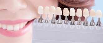
According to various studies, the majority of teeth belong to shade – A on the Vita scale (Yamomoto 1992, Vanini 1994, Tuati 2000). Due to the fact that the enamel of children is brighter than that of adult patients, the color of their teeth most often corresponds to shades A1, A2 (according to Vita, since the most common lesions in childhood are injuries to the frontal group of teeth, accompanied by a violation of the integrity of the crown angle or the entire cutting edge, pediatric dentists need a material that reproduces all the optical characteristics of the cutting edge of the tooth.
At the moment, the restoration material that best meets all the requirements of pediatric dentistry is Enamel plus.
Enamel plus is a radiopaque, microhybrid, light-curing composite that allows you to achieve excellent results in aesthetic restorations of anterior and posterior teeth, using direct and indirect methods. Еnamel plus is a unique restoration system that makes it possible to reproduce all the optical effects of the cutting edge.
Its creator is Dr. Lorenzo Vanini (Italy). When developing this material, L. Vanini took into account all the components of tooth color. His main task was to create a material, using which it would be possible to obtain a predictable result, which is so important in the daily practice of a dentist. The Enamel plus set includes three basic enamels, seven universal fluorescent dentins, two intensive enamels (for personalizing the enamel on the surface) and opalescent enamels, which can be used to highlight internal incisal opalescences and mamelons. (Fig. 2) In addition, the set includes a Glass Connector . It is a flowable composite that mimics the protein layer of natural teeth and six colors to replicate the characteristics. To determine the color, it is proposed to use the Enamel plus scale, made entirely of composite. (Fig. 3) The set also includes a special color card. This card remains in the medical history, and you can use it in further work. (Fig. 4, 4a)
Rice. 2.
Rice. 3.
Rice. 4.
Rice. 4a.
To obtain maximum results when using the Enamel plus HFO system, it is proposed to use the anatomical layering technique developed by L. Vanini. The anatomical stratification technique involves the construction of lingual enamel, internal dentinal body and vestibular enamel.
Before moving on to consideration of the stratification technique, I would like to note some features of cavity preparation under Enamel Plus. The fact is that preparation for this material is distinguished by the possibility of maximizing the preservation of healthy tooth tissue and does not require modeling a fold on the enamel. It is by increasing the width of the rebate and covering a larger surface of the enamel with a composite material that doctors often try to improve the aesthetics of their restoration (make the transitions of the material to the tooth tissue less noticeable and avoid the appearance of a gray stripe at the border of the filling with the tooth). At the same time, sometimes restorations of large cavities of classes III and IV turn into the production of veneers using the direct method, which is absolutely incorrect in pediatric dentistry, especially in cases where the tooth has not yet fully erupted. When preparing under Enamel plus HFO, a groove is formed on the vestibular enamel and proximal surfaces, along the edge of the cavity being prepared, with a spherical bur, the palatal side is processed at 90 degrees. This preparation technique is very gentle. (Fig. 5, 5a)
Rice. 5.
Rice. 5a.
Rice. Defect in the cutting edge of the 21st tooth.
Rice. Temporary restoration with composite without adhesive preparation.
Rice. Taking a silicone impression.
Rice. Example of a silicone key.
Restoration of dental injuries without opening the pulp.
The most common defect requiring restoration in children is trauma to the frontal group of teeth without opening the pulp. The break line is located parallel or diagonally to the cutting edge. In this case, the medial angle is most often affected.
After completing the color card, preparation and adhesive surface treatment, we begin to restore the lingual enamel. Because enamel in children has a high brightness; most often, we take the shade of enamel GE3. (Fig. 6, 6a)
Rice. 6
Rice. 6a
To simplify the task in case of extensive defects, a silicone block is made, which allows a thin layer to distribute the material and avoid inaccuracies in the formation of the macrorelief. (Fig. 7) When modeling, to create a more regular surface, in addition to conventional trowels, silicone trowels (micerium) are used, which give “finger effect” (Fig. 8).
Rice. 7
Rice. 8
Using a special brush, a thin layer of Glass Connector is applied to the inner surface of the enamel sheet, and it should be located strictly at the enamel-dentin border. (Fig.9)
Rice. 9
After applying the Glass Connector, we begin modeling the dentinal body. To achieve optimal saturation of the restoration, 3 base dentin colors are used. For example, if we want to end up with color A2 (according to Vita), we should start with UD4, then layer UD3 and UD2 - lighter ones.
At the stage of applying the last dentin, mamelons are modeled (Fig. 10, 10a, 11, 11a, 12,12a)
Rice. 10
Rice. 10a
Rice. eleven
Rice. 11a
Rice. 12
Rice. 12a
The finished dentin body is covered with a thin layer of Glass Connector.
To recreate the opalescence of the enamel, opalescent enamel (OBN) is applied between the mamelons and in the incisal area. After this, if necessary, intense white enamels (IM, IW), opalescent enamels (AO, OW) and paints for characterization are applied (Fig. 13, 13a, b)
Rice. 13
Rice. 13a
Rice. 13b
The final stage is the application of the main enamel (GE3) - vestibular enamel sheet. (Fig. 14, 14a, 14b, 15, 15a)
Rice. 14
Rice. 14a
Rice. 14b
Rice. 14c
Rice. 15
Rice. 15a
Thus, with strict adherence to the anatomical stratification technique, the use of Enamel plus material allows one to achieve a natural appearance of teeth in the restoration.
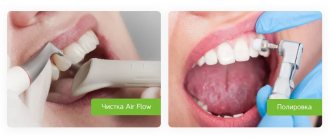
Surface treatment
Includes final modeling of the tooth shape (macro- and microrelief) and surface polishing. To simplify the task, when creating a vestibular convexity, transition lines, and Recius lines, guidelines can be drawn on the surface of the tooth with a slate pencil. Modeling of macro- and microrelief is recommended to be done with diamond burs. After which we begin polishing the surface. To do this, use the polishing system included in the Enamel plus HFO set, which includes three pastes and polishers with a silicone head, goat bristles and a felt disc. (Fig. 16)
Rice. 16
Recommendations for doctors
The requirements for restorations made from Enamel plus are no different from those for any other composite.
We must remember that before starting work, it is necessary to establish individual oral hygiene. After all, good hygiene will prolong the life of any restoration.
The key to the success of your work is high-quality insulation of the working area. From the age of 7-8 years, children can easily tolerate the rubber dam. It is important not to forget that what frightens patients most of all (and, it should be noted, not only children) is the unknown. Therefore, before starting treatment, we show and tell you what it is and why. Let's compare the rubber dam to an umbrella or a cloak for a tooth. Rubber dam is used both for direct restorations and for fixation of indirect restorations.
High-quality finishing and polishing of the surface will not only improve the appearance of your restoration, but will also make it more durable. Despite the fact that we recommend polishing fillings once a year, our foreign colleagues have excellent results 9-10 years ago. Moreover, during this time the patient never came for polishing or simply for a medical examination. It was a completely different tooth that brought him to the clinic. Neither the esthetics nor the marginal fit of the Enamel plus restoration performed for trauma was compromised (Dr. F. Mangani, Italy).
Conclusion
Using Enamel plus HFO, the pediatric dentist will receive the final result of the restoration immediately after a dental injury, detection of caries or any other destructive process.
Due to the high adhesion force of the material (30 mPa), excellent strength characteristics, and the degree of abrasion as close as possible to natural enamel (Fig. 17 diagram). Enamel Plus HFO makes it possible to restore teeth even with partial eruption. Moreover, after complete eruption, after a year, five or even ten years, the restored tooth will look natural.
Rice. 17 diagram
Author:
Timoshenko O.A., dentist, Department of Pediatric Dentistry, Sechenov Moscow Medical Academy
Why do you need dental filling?
The first experiments with the installation of fillings were carried out before our era. Holes in the teeth were filled with resin and various mixtures. However, the archaic technique of filling teeth brought almost no positive results. Fortunately, today everything has changed dramatically. Modern fillings are successfully used to perform a variety of tasks, in particular for:
- functional filling
- aesthetic dental filling
- filling root canals
- temporary sealing of a damaged tooth
Types of dental fillings
Classic filling of carious teeth involves the installation of a dental filling without depulpation of the root canals. This technique is used for minor mechanical and carious damage to the tooth (superficial and medium caries) and is carried out in several stages.
When the dental pulp is damaged, in most cases it is necessary to fill the root canals of the teeth. In the case of milk and young molars, there are methods for preserving the functionality of the root - a biological method of treatment using calcium-containing preparations (tooth filling with calcium) or partial amputation of the pulp. This allows you to maintain the blood supply to the tooth and its vitality, which is especially important when it is in the development stage.
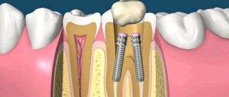
Depulpation involves removing the tooth root, cleaning and developing the canals with special instruments and filling them with artificial materials in order to eliminate the risk of developing inflammatory processes. There are various methods of root canal treatment. There is no absolutely ideal method, so the doctor focuses on a specific clinical case.
Where to go
You can make an appointment with a dentist in Ivanteevka at our Sanident clinic. We provide all types of dental care to patients from Shchelkovo and the urban district of Ivanteevka.
In case of significant damage in the oral cavity, our specialists:
- install pins of various types;
- restore the angle of the tooth;
- carry out complete anatomical restoration of the coronal part and other types of work.
High-quality filling and anesthetic materials are used for treatment.
A consultation with a dentist at the Sanident clinic is your first step to a healthy smile!
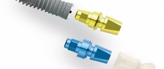
Installing a seal on a pin
More recently, it has become common practice in dentistry to install a filling on a post if the tooth is more than 50% destroyed. Thus, the technique made it possible to avoid prosthetics. Filling a tooth with a pin must be preceded by removal of the nerve. Then a special screw is inserted into the root canal and fixed with cement. The procedure is completed by layer-by-layer application of light-polymer composite materials. However, installing a pin does not guarantee reliability and safety. Over time, the cement breaks down, causing microbes to get under the filling, causing the formation of caries. Having chosen modern dentistry to install a filling, you are unlikely to encounter such technology.