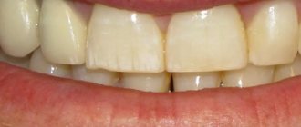
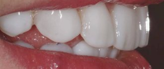
The Smile-at-Once clinic uses new generation composites - increased strength using nanoparticles. Despite the fact that they are stronger than classical materials, the patient in any case needs to follow the rules for caring for the extended teeth.
Extensions are very similar to regular fillings, since the same materials are used. Therefore, the rules of care are largely similar. However, there are also differences - artistic restoration is carried out in a more elaborate manner, since a small piece or even a whole crown is built up. The composite is fixed on top, and not placed inside the cavity, so the absence of walls imposes certain obligations in terms of caring for such teeth.
How much should you not eat after teeth extensions?
Since anesthesia is used for recovery, you should avoid eating for 2-3 hours after the end of treatment. You need to wait until the numbness goes away completely, otherwise you may accidentally bite your lip, tongue or cheek while chewing.
Modern photopolymer dental fillings harden under the influence of a special lamp almost instantly, so the load on them will not lead to shrinkage of the material - if anesthesia was not used, you can eat right away. But it is better to give preference to softer and warmer foods.
If the tooth has been significantly damaged and a pin or stump insert was used as additional support, you should avoid eating hot or very cold food for the first 24 hours, since special dental cement was used - its fixing properties may become worse under temperature stress.
Installation of crowns
Prosthetics with crowns is a reasonable way to restore quality of life and restore the functionality of a severely damaged unit. Today it is possible to make beautiful and comfortable dentures at an affordable price.
Metal-ceramic crowns on teeth
If chewing teeth have been damaged, then the most affordable way to restore them is the use of metal-ceramics. Such crowns for sixth teeth or other molars will be strong and aesthetically pleasing. The ceramic coating visually brings them closer to natural teeth, and the solid metal base allows you to chew familiar foods without fear.
Metal ceramics
– a compromise look that combines comfort, quality and attractiveness. Chewing teeth actively work under significant load; the metal will be stronger than its own enamel.
The method of making such crowns:
- An impression is made.
- Based on the wax model, a one-piece structure is created in the dental laboratory, repeating all the shapes of the recreated dental crown.
- Layers of ceramic are applied to the model, each of which is baked at high temperature.
It is recommended to follow the usual precautions - do not eat hot and ice-cold food at the same time. With a sharp change in temperature, the ceramic surface may crack. This way of eating is also harmful to the “native” enamel.
How much does it cost to make a metal-ceramic crown on a tooth? For me – from 17,900 rubles for 1 tooth. If you need several prostheses, then flexible pricing applies. Accordingly, if the patient wants to restore a tooth not with metal-ceramics, but with ceramics, then there are no problems, he will be offered only the best materials e-max and zirconium dioxide.
Previously, in case of serious damage, a pin and a filling were used to recreate the stump in order to place a crown on the tooth on such a support. Nowadays, more and more people are switching to Cerec technology and restoring teeth using single “crown + root” modules, which is stronger and more reliable. And most importantly, it is much faster in time and costs less in total:
Come to me, Dr. Sergei Samsakov, and we will assess the condition of the problematic tooth and choose a method to restore it. If a tooth is broken, let's see if it is possible to put a crown and what material is better to choose. All manipulations are performed without pain, in a comfortable environment.
What dietary restrictions are there after dental restoration?
Modern composites, even despite their high performance characteristics, are susceptible to staining, which is why even if the filling is in good condition, after a few years it becomes darker compared to natural enamel. Over time, a clear contrast will be felt between the living tooth and the extended piece.
Extended teeth are quite weak - under load, a piece of filling material can break off. Especially when carrying out restoration of the frontal zone. All this requires following a certain diet.
- coffee, tea, red wine, sweet colored soda, beets, sauces (especially tomato and soy), berries, candies and caramel are not recommended - that is, all products that over time will lead to a change in the shade of the extension,
- Avoid eating solid vegetables and fruits whole - it is better to cut them into pieces and chew them with unextended teeth. This is especially true if the front teeth have been restored - they should not bite off hard foods, gnaw seeds,
- give up the habit of cracking nuts or cracking seeds, gnawing crackers with extended teeth,
- Chew food slowly, especially if it is hard or fibrous,
- Avoid food that is too hot or cold - it can cause cracks and chips in the material.
If women have extended front teeth, it is better to avoid bright lipsticks, since composites can again become stained.
To whom is the technique contraindicated?
Artistic restoration of teeth with composite material is not possible for all clients, since the technique has a number of contraindications (some of them are temporary and correctable):
- Allergic reaction to the composite;
- Installed pacemaker;
- Some individual characteristics that prevent the sealing of a carious cavity;
- Poor oral hygiene;
- Night grinding of teeth (bruxism);
- Pathological bite;
- Excessive abrasion of tooth enamel;
- Frequent stay in traumatic conditions (athletes, rescuers, etc.).
By registering with Nurimed, clients can be calm about their own safety and the results of the procedure, since the clinic’s doctors individually assess the risks and benefits for everyone.
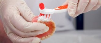
How to do oral hygiene
If it is impossible to refuse drinks and foods that stain filling materials, you need to pay increased attention to hygiene. In such situations, cleaning after every meal is mandatory.
- cleaning in the morning and evening (before and after breakfast, as well as after the last meal) with a medium-hard brush,
- irrigator, dental floss, brushes and rinse for cleaning the interdental spaces after each meal and even after a regular snack.
Research results
The patient, 46 years old, complained of an aesthetic defect in her anterior teeth (Fig. 1). Objectively: in the 11th and 21st teeth, extensive partial restorations with signs of secondary caries and a violation of the marginal seal are determined. The central incisal line is shifted to the left. Pathological abrasion along the cutting edge. Color 21 slightly changed. According to the patient, the tooth darkened after primary endodontics many years ago. After repeated endodontics, the color did not change. The tooth doesn't bother me. The radiograph shows obturation of the canal to the apex, the periodontal fissure is not widened.
Rice. 1. Defects in the fillings of the 11th and 21st teeth.
In accordance with the clinical picture, a diagnosis was made: secondary caries of the 11th and 21st teeth. The patient was informed about alternative methods of orthopedic and therapeutic restorative treatment of anterior teeth. She chose the method of complete direct restoration of 11 and 21, which is consistent with the opinion of the dentist. This choice is justified by the following factors:
- a large volume of lost tooth tissue dictates the need to correct the crown height and color, which excludes partial restoration;
- the relative cheapness of the method, treatment in one visit is attractive for the patient, in contrast to prosthetics or indirect restoration;
- slight color change 21, time and economic factors excluded intracanal bleaching 21.
The patient signed a preliminary consent to perform the necessary manipulations. Teeth were cleaned of plaque mechanically using a product that did not contain fluoride and oil. For this purpose, Clint paste (VOCO) was applied to a special brush rotating at low speeds. To avoid heating the tooth, an excessive amount of paste was used. Then the tooth was thoroughly washed with a stream of water.
Next, the size, shape, and relief were planned, which is a strictly defined sequence of actions, including odontometry and odontoscopy of the central incisors. Based on visual assessment and measurement results, a rectangular crown shape was planned: the side surfaces are located almost parallel; the horizontal dimensions in the middle and lower thirds are close in value.
Evaluation of signs of teeth belonging to the side showed a predominance in size of the distal angle of the crown. The sign of crown curvature is not expressed. The description of the individual characteristics of the tooth consisted of assessing the type of macrorelief of the vestibular surface.
The selection of the desired shades of the Admira Fusion (VOCO) photocurable composite was carried out in natural light using special standards by comparing them with the intact lateral incisor before installing the rubber dam. The color of the composite was specified by applying a “drop” of the composite to the vestibular surface of the incisor directly from a syringe (Fig. 2, 3). The following syringes were selected: cervical region - A3.5, body - A2, transparent enamel - Incisal. The material allows intuitive selection of shades and makes it possible to create colors using both opaque and enamel shades.
Rice. 2. Selection of enamel shades of the composite.
Rice. 3. Selection of dentin shades.
Considering the slight difference in the color of the 11th and 21st teeth, it was decided to carry out a correction using enamel shades of the material. Thus, for the cervical area of the 21st tooth, a syringe GA3.25 was chosen (the original shade between A3 and A3.5 on the Vita scale), and shade A1 was used for the body of the veneer.
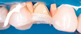
The preparation was performed with medium-grain diamond burs and began with the removal of low-quality fillings, protruding enamel edges, and dentin necrotomy (Fig. 4). The contours of the veneer coating were marked using a small spherical bur in the form of grooves no more than 0.5 mm deep.
Rice. 4. A set of burs for preparing a tooth “for veneer”.
In the proximal areas, to hide the edge of the veneer, the boundaries extended to the contact surfaces. The cervical border of the veneer is located along the border with the gum to avoid the transmission of pigmented tooth tissues. Cervical ledges are formed, internal angles are smoothed. The vestibular surface of the incisors was processed with a fine-grained cylindrical bur (Fig. 5, 6).
Rice. 5. Marked bur in sterile packaging.
Rice. 6. A diamond bur is placed in the turbine tip.
A rubber dam was installed with exposed teeth 14 to 24, which will allow you to focus on the dentition when creating the shape and distributing color areas. The quality control of preparations 11 and 21 was carried out (Fig. 7). Next, the technique of total etching of the prepared surfaces of the central incisors was carried out (Fig. 8).
Rice. 7. The central incisors were prepared.
Rice. 8. Total acid etching.
The gel used was Vococid (VOCO) - 35% phosphoric acid. The gel was washed off with a stream of water, and the teeth were dried with a reflected stream of air. Adhesive preparation was carried out with a universal bond, suitable for any adhesive technique (Fig. 9). A thin layer of Futurabond was applied to the etched surface of the teeth with a brush and cured with the light of a halogen lamp (Fig. 10). Next, the veneers were modeled using the following Grandio dentin shades for the 11th tooth: cervical area - color OA3.5, body - OA2 (Fig. 11).
Rice. 9. Futurabond U adhesive system.
Rice. 10. Adhesive preparation of the 11th tooth.
Rice. 11. Photo-curable material.
The production of a classic veneer began with the application of an opaque composite to the prepared surfaces. A restoration base is created that includes a dentin geometric outline to define the lateral and inferior boundaries of the veneer. The sign of the crown angle is modeled. The first layer of Grandio opaque filling material was placed on the mid-gingival area and moved towards the neck of the tooth. The next layer was placed on top of the previous one and distributed evenly over the entire area. The lateral surfaces of the veneer were not brought into contact with adjacent teeth by 1.0 mm, which is due to the type of transparency of the enamel in this area (Fig. 12).
Rice. 12. Opaque layers of the composite are applied.
In accordance with the instructions for using the composite material, the latter was added in layers no more than 2 mm thick, compacted to the tooth tissue and cured with halogen light. The prepared opaque base, which replenishes the shape and volume of the tooth dentin, was covered with enamel shades of the material. To form a periodontal contour, a portion of enamel composite was applied to the central gingival area of the tooth and smoothed from the center to the periphery.
When modeling the enamel layer, shades A3.5 and A2 were used, respectively. Space is left for the transparent Incisal shade in the area of the incisal edge and side edges (Fig. 13). The formation of contact surfaces was completed by applying a transparent composite, which was distributed taking into account the individual degree of transparency of the enamel. The same transparent material was used to model the cutting edge and cover the corners of the veneer.
Rice. 13. Modeling the enamel layer.
For the restoration of the 21st tooth, dentin colors were chosen similarly to veneer 11. Syringes of the enamel composite are presented in shades GA3.25, A1, Incisal (Fig. 14). Dentin shades for the formation of a veneer on the 21st tooth were applied in layers up to 2 mm thick, which ultimately filled the volume of missing dentin without involving the boundaries of the enamel layer (Fig. 15). Using individual tint enamel syringes, the effect of neutralizing color accents was achieved (Fig. 16). The surface of the restoration was covered with a transparent layer in accordance with the type of enamel transparency.
Rice. 14. Tint composite syringe.
Rice. 15. Opaque layer of the 21st tooth.
Rice. 16. Color neutralization has been completed.
Immediately after the aesthetic structure is manufactured, it is processed: the surface hybrid layer is removed and the relief is enhanced. Contouring and polishing of the vestibular surface of the veneer was carried out using Dimanto diamond instruments (VOCO) (Fig. 17). After removing the rubber dam, adaptation to the bite, grinding, and polishing are controlled (Fig. 18).
Rice. 17. Polishing system.
Rice. 18. Ready-made aesthetic restorations.
The final stage of treatment is coating the enamel surrounding the restoration with fluoride-containing varnish Bifluorid 12 (Fig. 19). Long-term quality control of the work performed shows the high efficiency of using the method of producing direct veneers from a photocurable composite.
Rice. 19. Fluoride varnish.
Does the restoration need to be changed and when might it be necessary?
On average, the service life of composite extensions is about 3-5 years. Such a filling may last longer, but it will stand out on the front teeth after 3-4 years. Composites shrink, albeit slightly, so over time a small step will appear between the tooth and the filling material. This is especially noticeable on the front teeth if they are partially restored. In such a situation, you can update the restoration, that is, completely grind it down and replace it with a new one. Or simply re-build it up with a composite on top - in certain situations this is also allowed.
Cost of procedures
Prices for dental extensions vary depending on:
- Number of units restored.
- Severity of the defect.
- The chosen extension technique.
- Materials used.
The cost is also affected by the condition of the oral cavity, the preservation of the tooth root, the need for preliminary sanitation and other procedures. The final price of services is determined after examination by the dentist. The patient can influence the final cost by choosing more affordable composites and restoration technologies.
Regardless of the chosen technique, the doctors at the Ulybka dental clinic guarantee their patients beautiful and long-lasting results. Our specialists have extensive experience in building extensions of varying complexity, have modern tools and use advanced materials to ensure a highly aesthetic result.
You can find out the cost of extension of front and back teeth in Yoshkar-Ola in our price list.
Why do we most often recommend removing teeth with periodontitis?
Our natural teeth are a hotbed of inflammatory processes. They have a porous structure, so it is on them that all microbes, which have a destructive effect on periodontal tissue, first settle. If implants with an active surface are placed next to such teeth, even the most resistant ones, which contain chemical elements that promote the healing of bone cells (such as SLActive® from Straumann and TiUnit® or TiUltra® from Nobel Biocare), they will still are unable to cope with a massive attack from all sides and bone tissue atrophy. That is, by preserving our natural teeth, we risk losing all the results of implantation.
So if there are only a few teeth left, they are destroyed and affected by diseases, then there is no point in preserving them - it is much easier to carry out removal with simultaneous replacement with implants, after which it will be enough to carry out regular oral hygiene (on your own and with a dental hygienist) and actually forget about dental problems forever. After all, if there are no teeth, there is no inflammation.
We categorically do not recommend implantation on one jaw if there are foci of generalized periodontitis of a severe and acute stage on the second - even though there is no direct contact, it is still dangerous. Our body is a single whole, let alone the oral cavity. Pathogenic microorganisms will circulate along with saliva, so inflammation of the tissues around installed implants is only a matter of time. It is impossible to talk about any lifelong service in such a situation.