Each person goes through the stages of the eruption of the first teeth, the development of milk teeth and their subsequent replacement by permanent ones. Despite their similar appearance and function, temporary and permanent teeth have differences, which we will talk about, at the same time we will consider the timing of the appearance of the main teeth, possible problems with them in the process of their development.
The photo shows a diagram of the structure of human teeth
The structure of human teeth
Teeth are not only intended for mechanical processing of food, but are also necessary for the formation of speech, breathing, and influence facial features. To navigate what dentists advise, how to take care of your teeth, and what the risks of disease are, it is useful to know how they work.
Anatomical structure
3 parts that make up a tooth:
- Crown. The visible part of the tooth used for chewing. The outside is covered with durable enamel, which protects it from bacteria, chemicals contained in food, water, and saliva. The surfaces have their own names: Facial (vestibular) - in contact with the lip or cheek.
- Lingual (lingual) – the opposite of the facial, involved in the formation of speech.
- Occlusion – the upper surface in contact with the tooth of the opposing jaw.
- Contact (approximal) – contacts with adjacent teeth.
Milk teeth, having a largely similar structure, also have differences in anatomy:
- They are noticeably smaller in height than permanent ones.
- The crown is much wider than the root.
- Enamel is thinner and more fragile.
- The roots are more round.
- The wear of baby teeth, as well as their spontaneous loss, is a normal physiological process.
Histological structure
The structure has several layers:
- Enamel is the most durable fabric. When a tooth just erupts, a cuticle is located on it, which is gradually, under the influence of saliva, replaced by a pellicle.
- Dentin is a highly mineralized tissue that resembles bone, but has better mechanical strength. Instead of enamel, the root part of the dentin is covered with cement.
- The pulp, the central part of the tooth, is a soft connective tissue containing a large number of blood vessels. Caries and inflammatory processes “owe” pain to the pulp with its large number of nerve endings.
Milk teeth are distinguished by dentin with a lesser degree of mineralization, which weakens their protection against caries. The volume of pulp occupies most of the tooth, and small protective layers (enamel and dentin) provide less protection against the penetration of bacteria and the development of inflammatory processes.
Types of teeth
There are 4 groups:
- Incisors. 4 chisel-shaped cutters. The largest are a pair of upper central incisors, and the situation is opposite from below - the lateral incisors are slightly larger than the central ones.
- Fangs. 2 on the upper and the same number on the lower jaw. Their length is longer than the others, the front wall is convex.
- Premolars. There are 8 in total, prismatic in shape, the upper surface with two tubercles (buccal and lingual). Premolars have 2 roots. The second premolar has a larger buccal surface. There are no primary premolars.
- Molars. The first molar (molar) is the largest tooth in the upper jaw. The chewing surface has four tubercles, 3 roots. The cubic-shaped second molar is smaller, and the buccal tubercles are larger than the lingual ones. The third (“wisdom tooth”) is in many ways similar to the second, but not everyone has it.
Where are the molars located?
Man is an omnivore. For normal metabolism, we need different foods - rough and soft, plant and animal. In order for the product to be well absorbed by the body, it must be thoroughly chewed. This is facilitated by molar teeth, that is, chewing or lateral teeth. They are distinguished by the largest chewing area - up to 1 cm2. Researchers estimate that each can withstand pressure of 75 kg.
They are located on the 6th, 7th and 8th positions in the row. There should be a total of 12 such teeth in the mouth of an adult – six on each jaw, three on each side of the row.
Interesting fact! Eights or “wisdom teeth” have shown a tendency towards retention in recent decades, i.e. are delayed in terms of eruption in the jaw bone or gum, and may never erupt at all.
Dental formula
In order to improve the convenience of describing each tooth, numbering them, and filling out cards, it is customary to record the order of the teeth using a special formula. There are several varieties of it.
Zsigmondy-Palmer system (quadratic-digital)
Arabic numerals are used, numbering starts from the central incisors in each direction:
- 1 and 2 – incisors.
- 3 – fang.
- 4, 5 – premolars.
- 6-8 – molars.
Milk teeth are designated differently - using Roman numerals:
- I and II – incisors.
- III – fang.
- IV and V – molars.
Two-digit Viola system
Teeth numbering uses 2 digits. The jaws are divided into 4 quadrants. The first digit shows its number.
For adults this is:
- 1 – upper jaw on the right.
- 2 – upper jaw on the left.
- 3 – lower jaw on the left.
- 4 – lower jaw reference.
For a similar description of baby teeth, numbers 5 to 8 are used.
So, there are 8 teeth in each quadrant, its number is shown by the second digit. Thus, the first molar of the lower jaw on the left is designated 35, and the child’s canine from the lower right is designated 43. Therefore, the phrase that “treatment of the 48th tooth is required,” or, for example, the 55th, does not indicate the doctor’s lack of qualifications or what - or pathology in your child, who suddenly acquired so many teeth.
First molars (sixes) of the upper jaw
The first molars, located on the upper jaw, are the largest in the mouth. In an adult, the length of such a tooth can reach up to 2.5 cm. There are 4 tubercles on the diamond-shaped chewing surface - 2 each on the side of the cheek and tongue. The buccal ones have a conical shape, while the lingual tubercles have a rounded shape. Moreover, on one of the anterior tubercles there is a small additional tubercle.
On the buccal and lingual surfaces of the upper sixes, one groove is located in the vertical direction. The surface on the tongue side is smaller than on the cheek side, but has a much more convex shape.
Top sixes usually have 3-4 channels and 3 roots:
- palatal: resembles a cone in shape,
- anterior and posterior buccal: slightly flattened. The rear one is similar in shape to the front one, but smaller in size.
Interesting! Sixes are the first to grow in children's permanent dentition. They usually erupt at 5-8 years of age. There are frequent cases when only a few days pass between the appearance of all 4 pieces.
Dental development
The differences between primary and molar teeth begin with their number - only 20 primary teeth, 8 incisors and molars, and 4 canines. This is explained by the fact that children simply have nowhere to fit more teeth. In this regard, there are no primary premolars. By the time the permanent teeth appear, the adolescent's jaws are already sufficiently developed for all teeth to appear.
The formation of tooth buds in humans begins at the 6th week of intrauterine development, and at the 14th week hard dental tissue appears. The crown develops first. The development of the rudiments of permanent teeth begins in the 5th month.
By the time a child is born, the formation of the rudiments of both milk and permanent teeth is almost complete. The process of development of permanent teeth, which have no analogues among milk teeth, begins a year after birth.
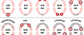
While the first teeth may appear at 4 months, and their eruption may be delayed for up to a year, permanent teeth erupt in everyone at approximately the same age. The sequence of their eruption is the same as in the case of milkweeds:
- 6-7 years. The central incisors appear from below.
- 7-8 years old. The central incisors on top and the lateral incisors on the bottom are replaced.
- 8-9 years old. The lateral incisors of the upper jaw appear.
- 9-12 years old. Canines and premolars are replaced.
- From the age of 12. From this age, molars begin to change, and from about 14 years of age, teeth appear, which were not among the milk teeth.
Features and difficulties of treatment
On the chewing side there are tubercles and grooves-fissures (cone-shaped, grooved, teardrop-shaped). These relief formations on the surface are formed during the formation of the tooth germ. They are involved in grinding food. But it is the fissures that are most susceptible to carious lesions. If sealing is not done in time, caries will penetrate into deeper tissues and into the pulp.
Treatment of pulpitis is a labor-intensive process. Here, options for biological treatment or depulpation are possible - they will be discussed further in the article. The main difficulty during depulpation is narrow and curved root canals, because molars have the largest number of roots - usually 3-4, but there are situations when there are even more of them - 5 or 6. Therefore, to assess the quality of canal filling and avoid the risk of secondary caries, a qualified dentist refers the patient for x-ray diagnostics.
Important! There is a habit of calling any chewing teeth “molars.” This is a bit of a misnomer in terms of terminology. There are two concepts - temporary teeth (baby teeth) and permanent ones. The latter should be called molars, because their roots do not dissolve, like those of milk teeth during a change in bite.
Signs of the imminent appearance of molars
You can determine when you should soon wait for the baby teeth to begin replacing with permanent teeth based on several signs:
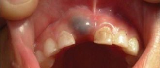
- The gradual growth of the baby's jaws leads to increasing gaps between the teeth.
- The tooth begins to wobble. This is due to the fact that the already small root begins to gradually dissolve, causing the fixation of baby teeth to be significantly weakened.
- A fallen tooth indicates that the formed permanent one, which is about to appear, pushed it out.
- Swelling and redness may appear on the gums at the site of the eruption of a permanent tooth.
- Pain in the gums, where the permanent tooth erupts, increased temperature, and poor health of the child indicate problems have arisen, and it is necessary to see a doctor. The process of erupting molars should be painless.
The main differences between baby teeth and permanent teeth
Unlike 28 permanent teeth, the primary dentition requires the presence of 20 units. At the same time, they have a number of characteristic features.
- Smaller in size compared to permanent teeth.
- White with a slightly blue tint (permanent units have a slightly yellowish tint).
- Less developed and slightly short roots compared to permanent teeth.
- The enamel of primary teeth is poorly formed - thinner.
- Milk units can be erased (permanent ones can too, but this is considered a pathology).
As the child grows, baby teeth fall out on their own - this is the norm. The permanent dentition units should not fall out on their own.
Possible problems
At the moment the molars appear, certain dental problems are possible. In order to take timely measures to eliminate them, parents must have an idea about them.
Molars do not erupt
A situation is possible in which baby teeth do not fall out in a timely manner, or they have fallen out, but molars have begun to appear in their place. The reason for this must be determined by the dentist, who must be visited without delay. A general x-ray is usually taken to show the degree of development of the molars.
Among the options for the lack of eruption of molars in due time can be indicated:
- Hereditary predisposition, which is the cause of a possible delay in the appearance of molars. If the x-ray shows that the process of forming the rudiments of teeth is underway, then you will just have to wait a little for their appearance.
- Adentia. Disturbances in the processes of formation of tooth germs during the intrauterine development of a child, inflammatory processes can lead to a similar pathology - the absence or death of tooth germs. The solution is prosthetics.
Pain
The first time after teething, the tooth is poorly protected from caries and the effects of various bacteria. This is explained by the low degree of enamel mineralization at the initial stage. Almost nothing interferes with the development of caries; tooth tissue is destroyed, pulpitis occurs, with the subsequent risk of its transition to periodontitis. Severe pain, changes in body temperature and deterioration in well-being may occur.
It is highly advisable not to let the situation get worse, not to cause severe pain, but to visit a dentist as soon as painful sensations appear. If a child is predisposed to caries, it is better to carry out preventive procedures, for example, fissure sealing. The folds on the chewing surface are covered with a composite material that protects such natural cavities from the accumulation of food debris in them, the development of bacteria, and inflammatory processes.
In the worst case, you can lose a tooth.
Teeth grow crooked
A common situation is when the molar has already begun to erupt, but the baby tooth does not want to fall out. The result is that the new tooth seeks alternative growth paths, which leads to its displacement and change in the direction of growth. Hence the malocclusion and the alignment of the dentition. Treatment by an orthodontist will be required.
If this situation occurs, you should not remove or loosen a baby tooth yourself; you should visit a doctor.
Loss of molars
An alarming symptom of the presence of diseases (caries, etc.) in the oral cavity, or there are problems with the entire body (connective tissue diseases, diabetes, etc.). A visit to the doctor is mandatory.
This is necessary to develop a strategy for restoring a lost tooth. This is necessary for the proper growth of the remaining teeth and the formation of the maxillofacial system. Considering that the jaw tissue is still in the process of growth, prosthetics are only possible temporary, which must be adjusted as the jaws develop. Permanent prosthetics will be available only after their formation is completed.
Injuries
The first few years after teething, teeth are at increased risk of injury from impact. Sports injuries, falls, and blows can lead to chipping of parts of the tooth and cracks. Be sure to contact a dentist who will restore the lost part with modern materials.
When do primary molars become molars?
To keep the chewing organs strong, fluoridation of the teeth is often required. This technology ensures the prevention of carious lesions due to additional mineralization of the enamel layer. Fluoride ions create an antibacterial environment in the oral cavity, prevent acid damage to crowns, reduce the vulnerability of units, and reduce the formation of soft plaque and stones. After the procedure, patients develop gum disease much less often.
When the milk organ is replaced by the root organ, children experience slight discomfort. The first permanent posterior tooth appears after six to seven years. New units grow under the temporary ones, appearing above the gum after they fall out. You should not speed up the process of hair loss, as this can cause:
- Incorrect growth of permanent units;
- Malocclusion;
- Disproportional growth of the jaw bone;
You should visit the dentist regularly during this period. If the units grow crookedly, the doctor will immediately respond to the problem, correcting the child’s bite in a timely manner. Scheme for cutting through constant molar units:
- In the period of 6-8 years, permanent sixes appear;
- When the child turns 12-13 years old, sevens, both upper and lower, are cut through;
- After 17 years, only a few wise people make it through. Their growth can last up to 30 years or more;
Eights may not erupt at all, remaining under the gum tissue or in the jawbone. It is important to visit the dentist regularly to prevent serious complications.
Molars (6, 7 and 8 teeth) – where they are located, what structural features they have and nuances in treatment
A healthy adult should normally have 32 teeth - 16 each in the upper and lower jaws.
Moreover, they all differ in shape and structure from each other, depending on the location and their immediate tasks. Molars are the sixth, seventh and eighth teeth located on both sides of both jaws. Their main purpose is to ensure thorough chewing of food. They must be strong and hardy, and these requirements partly explain their special structure. Further in this article, read more about molars – what kind of teeth they are, when they “come out” to the surface in children, and whether there are any nuances associated with their treatment.
Why do teeth erupt with irregularities?
The condition of a child’s teeth in the first years of life depends on the health of the mother during pregnancy. The formation of hard dental tissues may be disrupted if a woman:
- suffered from toxicosis at the initial stage of pregnancy;
- experienced severe prolonged stress;
- was treated for kidney disease;
- suffered from rubella.
In addition to maternal diseases, the maturation of teeth and the development of the jaw system can be affected by:
- fetal prematurity or, conversely, delayed birth;
- conflict of Rh factors;
- sepsis suffered by a child in the first month of life;
- frequent colds, pneumonia;
- convulsions;
- toxicosis;
- refusal to breastfeed.
Often, parents, driven by the best intentions, want to alleviate the baby’s suffering by giving him medications. You must understand that any medicine, even if it seems completely safe, cannot be taken without consulting a pediatrician!
Features of premolars
Premolars are the lateral teeth that are located next to the canines. There are no premolars in the primary dentition. In their place, children have baby molars. After their loss, space is freed up for both molars and premolars.
The coronal part of the maxillary premolars has a convex surface. The first premolars often have two roots, the second, as a rule, one. Sometimes there are three-rooted premolars. The premolars of the lower jaw have one root, which has an oval or slightly pointed conical shape. There are two tubercles on the surface of the first dental unit. The second feature is its size. The second premolar is larger in size than the first.
Premolars are much less susceptible to caries than molars.
When is there a reason to visit the dentist?
The formation of the dentition requires constant monitoring and competent assistance from the parents to the child, so that in case of developmental deviations, pathology is noticed in time.
- Teething in children: how to relieve pain
It is good if, when the first permanent teeth appear, the child visits the pediatric dentist for preventive purposes.
Such an examination will help identify a number of problems:
- malocclusion;
- gum disease;
- inadequate mineralization of enamel;
- curvature of the dentition;
- milk caries.
Malocclusion
Caries of baby teeth
Insufficient attention to teeth in childhood means not only excruciating pain, tears and insomnia for the whole family, but also painful treatment and fear of the dentist for life. Therefore, it is important to constantly maintain contact with your doctor and devote enough time to the health of your children.
Losing the first teeth is a natural process for all children. And you only need to worry when problems arise with the formation of adult teeth. They can be prevented if the eruption of the first tooth is controlled. Jaw hurts after tooth extraction, you will find the answer in the link.
Treatment methods for chewing teeth in adults and children
The whole difficulty lies in the difficult passage of the channels of the masticatory elements. When processing them, there is a serious risk of injury to the pulp, which can provoke pulpitis. In this case, two treatment options can be distinguished:
- depulpation – partial or complete removal of the pulp with further endodontic treatment, that is, cleaning and filling the canals. This method is more often used in adult dentistry,
Surgical method of pulp treatment - biological method - a medicine, usually an antibiotic, is applied to the affected area of the pulp, after which a temporary filling is placed on top. The active substances of the drug destroy pathogenic microorganisms and stop the inflammatory process. It must be admitted that pulpitis on baby teeth progresses much faster than on adults, so quite often the doctor has no choice and removes the affected temporary element. If necessary, prosthetics can be performed before a permanent one appears.
This method involves killing the pulp with medication.
Molars are directly involved in the process of chewing food. Like other teeth, they require timely diagnosis and treatment. And in case of their early loss, it is imperative to take care of their correct replacement - the best option for this would be implantation.
Regular cleaning is the basis of hygiene. But even if we follow this rule responsibly, we cannot completely clean out all food debris and bacteria in one such procedure. Subsequently, they transform into plaque and hard stone, which causes gums to become inflamed, teeth to decay and rot. Therefore, it is so important to regularly visit the dentist for a preventive examination and professional cleaning of plaque and deposits. It is also important not to forget to rinse your mouth after each meal, use floss and, preferably, an irrigator. Proper care of your oral health will be the best prevention of any dental diseases.
1Dmitrienko, CB Anatomy of human teeth, 2000.
First aid for teething
When baby teeth appear, the baby may require not only parental care, but also medical attention. The dentist may recommend that parents use anesthetic dental gels and treat the affected areas of the gums with decoctions of sage, oak bark or soda solution. If the pain is severe, the baby may be prescribed paracetamol, ibuprofen and other systemic painkillers.
To ease the discomfort that a child experiences when teething, it is necessary to use teethers - specialized devices made of rubber or plastic that the baby can bite and gnaw on without risking damage to the soft gum tissue. In addition, it is advisable to regularly massage the child’s gums with a finger wrapped in a clean, damp bandage.