Performing syndrome diagnosis
To properly check for the presence of a disease, it is necessary to perform a full range of various procedures. In particular, diagnosis of stylohyoid syndrome includes a number of actions and manipulations.
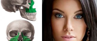
First of all, a professional examination of the patient is performed, as a result of which the doctor examines and identifies the compaction of the bone growth in the area of the anterior zone of the cervical spine of the patient. If you press on this part, the person should experience pain, and his health will deteriorate greatly.
Secondly, an X-ray of the facial skeleton, skull bones, and neck is taken.
At the time of diagnosis, you should carefully approach the procedures, because this disease can easily be confused with other similar pathologies, the symptoms of which are very similar. As an example, suppuration of the tonsils can be cited.
Causes and risk factors
The area where this compression occurs is called the superior thoracic outlet or thoracic outlet. The arm and neck are in constant motion, therefore, with the congenital narrowness of the gap between the collarbone and the first rib, constant trauma to the nerves of the cervical brachial plexus, subclavian arteries and veins occurs. Additional cervical ribs and shortening of the scalene muscles can lead to the development of compression of the vessels and nerves of the shoulder girdle. Common causes of superior outlet syndrome can be work-related or sports injuries.
The cause of compression of the neurovascular bundle of the arm is most often a congenital narrowing of the space between the first rib and the collarbone. However, thoracic outlet syndrome develops only under certain conditions:
- Anatomical defects
Congenital accessory cervical ribs located above the first rib or a shortened anterior scalene muscle can compress the neurovascular bundle. Sometimes the subclavian bundle can be compressed by the tendon of the pectoralis major muscle due to the anatomical features of the shoulder girdle.
- Poor posture
The habit of sitting and standing with drooping shoulders can cause compression of the vascular bundle at the exit from the chest.
- Injury
A traumatic event, such as a car accident with a fracture of the clavicle or sternum, can cause changes in anatomical relationships and lead to compression of blood vessels and nerves.
- Same type of movements in the shoulder girdle
Sports activities, work on a conveyor belt and other activities associated with constant raising of arms above the shoulders can contribute to the development of clinical signs of compression of the neurovascular bundle.
- Pressure on the shoulder joints
Carrying heavy bags on the shoulder or backpacks can be a provoking factor for the onset of the syndrome.
Clinic
In the presence of an elongated and/or curved styloid process, or a calcified stylohyoid ligament, or a calcified stylohyoid fold, or elongated horns of the hyoid bone, excessive pressure of these structures occurs on the internal and external carotid arteries. Due to this, in the areas fed by the carotid arteries, many seemingly unrelated clinical symptoms appear, such as:
- sensation of a foreign body in the throat;
- chronic inflammation of the pharyngeal mucosa;
- pain in the maxillary joint;
- pain and noise in the ears;
- unilateral and bilateral orbital or headache;
- “shooting” pain when turning the head.
According to the literature, patients with stylohyoid syndrome most often complain of poorly localized pain with unilateral localization in the upper anterior part of the neck and irradiation to the pharynx, root of the tongue, and ear. In this case, pain can spread to the temporomandibular joint, lower jaw, temporal, buccal areas, and submandibular triangle. In some patients, pain occurs in the teeth of the upper and lower jaws. M. Kiely et al. (1995) described the spread of pain to the sternocleidomastoid muscle, and D. Savic (1987) - to the supraclavicular fossa, shoulder girdle and anterior chest wall. Patients usually characterize the pain as dull, constant with periods of intensification and weakening. Its intensity increases towards the end of the day, intensifying when turning or throwing back the head, after a long conversation or singing, or changes in weather conditions.
Patients usually complain of pain in only one organ, most often in the pharynx, ear, temporomandibular joint, and only a detailed survey allows us to clarify its localization and areas of distribution. Some patients develop glossopharyngeal neuralgia with its characteristic painful paroxysms. Dysphagia is a common symptom of the disease, and difficulty swallowing is usually associated with a significant increase in pain in the throat and ear. Some patients are bothered by the sensation of a foreign body in the throat, others are worried about a “constantly sore throat”; they may experience pharyngeal spasm, persistent dry cough without objective signs of inflammation in the upper respiratory tract.
Stylopharyngeal syndrome
As a rule, it is right-sided, since the right styloid process is normally longer than the left by an average of 0.5 cm. Pain occurs as a result of pressure, for example, by an elongated and inwardly curved styloid process, on the tissue in the area of the tonsillar fossa and irritation of the nerve endings of the glossopharyngeal nerve. The intensity of pain varies greatly - from minor pain or the sensation of a foreign body in the throat, especially when swallowing, to sharp, severe constant pain in the throat and tonsils, radiating to the ear. Some patients also note pain on the front surface of the neck, in the area of the hyoid bone. Occasionally, these sore throats are mistakenly associated with pathological changes in the tonsil and it is removed, and ongoing pain is explained by irritation of the nerve endings by a postoperative scar. However, stylopharyngeal syndrome should be differentiated not only from damage to the tonsils, for example by a chronic inflammatory process, but also from neuralgia of the glossopharyngeal nerve. Neuralgia of the latter is characterized by paroxysmal burning or shooting pains in the pharynx, tonsils, root of the tongue, radiating to the ear, which occur during talking, laughing, coughing, yawning and eating.
Styloid-carotid syndrome (“carotid artery syndrome”).
The pathogenesis consists of pressure from the tip of the elongated and outwardly deviated styloid process on the internal or external carotid artery near the bifurcation of the common carotid artery, irritation of the periarterial sympathetic plexus and pain receptors. When the internal carotid artery is irritated, the pain is constant and is felt in the frontal region, orbit, eyes, that is, in the area of branching and blood supply of the internal carotid artery or its branches, in particular the orbital artery. Due to the pressure of the appendage on the external carotid artery, pain radiates along its branches to the temple, crown, face (below the eye).
- Why does the jaw hurt on one side when chewing near the ear: what does pain on the right and left mean, how to treat it?
Myofascial syndrome - symptoms and treatment
An integrated approach is used in the treatment of myofascial pain. It involves eliminating pathological muscle tension and trigger points.
In case of primary syndrome, a local effect is applied to the affected structures:
- rest and moist hot wrap of the affected muscle;
- application of anti-inflammatory gels and ointments to the area of trigger points;
- ischemic compression (squeezing) of trigger points;
- post-isometric relaxation - relaxation of muscles after volitional tension;
- stretching muscles through exercises, gentle muscle relaxation techniques and relaxing massage;
- using lidocaine patches (local anesthesia);
- trigger point injections.
In case of secondary syndrome, treatment of the underlying disease comes to the fore.
Drug treatments for myofascial pain include:
- NSAIDs—nonsteroidal anti-inflammatory drugs;
- muscle relaxants;
- tricyclic antidepressants.
NSAIDs relieve pain, inflammation and lower body temperature. The mechanism of their action is based on suppressing the activity of substances that participate in the cascade of inflammatory reactions.
For severe pain, low doses of tricyclic antidepressants are prescribed. They reduce muscle pain and have a sedative effect.
Ischemic compression of trigger points is aimed at stopping or significantly reducing muscle tension and reducing pain. The trigger point is compressed with your fingertips and held for 60-90 seconds with a gradual increase in pressure.
Simultaneously with squeezing the trigger point, the affected muscle is stretched. This allows you to reduce the time of the procedure: the more the muscle is stretched, the more it relaxes, hypertonicity is relieved, and pain relief occurs faster.
Trigger point injections are sometimes performed with bupivacaine, etidocaine, lidocaine, saline, or water for injection combined with passive muscle stretching and/or coolant spray over the trigger point and area of referred pain [13][14][15][16][ 17]. All this is done for local anesthesia. In some cases, a “dry” puncture is performed - an injection into the trigger point without the introduction of any substance [16][17].
Botulinum toxin type A, injected into trigger points, reduces muscle spasm for 3-6 months [18][19][20][21][22][23]. It inhibits the release of the neurotransmitter acetylcholine, which mediates neuromuscular transmission.
The effectiveness of injection therapy with botulinum toxin type A in combination with physiotherapy was confirmed by a retrospective study by Spanish scientists. During the experiment, medical records of 301 patients with persistent myofascial pain syndrome were studied. Scientists found that after 6 months, as a result of this combined treatment, pain decreased in 58.1% of patients, including 82.9% of patients with primary myofascial pain syndrome and 54.9% of patients with secondary syndrome [23].
Complications associated with trigger point injections are rare and depend on the area of injection. They include pain at the injection site, bleeding, bruising, intramuscular hematoma formation, infection, damage to nerve or vascular trunks.
Local therapy in the form of an anesthetic patch relieves myofascial pain and does not cause discomfort, unlike injection techniques. This conclusion was made by scientists from Italy during a randomized controlled trial [24].
The patients who took part in the experiment were divided into three groups of 20 people: the first group was treated for 4 days with a lidocaine patch glued to the trigger point; the second group received a placebo patch; and the third group received injections of 0.5% bupivacaine hydrochloride.
The researchers found that in the first and third groups, subjective symptoms decreased significantly and the pain threshold increased. Moreover, the effectiveness of therapy was higher in patients of the third group, who received anesthetic injections, but they experienced more discomfort from the treatment than patients from the first group.
The patch is used only once a day. It is glued to dry, intact skin in the area of pain for no more than half a day, after which a break is taken for at least 12 hours. Before gluing the patch, the hair on the skin should be cut with scissors, not shaved.
No more than three patches can be used at a time. If necessary, the patch can be cut into pieces, but only before removing the protective film. The removed patch should not be reused.
It is important to regularly evaluate the effectiveness of such local therapy. This will determine the optimal number of patches that can be used at once to cover the area of pain or to increase the time between applications.
After 2-4 weeks from the start of treatment, the effectiveness of the applications should be re-evaluated. If during this time the response to therapy was insufficient or the therapeutic effect is determined only by the protective properties of the patch, treatment should be discontinued.
Other effective procedures that reduce pain , according to research, include phonophoresis [25][26], massage and exercise [34], stretching (work with an instructor), ultrasound [30], electroacupuncture, electrical nerve stimulation [28][29 ], a combination of the above methods [25] and EMG-BFB - biofeedback based on the electromyogram [32].
A systematic review by Canadian scientists suggested that electroacupuncture is more effective than transcutaneous electrical nerve stimulation [33]. In electroacupuncture, a needle is inserted into a trigger point and an electrical current is applied. With transcutaneous electrical stimulation, electrodes through which current is supplied are applied to the skin without damaging it.
Acupuncture and manual therapy may also be effective [31][35][36]. But, like any other non-drug treatment, they work differently for each patient.
A study conducted by scientists from Taiwan showed that a self-massage and regular exercise therapy at home improved the results of treatment of myofascial pain and significantly increased the threshold of pain sensitivity when pressure was applied to trigger points [34].
The treatment complex should include exercises for restructuring the non-optimal motor stereotype. They correct postures and movements performed in everyday life and during work.
At the University of Michigan, a study of various massage and manual therapy techniques was conducted, as a result of which a special method of influencing myofascial structures was developed - “ myofascial release ”. This technique involves performing exercises independently, without the help of a doctor or massage therapist, which makes it possible to regulate the degree of pressure on the muscles and their stretching, guided by your own sensations.
Myofascial release can be performed using a variety of tools: foam rollers, hand rollers, latex balls, or other assistive devices. They allow you to relieve excess tension in trigger points, relax muscles and ligaments by influencing the fascia. The result is complete relaxation of one or a group of muscles.
The mechanisms underlying myofascial release are not well understood. Studies attempting to illustrate the effectiveness of this technique are often poorly designed and do not answer questions about how long the procedure should be performed, how much pressure should be applied to the affected muscle, and what type of exercise device is best.
The rehabilitation program involves the use of orthoses: corsets, bandages, special shoes, insoles, etc. Orthopedic insoles, special orthopedic shoes and heel supports, for example, can be useful for correcting leg length.
Diagnostics
To make a diagnosis, it is very important to carefully collect complaints and medical history. So, it is necessary to establish when the painful sensations appeared, what nature they are, what provokes an increase in pain, and what causes a weakening. It is necessary to accurately determine the areas where pain occurs and where it radiates. This is important for differential diagnosis with neuralgia of other nerves (occipital, glossopharyngeal, upper laryngeal, trigeminal, auriculotemporal).
It is mandatory to examine the throat to rule out pharyngitis and tonsillitis.
An additional examination method is skull radiography (or targeted radiography of the stylohyoid process).
Diagnosis of stylohyoid syndrome in Pirogov
At the National Medical and Surgical Center named after N. I. Pirogov, the diagnosis is made by studying the anamnesis in conjunction with studies carried out in the clinic.
The disease is most often detected by the following method: palpation of the cervical spine or the fossa of the tonsillar part of the oral cavity occurs, resulting in pain.
Then the patient receives a referral for fluoroscopy, which confirms the diagnosis. There have been cases when an increase in the size of the stylohyoid process did not have a painful effect on patients. Before the pathology was discovered, they felt quite comfortable.
