Features of Dental Science
Endodontics is a branch of dentistry that specializes in therapeutic diagnosis and treatment of the inside of teeth. Endodontics does not deal with improving the appearance of the oral cavity, but treats the incisors from any infections, which helps to preserve them.
Over time, the dental canals become covered with microcracks, various chips, and holes may appear in them. Even if the supragingival segment remains intact, it needs to be restored or removed - inflammatory processes sometimes develop in the area of damaged areas. Endodontics can help solve this problem.
Symptoms of root canal damage include aching pain, sensitivity when touching hot and cold food, and pain when chewing. If the pulp (connective tissue in the incisor cavity) is inflamed, the gums may swell and turn red, and the enamel may darken. The most common procedure in endodontics is root canal treatment.
Endodontist's tasks
The patient is referred to a doctor who specializes in endodontics if a diseased incisor or trauma causes pulpal or root complications. Such a specialist is well trained and has sufficient experience in root canal therapy.
The endodontist deals with unfilling and re-treatment of incompletely cured units. He also performs an operation to resect the apex of the tooth root.
Endodontics is now at a high level, and this makes it possible to carry out therapeutic procedures in dental clinics of any size.
There is also advanced endodontics to help treat the most serious cases. It uses high-tech instruments (special tip, microscope and other devices).
Endodontics makes it possible to open the problematic element, extract the pulp, treat it with antibacterial agents and seal the entire length of the canal. Next, you can begin the restoration of the dentition.
What does a dental microscope do?
The dental microscope has completely changed dental treatment, because a 25-fold magnification allows one to see the slightest and most insignificant nuances in the structure of the canal walls, which can affect the health of the teeth. Think for yourself: previously, the doctor could have missed a canal or its branch, not noticed cracks in the walls of the tooth and canals, or not seen perforation of the root. All this led to improper treatment of dental canals and was fraught with complications. Now, under directed light and with a magnification of 25 times, the endodontist will not miss anything! Also, a dental microscope is used for filling canals and for dental prosthetics.
The main advantages of dental treatment with a microscope:
- Improved diagnostics due to an illuminated and enlarged working field. Possibility of photographic documentation of treatment stages.
- Improving treatment by increasing accuracy: the doctor will not miss a single canal, will remove only truly affected tissue, will accurately determine the width and length of the canal, and will fill it correctly along its entire length.
- Patient comfort and convenience during treatment - the patient lies on a chair, and the doctor sits behind his head.
- The cost of treatment is optimal, given its high quality.
When is endodontic therapy performed?
Endodontics is indicated in the following cases:
- deep caries;
- chronic or acute pulpitis (inflammation of the connective tissue, accompanied by sharp or acute pain, sensitivity when touching cold and hot food, darkening of the enamel, as well as swelling of the gums);
- periodontitis, when the tissues surrounding the tooth root become inflamed and a purulent formation appears.
- preparatory work before prosthetics. Destruction of the nerve will be required when installing orthopedic elements.
Description of endodontic treatment
Therapeutic techniques are determined by the severity of the pathology and diagnostic data. Endodontics involves performing manipulations in one session, or in two approaches, and it involves the use of anesthesia.
Scheme of performing dental manipulation in endodontics:
- The dentist removes the carious masses and clears access to the inside of the tooth.
- Then the canal is processed and expanded using special dental devices.
- The walls of the root hole are treated with chemical compounds to kill bacteria and remove damaged tissue.
- The further course of the procedure depends on the severity of the inflammation. If radiography shows that the inflammatory process has not reached the jawbone, then one visit to the doctor is enough. The dental canal is disinfected and filled with gutta-percha. The dentist completes the treatment and can continue further restoration of the coronal part. If radiographic diagnostics determine that the inflammation has affected the bone and is severe, then disinfectants are first introduced into the dental space and the hole is closed for 2 weeks with a temporary filling.
- After 2 weeks, when the signs of the inflammatory process have disappeared, the specialist removes the filling and previously administered medications. The root canal is re-processed, filled with gutta-percha and the pulp cavity is filled.
What does an endodontist do during treatment?
Let's first understand what endodontics is. This is a narrow specialty in dentistry that is aimed at studying problems inside the tooth, namely in the root canals. In many countries, endodontists are a kind of elite among dentists. This is, of course, due to the complexity of the work and the level of responsibility for treatment.
An endodontist is usually the specialist who saves teeth from removal. His help is necessary when the infection penetrates inside the tooth and affects the pulp or when the infection extends beyond the apex of the tooth root and causes inflammation of the periapical tissues.
A doctor working in the canals deals with microstructures that are difficult to see with the naked eye. To do this, you need an operating microscope, the work with which is honed through hours of practice, because all manipulations take place in a mirror image and it takes time to feel the hand in space. Photo 1 as seen without magnification
Photo 2 as seen through a microscope
The main task of the specialist is to find all the root canals and remove necrotic tissue from them, since it is this that is the source of infection and further complications.
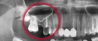
Before treatment, it is recommended to do a computed tomography to visualize the tooth, its root system and see the number of root canals; on CT they are visible as dark stripes. Photo 3 root canals
In practice, it is not always possible to accurately determine the number of root canals from an image. I have come across descriptions of radiologists (professionals in reading images), in whose conclusions the number of channels did not correspond to reality. It is possible to clearly determine only with the help of a microscope, after careful mechanical and medicinal treatment of the tooth. This is due to the complex structure of the tooth root system. Photo 4 is what the entrances to the main root canals look like, but there is an additional one and we found it after special scouting in the next photo
Photo 5 Here is the mouth of an additional root canal in tooth number 16, which often causes inflammation if undetected.
The doctor’s next question is preparing a clean surgical field so that unnecessary bacteria do not enter the tooth cavity. This requires a rubber dam, which isolates the tooth from the oral cavity, from saliva and gum fluid. It is also necessary to remove all caries (this is a bacterial zone) and all old fillings and restorations before treatment. It is necessary to create a sealed, clean core section. Photo of tooth 6 before filling replacement
Photo 7 tooth is fully prepared for endodontic treatment
Next, the specialist dives into another floor of the tooth - this is the area of the coronal part. If this is primary inflammation of the pulp, then the entire main infection is concentrated here. Therefore, before moving it through the root canals, it is necessary to carry out treatment using ultrasound and an antiseptic. If a tooth is re-treated, then often this area is filled with softened dentin and necrotic tissues; everything is cleaned using an antiseptic, ultrasound and special burs. Photo of tooth 8 before cleaning
Photo 9 clean tooth after treatment
Next begins the most difficult, but at the same time interesting, work of the doctor. It must be said that endodontists who like to “rake” / “digging” in root canals cause misunderstanding among colleagues. You need to have a certain character. These are often pedantic people who love to put everything in its place and clean out their spaces, get to the bottom of things and solve all sorts of difficult problems. Only with such data can you be an excellent specialist in root canal treatment. Actually, you already understand what the doctor does at this stage :) Yes, he finds, clears, passes through, washes out, expands. Makes the root canal as clean as possible. This process takes a lot of time, since you need to work with small structures carefully. Therefore, you often need to spend about two hours in the dentist’s chair, and there may be 2 or more visits.
Also at this stage, all modern instruments : files, machine tools, antiseptics, special disposable needles for irrigating solutions and, of course , ultrasound . In our clinic we use the most modern apparatus for irrigating root canals. The unique features of ultrasound, such as cavitation, microstreaming and heat generation, make it possible for the irrigant to penetrate the deep structures of root microdentin and microtubules. All this helps to remove bacteria from the tooth root as much as possible. In addition to ultrasound, we need manual nickel-titanium files for primary streaming. Going through root canals puts stress on the metal, so these instruments are often disposable and thrown away.
Next, the working length is determined using an apex locator . A mouthpiece is hung on the patient's cheek, and we hook it to the file; this method allows us to determine the length of the root canal. An x-ray is required with the files, thanks to which we confirm the working length. And only after all the checks does the root canal expand.
To do this, we use machine tools; each canal is processed with about five machine files; between sizes, the canals are thoroughly cleaned using an irrigant and ultrasound. So let’s imagine the upper sixth tooth, a molar, 4 root canals. In total, 20 dives with mechanical files, about 8 manual dives and many dives with an ultrasonic tip and endo-needle, until the doctor sees through a microscope that the irrigant in the canal is clean, without chips and reactions. All these manipulations require concentration, time, and most importantly patience and responsibility. After all this work, medicine is often given to create an alkaline environment that is harmful to many bacteria. The tooth is closed with a dense filling, and the patient leaves for 2 weeks. There is an important point here: a good temporary filling will never pop out of a tooth. And we have already understood that it is important to create clean conditions inside the root. Therefore, if your temporary filling does not hold or chips, think about whether your treatment is truly airtight. After all, the success of treatment depends on this.
After a break of 2 weeks, the patient and doctor meet again to continue the work. On the second visit, the doctor again rinses and cleans the root canal, dries everything out and seals the passage tightly. We are guided by the principle - nature abhors a vacuum! All root canals that previously contained neurovascular tissue (pulp) must be tightly sealed with gutta-percha pins and a special sealer. There are many filling methods. In our clinic we often use one of them - the vertical condensation method. A master pin is inserted into the root canal, the excess is cut off with a hot plugger, and the apical zone is compacted with plasticized gutta-percha using a continuous wave.
photo 10 view of a filled tooth
The final stage is tooth restoration. The endodontist makes a build-up, the foundation for the future crown. It is of course completely sealed. And transfers the patient to an orthopedist to restore the coronal part using indirect restoration. The choice of design will depend on the degree of tooth decay.
Medicine is moving forward and now a device is being developed that would be able to carefully pass, expand and rinse the root canals, without using so many tools and time. The introduction of laser and vacuum systems is taking place at a slow pace. Perhaps, if we obtain positive long-term results, we will soon use such methods in practice and will be able to save more teeth from removal, reduce the treatment time, making it high-quality and at the same time fast.