Causes
Etymologically, odontoma is associated with anomalies in the development of hard dental tissues. It affects the bone material of premolars and molars and can be localized in both the upper and lower jaws.
Despite the fact that the causes of odontoma are not fully understood, the most significant factors are:
- genetic predisposition;
- specific infections;
- jaw tissue injuries.
The period of maximum activation of the tumor occurs in childhood (from 6 to 11 years), when the main (permanent) teeth begin to erupt, or later, when the third molars erupt. The tumor is detected in most cases somewhat later, about 20 years, and most often it is discovered by chance on an x-ray or due to the development of an inflammatory process in the tumor tissue. Timely diagnosis of odontoma helps prevent the development of complications.
Treatment of complex mixed odontoma
Odontomas are the most common odontogenic tumors of the jaws, which are characterized by slow and non-aggressive growth. The term "odontoma" was first introduced by Broca in 1866, who defined it as a tumor that grows from mature dental tissue.
Odontomas are considered malformations resulting from the growth of fully differentiated epithelial and mesenchymal cells, from which amelo- and odontoblastomas can also develop. They consist of enamel and dentin, but may also contain inclusions of cementum and pulp tissue.
Odontomas are more likely to be hamartomas than true neoplastic formations, since both epithelial and mesenchymal cells can be found in small numbers in the structure of healthy tissues.
The etiology of odontomas is still not fully understood. Local trauma or infection can lead to the formation of odontomas. Radiologically, odontomas are diagnosed as dense radiopaque lesions with clear outer boundaries, surrounded by a thin radiolucent zone.
The World Health Organization (WHO) in 2005 classified odontomas according to histopathological features: complex odontomas (dental tissues are formed, but amorphous and randomly arranged) and compound odontomas (dental tissues correspond to those normally, are located in a certain order, but their size and the ratio is variable, which can lead to the formation of several small teeth, or elements called odontoids or denticles).
Most odontomas that are located in the anterior part of the upper jaw are composite, while most odontomas located in the posterior part, especially the lower jaw, have a complex structure.
Odontoma treatment involves their removal. The prognosis after treatment is favorable, relapses are rare.
Clinical case
A 13-year-old female patient contacted the Department of Pediatric Dentistry with the main complaint of pain in the right posterior region of the lower jaw. The medical history and life history did not allow us to make a final diagnosis. During an intraoral examination, foci of carious lesions were discovered on the chewing surface of the primary second molar of the lower jaw. The results of the radiographic examination helped to diagnose the presence of caries of the occlusal surface progressing towards the pulp, affecting the enamel and dentin (photos 1 and 2).
Photo 1
Photo 2
Multiple small teeth in the form of radiopaque structures were found at the apex of the distal root of a mandibular primary second molar. These lesions were surrounded by a thin radiolucent zone measuring approximately 1.5 × 1.0 cm.
The orthopantomogram revealed the same pattern as the intraoral periapical radiography (Figure 2). Based on clinical and radiological studies, a preliminary diagnosis of compound odontoma was made. During the treatment, surgical excision of the lesion was performed by curettage, after which X-ray control was performed.
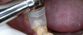
After adequate local anesthesia, a mucoperiosteal flap of tissue was folded in the area of the distal surface of the first right primary molar of the mandible to the medial surface of the first right permanent molar from the side of the lip. The layer of bone overlying the odontoma was removed using a round surgical bur under continuous saline irrigation (Figure 3).
Photo 3
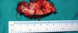
Calcified teeth were removed along with the fibrous capsule without disturbing the integrity of the unerupted permanent premolar (Figure 4).
Photo 4
The surgical site was cleared by curettage and irrigation with saline povidone-iodine solution.
Once complete hemostasis was achieved, the flap was placed in place and secured with 3.0 silk sutures. For control, a postoperative x-ray was taken (photo 5).
Photo 5
The sutures were removed one week after surgery. Histopathological examination confirmed the preliminary diagnosis of compound odontoma, demonstrating the presence of dentinal tubules in transverse and longitudinal sections.
Amorphous basophilic masses resembling dental cement were also identified on sections. The dense fibrocellular connective tissue in the sections resembled the periodontal ligament.
Some sections also showed numerous trabeculae with the presence of osteocytes in the lacunae (Figure 7). Such signs are also characteristic of those odontomas that consist of varying amounts of enamel and pulp tissue, enamel organ and cement. Odontogenic epithelium and mesenchymal pulp tissue can also sometimes be present in odontomas. The connective tissue capsule is similar to the structure of the dental follicle. The cell remains in their structure often had spherical foci of dystrophic calcification, enamel and several layers of dysplastic dentin.
Photo 7
Six months after surgery, it was recommended to perform an orthopantomography to monitor the process of eruption of the second permanent premolar (photo 6). In the picture, tooth eruption is physiological in nature without pathological changes.
Photo 6
Discussion
Definition
Odontomas are non-aggressive hamartomatous developmental malformations or malformations of odontogenic origin that occur as small, solitary or numerous radiopaque lesions that are detected as a result of routine radiographic examinations.
They are the most common odontogenic tumors, accounting for 22% of the total number of all odontogenic neoplasms affecting the jaws.
Etiology
The etiology of odontomas still remains unknown. The occurrence of tumors is associated with various pathological factors, such as local trauma, inflammatory and/or infectious processes, mature ameloblastomas, and remnants of dental plate cells. Odontomas can also be caused by hereditary anomalies (Gardner's syndrome and Hermann's syndrome). Odontoblastic hyperactivity and changes in the genetic component responsible for the process of tooth development play an important role.
Classification
In 1914, Gabell et al grouped odontomas according to their development and origin into several groups: epithelial, combined (epithelial and mesodermal), and connective tissue odontomas. According to the 2005 WHO classification of odontogenic tumors, there are two types of odontomas - compound and complex. Clinically, they are classified as intraosseous (central), peripheral (located in soft tissue or extraosseous tissue), and erupting odontomas. Central (intraosseous) odontomas are the most common and account for 51% of all odontomas. More often they occur in the anterior part of the upper jaw (compound odontomas), as well as in the molar area of the lower jaw (complex odontomas). Peripheral odontomas are rare and occur in soft tissues at the border with dental tissues. The most common among them are compound odontomas.
Odontomas of teeth that erupt are located above the coronal part of the tooth, or superficially in the bone above the impacted tooth, which excludes the possibility of its eruption into the oral cavity.
WHO has defined a compound odontoma as a malformation in which the dental tissues are presented in some order in the form of small tooth-like structures. Most of them are not morphologically similar to ordinary teeth, but each of them contains tissues of enamel, dentin, cementum and pulp, which are arranged in a similar way to a regular tooth. Complex odontomas are less common compared to compound ones in a 1:2 ratio.
Prevalence
The prevalence of compound odontomas ranges from 9% to 37%. The majority of odontomas in the anterior jaw are compound combined (61%), while the majority of odontomas in the posterior are complex combined (34%). It is noteworthy that both types of odontomas are more common on the right side of the jaw (compound - 62%, complex - 68%). Compound combined odontomas most often occur in the area of the maxillary incisors, in contrast to complex odontomas, which usually occur in the area of the molars and premolars of the mandible. In this case, the compound odontoma is located in the posterior region of the mandible, which is a rather rare topography for this form.
X-ray examination is the most effective clinical method for differentiating the two types of odontomas. Compound odontomas, which have relatively well-organized malformed teeth or dental structures on radiographs, are similar to radiolucent cysts, whereas compound odontomas have an irregular oval radiopaque shape that is surrounded by a distinct, thin radiolucent border.
In the case described, a diagnosis of compound odontoma was initially made based on radiographic studies. This diagnosis was later confirmed by the results of histological examination of the lesion (Figure 7).
Odontomas, both compound and complex, must be examined under a microscope to make a correct definitive diagnosis.
Treatment
Kaban states that odontomas can be easily enucleated, and damage to adjacent teeth is rare during this operation because the affected area and healthy teeth are separated by a bony septum.
Odontoma has limited growth potential, but it must be removed because it contains various constituent dental tissues that can initiate cystic changes.
Therefore, careful visual, manual, and radiographic examinations are mandatory for all pediatric patients with clinical signs of delayed, absent, or temporarily displaced teeth, despite the presence or absence of a history of trauma.
Early diagnosis of odontomas helps:
- provide less complex and less expensive treatment;
- provide a better prognosis;
- avoid recurrence of lesions;
- Avoid displacement or devitalization of adjacent healthy teeth.
Authors: Morawala Abdul, Kapila Pragati, Chunawala Yusuf
Classification of odontomas
There are several types of odontomas:
- soft (at the initial stage it consists of epithelium and connective tissue);
- hard (calcareous deposits from hard dental tissues, most often localized in the corner of the lower jaw, or in the central region of the upper jaw);
- cystic (parts of the tooth germs are located in the cavity of the follicular cyst);
- simple (includes one follicular primordium);
- complex (affects combinations of various tissues and several parts of the teeth);
- composite (filled with tooth-like neoplasms).
Symptoms and course of the disease
Depending on the type of tumor, its symptoms also vary. In the early stage, soft odontoma has similar symptoms to ameloblastoma, but it is characteristic of younger patients.
The tumor grows slowly and then stops developing due to the end of the formation of the dentition. A constant and continuous growth process is observed only in hard complex forms of odontoma. As a result of tumor growth, the bone increases in size, then the cortical plate can be destroyed, and the tumor can penetrate into the periosteum and further.
Despite the fact that the hard type of odontoma is characterized by expansive growth, the development of the pathology remains painless. The first sign is the appearance of a swelling, the surface of which is uneven and lumpy, and feels hard to the touch. Often, during the formation of odontoma, it is accompanied by infection and ulceration of the soft tissues of the oral cavity.
In later stages, odontoma is accompanied by severe inflammatory complications, changes in jaw size, shift and damage to the dentition.
Diagnosis and treatment
To diagnose a tumor, radiography is mandatory; histological examination is used for clarification (during which it becomes clear what the nature of the tumor is).
Treatment of odontoma involves surgical removal of the capsule and tumor tissue, followed by curettage of the tumor bed and sanitation of the intervention area. In severe cases, partial or complete removal of the jaw with subsequent prosthetic replacement is possible.
The earlier the pathology is detected, the less tissue loss is accompanied by surgical intervention, so timely diagnosis in most cases has a favorable outcome. In addition, despite its benign nature, odontoma can also degenerate into a malignant tumor (this phenomenon is observed in 4% of cases).
Regular visits to the doctor and a thorough check of the health of the oral cavity are the key to timely diagnosis of any dental diseases.
Advantages of treatment in Israel
- First-class service: Israel is known for its friendly customer service. It is in this country that you can find not only a talented and experienced dentist, but also a reliable professional with a high level of integrity.
- Materials: Dentists in Israel have access to all the advanced dental developments that are not available in many clinics in the world, as well as various high-tech advances such as intraoral scanners, digital radiography, guided surgery, etc.
- Cost: Dental surgery prices are high around the world, but quality dental care in Israel actually costs significantly less than in the US, UK or Canada.
- Reviews: patients who have already undergone treatment in the dental department of the Assuta clinic leave positive reviews about the quality and efficiency of service, emphasizing the sensitive and caring attitude of the staff.
If you or your child are showing signs of illness, there is no reason to delay going to the clinic. Do this today and our medical consultant will do everything possible to quickly find you the right expert and arrange a direct consultation.
- 5
- 4
- 3
- 2
- 1
(2 votes, average: 5 out of 5)