Publication date: 03/26/2020
The Axioma Dental clinic has its own visiograph and orthopantomograph - this is digital X-ray equipment that allows you to achieve high efficiency in diagnosis and treatment. A visiograph is an ultra-sensitive technique for creating clear, accurate images of teeth. You no longer have to wait for the X-ray film to be developed; the image is instantly visualized on the screen. This saves the patient’s time without harming health.
An orthopantomograph is a safe alternative to X-ray machines. The technique takes a panoramic photograph of the patient’s entire dental system. The equipment is indispensable in any dental field for developing a treatment plan. The dentist can quickly make a diagnosis and obtain detailed information about the condition of the dental system.
The principle of operation of the visiograph
A dental visiograph is a system of equipment that includes an ultra-sensitive touch sensor, an X-ray, and a computer for digitizing data. The principle of operation of the technique is similar to x-rays, but it is a safe technique. If the film unit directs rays onto the film, then the visiograph imaging device sends data to an ultra-sensitive matrix, a CCD sensor. The microcircuit chip consists of photodiodes that can capture and visualize the smallest details.
The main advantage is that to create an image you need an ultra-low dose of X-ray radiation, only 2-3 μSv. This is safe for humans, and the doctor has the opportunity to reduce the time for diagnosis.
How to take pictures using a visiograph:
- The lead apron is protection for the patient, who wears it before the procedure.
- Placement of the sensor in the oral cavity - it can be wireless or wired. An individual protective cover is put on the sensor and placed in the oral cavity. The sensor is applied to the area where pathology is detected, the condition of the bone tissue is clarified for successful implantation, treatment of caries, pulpitis.
- The sensor of a dental touch imaging device is connected to a digital device for data visualization, digitization and analysis of results.
- Snapshot.
Within a few seconds, the doctor sees the full clinical picture on the PC screen, makes a diagnosis and quickly prescribes treatment.
Why do you need a dental x-ray?
- 1. It is sometimes very difficult to treat a tooth “blindly”. Even a very experienced dentist cannot always fill curved and thin root canals perfectly without taking pictures. And patients’ tooth anatomy is often very different from the picture in the textbook. And if you treat a tooth poorly, then after some time it may get worse, and you will have to re-treat it. But just one visit and one photo won’t do the trick.
- 2. It is difficult to diagnose a cyst, the degree of inflammation of the periodontium (tissues around the root), a fracture of the tooth root and other troubles without x-rays. More often than not it is even impossible. If you start treating a tooth without taking a picture, it may become so worse during the treatment that you will have to take antibiotics or go to a maxillofacial hospital and be treated there, which is not useful for a pregnant woman at all.
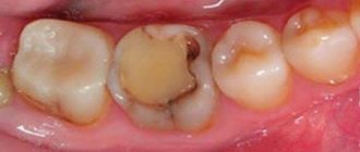
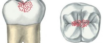
- 3. Carious lesions of teeth, invisible during a simple examination. Many hidden cavities are visible only in photographs.
- 4. Problematic wisdom teeth. When they have not yet erupted, how does the doctor know whether it is growing correctly, whether it has any chance at all, or whether it is only causing harm and needs to be removed?
- 5. Critical cases when there is a question of removing or preserving a damaged tooth. After all, after taking a couple of pictures and treating the root, the tooth can be restored with a core inlay and a crown. And without a picture, immediately sending it to a surgeon for removal, when there is at least some chance of saving the tooth, is a pity.
The difference between a visiograph and an x-ray
A visiograph is a device that does not independently emit rays, unlike an X-ray machine. Its task is to receive the rays onto an ultra-sensitive sensor and convert them into an accurate image for data analysis. At the same time, modern equipment does not need to produce a wide beam of radiation. A radiovisiograph is a very thin targeted beam of light that is captured by a sensor.
The system’s sensor is able to recognize and capture the smallest particles of radiation in just a fraction of seconds, which reduces the procedure and risks for humans. Modern digital equipment differs from x-rays in the speed of obtaining an image; shutter speed:
- visiograph - 0.06 seconds.
- film apparatus - 0.6.
Conclusion - innovative technology allows you to achieve maximum safety for the patient and reduce procedure time.
X-rays of teeth
What is a visiograph and how does it differ from an x-ray?
This is one of the most common questions, like what is the difference between a car and a traffic light. It seems that both concepts have some kind of connection, but it is quite difficult to compare them. Also here. A radiovisiograph is a system that receives x-ray radiation, transforms it into digital form and displays the image on a computer screen.
Roentgen (who was Wilhelm Conrad) was a German physicist who gained worldwide fame for his discovery of short-wavelength rays with enormous penetrating power. The physicist himself called these rays X-rays (in English today they are called exactly that - X-ray), but now we often call them X-rays, and in everyday life simply “X-rays”. The unit of radiation power was also called the x-ray. Now it is clear that a visiograph and an x-ray are completely different things. If you compare the visiograph with anything, it’s with x-ray film, which it is universally replacing from all areas of medicine.
X-ray of teeth
Is it true that a visiograph is safer than a regular film photograph?
When asked about such a comparison, they mean the radiation exposure that the patient receives when using different techniques. In this sense, indeed, a visionograph is better, since its sensor is much more sensitive compared to film. Therefore, to obtain a high-quality image using a visiograph, you need significantly less shutter speed. To take a picture on film, the shutter speed is 0.5-1.2 seconds. To obtain the same image using a visiograph sensor – 0.05-0.3 seconds, that is, 10 times less. As a result, the radiation exposure received by the patient when using a visiograph is reduced to an insignificant minimum.
How many pictures can you take at one time? And in general, isn’t it harmful that when treating a large number of teeth, you have to take a lot of X-rays?
This is the most pressing of questions. This is an echo of the Chernobyl tragedy and life safety lessons that emerge in memory. But in our society there is still a very strong phobia towards everything that is even remotely connected in our heads with radiation. Any extra photo often raises questions about radiation sickness, or “will I glow in the dark?” Therefore, we will try to explain in more detail here. First from a scientific point of view.
To measure the amount of radiant energy applied to living tissue, various units are used - joule per kilogram, gray, rem, sievert, etc. In medicine, X-ray procedures typically measure the dose received by the entire body during one procedure—the effective equivalent dose, measured in sieverts. According to SanPiN 2.6.1.8-38-2003, when carrying out preventive medical x-ray procedures and scientific research, this dose should not exceed 1000 μSv (microsievert) per year. Moreover, here we are talking specifically about preventive studies, and not about therapeutic ones, where this bar is much higher.
What is 1000 µSv? Is it a lot or a little?
Remembering the famous cartoon, the answer is simple - depending on what you measure it in. 1000 μSv is approximately:
- 500 targeted images (2-3 μSv) obtained using a radiovisiograph
- 100 of the same images, but using good X-ray film (10-15 µSv)
So, apparently, even if we take 1 picture on a visiograph every day for the whole year, and also a couple of 3D computed tomograms per year, and the same number of orthopantomograms, then even in this case we will not get out beyond the limits of safe permitted doses.
there is no need to be afraid of receiving a significant dose of radiation during dental procedures . Even if you want to go beyond the permissible values, it is unlikely to be possible. To be clear, here are the doses needed to cause any serious health effects:
- 750000 μSv - short-term minor change in blood composition
- 4,500,000 μSv - severe radiation sickness (50% of those exposed die)
All these figures are incomparable in their significance with the doses we receive in everyday life. So even if, for some reason, several pictures are taken at once in a row, and the day before you were already “exposed” by doing an orthopantomogram, you don’t need to panic and run to the store to buy a Geiger counter or search on the Internet for “the first symptoms of radiation sickness.” To calm yourself down, it’s better to “detoxify” with a glass of red wine. There will be no point in this, but the mood will immediately improve.
Let us clarify that, for example, with one-stage basal implantation, control targeted images are taken before and after installation of implants (when installing 1-3 implants). And also after final prosthetics (7-8 days).
Is it possible to do x-rays for pregnant women?
We will not remind you that before pregnancy it is better to take care of your health in advance, including “preparing” your own teeth at the dentist. In order not to run away later with acute pain and doubts whether this or that manipulation will harm the development of the fetus... Therefore, you should leave the lyrics and pay attention to the facts and common sense. Without phobias, prejudices, speculations and myths. So, is it possible to do x-rays for pregnant women?
Here's what they write to us about this in the documents (SanPiN 2.6.1.8-38-2003):
7.16. Pregnant women are prescribed for X-ray examination only according to clinical indications. Studies should, if possible, be carried out in the second half of pregnancy, except in cases where it is necessary to decide on termination of pregnancy or the need for emergency or emergency care. If pregnancy is suspected, the question of the admissibility and necessity of an x-ray examination is decided based on the assumption that there is a pregnancy...
7.18. X-ray examinations of pregnant women are carried out using all possible means and methods of protection so that the dose received by the fetus does not exceed 1 millisievert for two months of detected pregnancy. If the fetus receives a dose exceeding 100 mSv, the doctor is obliged to warn the patient about the possible consequences and recommend terminating the pregnancy.”
In general, the conclusion from these two main points is simple and clear. It’s definitely not worth taking pictures in the first half of pregnancy, but in the second half - 1 mSv for a visiograph - this is practically unlimited.
I would also like to add that one often encounters bellicose stubbornness: an X-ray at the dentist during pregnancy is an absolute evil. It’s better, they say, to screw up a tooth, to cure crooked canals... there are a lot of teeth, pregnancy is more important. Moreover, such sermons are given not only by lay patients who have little understanding of the essence of things, but also often by dentists themselves who have forgotten their school physics course.
To resolve this doubt, we must understand that sources of ionizing radiation are not only found in medical offices. And it doesn’t have to be near Chernobyl (and now Fukushima) to receive some doses from the environment. After all, every second we are influenced by both natural sources (sun, water, earth) and man-made ones. And the doses received from them are much greater than those received from an x-ray of a tooth.
For clarity, we can give one simple example. As you know from a school physics course, the sun emits electromagnetic energy in a wide range, not only in infrared (heat), visible (light), ultraviolet (tan), but also in x-rays and gamma radiation. Moreover, the higher the earth's surface, the more rarefied the atmosphere and, therefore, the weaker the protection from sufficiently strong radiation from the sun.
After all, while “fighting” radiation at the dentist, the same people often calmly fly south to bask in the sun and eat fresh fruit. Moreover, during a 2-3 hour flight in a “healthy” climate, a person receives 20-30 μSv, i.e. the equivalent of approximately 10-15 images on a visiograph. In addition, 1.5-2 hours in front of a cathode ray monitor or TV gives the same dose as 1 picture... How many pregnant women sitting at home, watching TV shows and surfing the Internet, think about how many pictures they “took”? while watching another program, and then discussing it with friends on the forum and social networks? Almost no one, because people do not associate all this with ionizing radiation, unlike a picture in a doctor’s office.
And yet, dear expectant mothers, prepare for pregnancy in advance. For many people, visiting the dentist still remains stressful. And it’s not so much that anesthesia or x-rays can be harmful during this period, but what is important is your peace of mind and the absence of unnecessary worries (of which many already have more than enough during this period). You can study more information on this topic in our article “Can teeth be treated during pregnancy.”
What is the best protection to use if you need to take a photo while pregnant?
The number of aprons does not matter. In contact radiography, the apron essentially protects not from direct radiation, but from secondary, that is, reflected. For X-rays, the human body is an optical medium, like a glass cube for a flashlight beam. Point the beam of a flashlight at one of the faces of a large glass cube and, regardless of the thickness and direction of the beam, the entire cube will be illuminated. It’s the same with a person - you can swaddle him completely in lead and shine only on his head - at least a little, but it will reach every heel. So even under two aprons with a good lead equivalent, a pregnant woman will, at most, simply have a harder time breathing.
Is it possible to do x-rays for breastfeeding mothers? And if possible, then what about feeding the child after the procedure?
Can. X-rays are not the same as radioactive waste. By itself, it does not accumulate in the biological environment. If you irradiate a loaf of bread with a lethal dose, it will not mutate, get radiation sickness, or begin to “foul.”
X-rays differ from light rays only in wavelength and have a direct damaging effect only under certain conditions. If you shine a flashlight into a bucket of water and turn off the flashlight, the light won't stay in the bucket, right? The same is true in a protein-fat solution, which is what many biological fluids are (including breast milk) - the radiation passes right through. So, with such a load that is necessary to work with a visiograph, it is unlikely that anything will happen to the milk itself.
As a last resort, to reassure yourself, you can skip one regular feeding. Another thing is that the breast tissue itself during lactation is, of course, more susceptible to the harmful effects of radiation. But, again, we are talking about doses much more powerful than is necessary for digital radiography (of course, subject to all protective measures and without “shooting” 20 times anywhere).
Advantages and disadvantages of a visiograph
Pros of sensory equipment:
- Minimum radiation dose.
- High image accuracy.
- Maximum safety for the patient.
- The procedure can be performed for pregnant women and children - consultation with a doctor is required.
- Compact device, able to work in a small office.
- Instantly visualize your photo without the need to develop film.
- The doctor gets the opportunity to see each tooth - multiple magnification of the image, storage of data on a PC, transmission via e-mail, local network.
- The manipulation takes several minutes.
- Editing a picture - the dentist uses different filters to improve the visual perception of the picture.
- The procedure is painless and does not require special preparation.
The disadvantage is the high price of equipment and procedures. But in modern dentistry, the priority is accurate diagnosis, effective treatment, and patient safety. Therefore, reputable clinics invest money in purchasing equipment.
Indications and contraindications for the procedure
List of main indications for use of the equipment:
- Prevention of pulpitis, caries, periodontitis;
- Viewing the condition of a filled tooth, decayed gums, damaged bone tissue, or installed implant.
Radiovisiographic examination allows complete control of the treatment process. It will help avoid many complications. This method is safer than x-ray examination. It does not pose a health hazard.
Radiovisiography can be performed during pregnancy and lactation.
The list of relative contraindications includes:
- Mental illnesses;
- Neurological disorders.
Features of the orthopantomograph
Dental orthopantomograph is equipment for creating high-resolution panoramic images. Devices of various types have an overview imaging system, which makes it possible to visualize the entire jaw, the patient’s teeth, and jaw joints. The principle of operation is that the X-ray emitter and sensor in digital modifications rotate around the head, forming a clear three-dimensional image of the jaws.
Digital orthopantomographs make shooting as safe as possible for humans - the radiation level is 2-3 times less than with film units. Innovative technology does not require laboratories to develop images; the picture will be ready quickly and visualized on the computer screen without waiting for results.
If the orthopantomograph has a cephalostat, the doctor can take a frontal profile photograph of the skull, which is needed to build orthodontic structures.
Innovative technology works with different shooting programs:
- Standard overview.
- Separately upper and lower jaw.
- Photo of the maxillary sinuses.
- Children's photography.
- Image enlargement mode.
- Other programs for different clinical cases.
Safety, speed, high-resolution image quality, and information content are the characteristics of equipment used by clinics all over the world.
How dangerous is this?
The most common question regarding a visiograph is how dangerous it is. In fact, due to its design, it is no more dangerous than a regular digital camera or office scanner. The radiation dose received by the patient during radiography of the teeth of the lower jaw will be 2 microsieverts and 5 μSv for the upper jaw. This amount of radiation is extremely safe for both the patient and the doctor. But only if the doctor follows safety standards and correct positioning. At the same time, individual forced ventilation must operate in the office, which will disperse ionized air, since it is hazardous to health.
Here is an example for comparison: when working with Soviet installations, the average radiation dose ranges from 20 to 30 μSv, and when using the 5D1 device, the values can reach up to 80 μSv.
Modern dentistry with visiograph and orthopantomograph
The technical equipment of the dental clinic is a guarantee of accurate diagnosis, effective treatment, and error-free diagnosis. Without the latest generation equipment, a medical institution cannot guarantee the patient a high level of service.
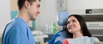
Axioma Dental Clinic is a center that invests money in purchasing innovative equipment. Doctors have a Sirona orthopantomograph and a visiograph at their disposal. This provides benefits to our patients:
- Examinations in one clinic - no need to waste time, sign up in advance for x-rays at clinics.
- Safety - there is no risk of receiving a high dose of X-rays.
- Treatment of children, adults, pregnant women - innovative equipment does not cause harm to health.
- Instant results - the photo will be ready in a few seconds.
The Axioma Dental clinic is the right approach to treating teeth, gums, correcting row defects, and implantation using safe equipment.
Sources:
- Yarnova E.A. Possibilities of radiation diagnostic methods in visualization of the temporomandibular joints in cases of dentofacial anomalies
- Goncharov I.Yu. Planning the surgical stage of dental implantation in the treatment of patients with various types of missing teeth, defects and deformations of the jaws, 2009
- Robustova Tatyana Grigorievna. Dental implantation: surgical aspects, 2003
Expert author:
Makanina Lina Viktorovna
Dentist-therapist
The information presented in this article is provided for reference purposes and does not replace the advice of a qualified specialist.
At the first signs of illness, you should consult a doctor.
Tell us about us:
How profitable is this?
And now the main question is why buy it. This is where elementary arithmetic comes to the rescue. The average cost of one package of film (1 package – 50 pictures) is 1250 rubles. The average daily throughput of the X-ray room is 15 patients. Taking into account the defect rate of 1/5, this is 20 shots per day. It turns out that one package of film is enough for 2.5 days. 144 packages are needed per year with a total cost of 180,000 rubles. And this does not take into account chemical reagents. With the average cost of a photo for a client being 120 rubles, the revenue will be 864,000 rubles. Thus, the net profit will be 684,000 rubles per year.
Let's now look at the visiograph. Its average price on the Russian market (with a resource of 400,000 images) is 130,000 rubles. Of course, there are obvious benefits. Add to this the simplest maintenance (you will have to clean it with soapy water or isopropyl alcohol), and a rather long service life.
You can purchase a visiograph by following the link.