Resistant teeth with problematic immature roots pose a serious problem. And external damage causes the condition of such complex teeth to worsen. If you postpone a visit to the dentist or receive poor-quality treatment, necrosis of the hard tissues of the tooth may develop, causing severe and rapidly progressing pulpitis.
The best treatment method in this case is apexification using calcium hydroxide. However, these materials have a number of disadvantages.
Through extensive laboratory research, a unique MTA material was developed. But its high price forces many dentists to refuse to use it in their practices, despite its excellent properties.
the innovative material Trioxident was created, which is an analogy of MTA and has practically replaced it in practical dentistry. It is much more affordable, and in quality is in no way inferior to its predecessor.
The new material is highly effective and improves the quality of retrograde fillings. Compact and airtight packaging allows you to work comfortably with the drug.
Composition and release form
Trioxidant consists of ideally selected active ingredients.
Increased alkalinity is obtained from the oxides of calcium, silicon and aluminum. These elements give the material strength, the ability to hermetically seal defects in the canal, prevent bacteria from entering the tooth cavity, and the solubility of the drug remains low, which allows for high biocompatibility. Copper and calcium hydroxide serves as a bacteriostatic additive. Bismuth oxide provides excellent radiopacity.
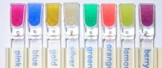
Release forms:
- powder;
- dropper bottle.
Additional equipment:
- mixing form;
- spatula for mixing;
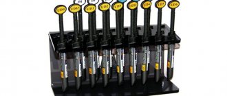
- set of tools for retrograde filling;
- tubes made of F-4D plastic, 4 and 8 mm;
- detailed instructions for use;
- box - case.
Application area
Trioxidant, in its own way, is a universal remedy:
- used for retrograde filling;
- used as a therapeutic pad for pulp isolation;
- used to close root canal perforations;
- The material fills the upper apex part of the dental canal.
Properties and capabilities
The material hardens in the canal for four hours, after a day it is completely hardened. This gives it advantages over all existing analogues.
The hardening process occurs in three stages:
- interacting with water, calcium oxide is converted into calcium hydroxide, which provides increased alkalinity;
- due to the saturation with calcium hydroxide, all elements are converted into a homogeneous elastic mass;
- calcium hydroxide saturates the mixture of the resulting calcium silicate, thereby increasing the plasticity of the mass.
Modern treatment methods involve retrograde root covering. To improve the quality of this procedure, Trioxydent material is ideal.
They fill and close the upper base of the dental canal, close the perforation of the root canal, and cover the tooth pulp. The drug has excellent bactericidal properties and hermetically covers drilled cavities.
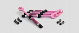
Trioxident - closing root canal perforations
Trioxydent is a dental water-soluble material for retrograde filling and correction of defects in the root canals of teeth.
PURPOSE
It is used for retrograde filling, for filling the upper apical part of the canal with incomplete root formation, for closing root canal perforations, and also as a therapeutic-isolating pulp coating.
COMPOSITION AND MAIN PROPERTIES
The main components of the water-soluble dental material Trioxident are oxides of calcium, silicon, and aluminum.
When mixing the powder with distilled water in a weight ratio of 3:1, an easy-to-use paste is formed that does not lose plasticity for 10-15 minutes at room temperature 18-23C and humidity 50-10%. To obtain a plastic paste, a plasticizer is added to the powder to prevent rapid drying of the material on the mixing plate.
The material hardens in the canal within 24 hours.
Curing of the material occurs gradually in three stages: first, calcium oxide reacts with water, turning into calcium hydroxide, which ensures high alkalinity of the material (pH 12.8). From a solution saturated with calcium hydroxide, the latter is released in an amorphous state and envelops the particles of the radiopaque filler, turning the mixture of components into a cohesive mass. The calcium hydroxide particles then compact the mass of calcium silicate formed, increasing the mechanical strength of the resulting cement.
The basis of the Trioxidant filling material is: - calcium hydroxide, which stimulates the processes of apex and osteogenesis in the treatment of teeth with unformed roots, stops bone tissue resorption;
when covering the pulp, it stimulates the formation of a dentinal bridge; has bactericidal properties. - copper-calcium hydroxide is an active bacteriostatic additive of a chemical nature common to the main components.
The material has high biocompatibility, low solubility and high mechanical strength, and also ensures tight closure of the channels and impermeability to bacteria.
The use of instruments for retrograde filling allows you to introduce paste into the canal, eliminating the entry of excess material into the periapical area, and also work with a small amount of material, directing all the material directly to the defect area.
RELEASE FORM
Powder 0.5 g x 10 pcs
Application diagram
The material is kneaded on a glass plate using a clean spatula. The required dose of powder and purified water are combined in proportions of 3:1. The mixture is brought to a plastic consistency. The resulting plastic paste is used to fill the defect area. As the material dries, additional water can be added (once).
When restoring perforation, it is recommended to carefully prepare the dental canals. After this, the channels are treated with sodium hypoporite and washed well. Having established the affected area, obturation of all canals is performed. The resulting paste is applied directly to the affected area and strengthened using an ultrasound attachment.
During root apexification, initial preparation of the affected canal is carried out. Having removed all contaminants, the canal is washed and dried with dry pins. The next stage is disinfection.
Then the potassium hydroxide-based paste is carefully applied. It should remain in the root canal for a week. After this period, the paste is removed and the canal is washed with sodium hypochloride.
When treating pulp and periodontal diseases, as well as as a result of repeated endodontic treatment, there is a risk of perforation of the roots and bottom of the tooth cavity. The reasons for errors in endodontic treatment can be different and are associated with the anatomical features of the tooth structure, iatrogenic factors, or caused by dentin resorption during the spread of the inflammatory process. The greatest difficulties in treatment are perforations in the area of furcations of the roots or the bottom of the tooth cavity, which can occur if the topographical features of the tooth structure are not taken into account (Maksimovsky Yu.M., Mitronin A.V., 2012; Bargholtz C., 2005). Closing a tooth perforation is a labor-intensive and complex process and largely depends on the doctor’s ability to use modern tools and materials. Modern endodontics places high demands on restorative materials for restoring defects in tooth root tissue, including biological compatibility, reliable marginal sealing, bactericidal and/or bacteriostatic properties, creation of favorable conditions for reparative processes, ease of clinical use and plasticity, radiopacity and insolubility in tissue fluids. , stability (Dubova M.A., Shpak T.A., Kornetova I.V., 2005; Mitronin A.V., Voronina K.Yu., 2008; Yoshioka T., Kobayashi C., Suda H., 2005 ). It should be noted that known conservative methods for closing furcation perforations do not always lead to the desired results - this also depends on the size of the defect and how long ago the perforation was made. And as a consequence of this - the impact of microorganisms and their toxins in this area and the development of an inflammatory-destructive process. Small perforations (less than 0.5 mm in diameter) can be sealed with flowable light-curing composites or glass ionomer cements. Perforations with a diameter greater than 0.5 mm are closed with special materials: mineral-based aggregate trioxide, (Pro Root USA) - a mixture of calcium silicates, calcium compounds of iron and aluminum, as well as hydrated calcium sulfate or gypsum, Super EBA - modified zinc oxide eugenol cement, IRM - strengthened zinc oxide eugenol cement (Rhodes D.S., 2005). More often these are foreign materials. However, there is a domestic filling material on the market, trioxident (Russia), a dental calcium aluminosilicate cement that contains copper-calcium hydroxide as an active bacteriostatic additive and is recommended by the manufacturer for filling root canals and perforations. However, there is insufficient data in the literature assessing their effectiveness in the endodontic treatment of teeth with defects in the hard tissues of the bottom of the dental cavity.
Purpose of the study
— evaluation of the use of dental cements Pro Root and trioxidant to eliminate hard tissue defects in the area of the bottom of the tooth cavity. Evaluation of the use of bioactive osteoplastic material kollapan in the destruction of bone tissue in the area of furcations.
Material and methods.
Laboratory studies were carried out on 18 samples of extracted teeth, divided into two groups (9 teeth each) for filling defects in the bottom of the tooth cavity with Trioxydent and Pro Root materials. In each tooth sample, an artificial perforation was made in the area of the cavity bottom. In accordance with the instructions for use of the materials trioxidant and Pro Root, the defect was filled.
To close the defect, the working area was prepared—cleaned of sawdust and half-life products, treated with sodium hypochlorite (Belodez 3%), washed with water, and dried. The materials were then placed into the defect area and compacted using a small amalgam plunger. The material can be condensed using a large ultrasonic nozzle without spraying water, at medium power. Using an x-ray, you need to make sure that the material is placed correctly. Then they isolated it with lining material and restored the tooth crown.
Before complete curing, the samples were kept in a humid environment to eliminate the effect of “burning” of cement, when, as a result of low atmospheric humidity, the material may lose the moisture necessary for complete crystallization, which will subsequently lead to its shrinkage or cracking. Samples with trioxydent material were cured for 72 hours, and with Pro Root for 4 hours, which corresponds to the curing times specified by the manufacturer.
Next, the tooth samples were sawed using a thin diamond cutter through the filling along the tooth axis parallel to the course of the roots. After this, the samples were treated for one minute with a saturated solution of EDTA, washed with running water, washed and dehydrated with a solution of ethyl alcohol at a concentration of 50%, and dried to remove any remaining alcohol in air.
Copper was sputtered onto the treated samples to impart surface electrical conductivity in a Bazer's SCD-040 apparatus in an argon atmosphere.
Using a Philips SEM-515 scanning electron microscope, images were obtained at various magnifications (accelerating voltage 18.9 kV).
The defectiveness of filling materials and the adhesion boundary with tooth tissues was assessed visually using the photographs obtained. The assessment took into account the presence or absence of voids (defects), the granularity of the material and the presence or absence of a tight fit of materials to the tooth tissues.
The clinical study also evaluated the restorative materials Trioxydent and Pro Root. After obtaining the voluntary informed consent of the patients, the work was carried out. Clinical studies were conducted in 58 patients in whom perforation of the hard tissues of the bottom of the tooth cavity was detected in 58 teeth. Two groups are defined: 1st group (24 teeth) and 2nd group (34 teeth). Defects—perforations—were closed using materials trioxident and Pro Root, respectively.
X-ray control was used to identify perforation, determine the size and location of the defect. If necessary, endodontic treatment was performed using standard techniques. If there was bleeding in the bifurcation area, a 3% hydrogen peroxide solution and a hemostatic collagen sponge were used. The defect was treated with 3% sodium hypochlorite and EDTA solution, washed with water, and dried. If necessary (if there is significant destruction of bone tissue in the underlying tissues due to long-standing perforation, there were 46 such cases in patients), biologically active bone material, for example, kollapan, can be introduced. There were 23 such patients where the kollapan material was used (23 teeth). The applied filling material was condensed (minor leakage of material into adjacent tissues is acceptable) under a temporary dressing, and X-ray control was performed. According to indications, usually after 2 days, at the second visit, the temporary filling was removed, the material was probed to assess the quality, the mouth of the canal was isolated with cushioning material, and the final restoration of the tooth crown was carried out. In order to identify dynamic changes in the area of bone tissue destruction, X-ray monitoring in both groups of patients was carried out every 3-6 months after endodontic treatment.
Results.
During a laboratory evaluation of thin sections of teeth filled with trioxydent material, signs of detachment of the material from the surface of the tooth tissue along almost the entire border were observed, and the material itself had larger granular inclusions, as a result of which it was more defective. The presence of defects, cracks and delaminations along the boundary suggests that both the edge permeability and the permeability through the material itself for liquid media will be quite high, and the strength of the material and its stability in the channel will be low. The long curing period of the trioxidant material can also be a negative factor when choosing this material for the purpose of eliminating perforations.
When examining samples of teeth with Pro Root material at the interface of the material with the tooth tissue, virtually no delamination was detected. The material provided a more uniform structure compared to the trioxidant material. According to the SEM results, the grains of the material were smaller in size, and defects were observed in isolation and quite rarely. The SEM results revealed a preference for the selection of Pro Root material for repairing perforations.
Assessing the results of the clinical use of trioxident Pro Root filling materials, it should be noted that the trioxident and Pro Root materials showed fairly high technological characteristics associated with the ease of their use in clinical practice. The pastes prepared on their basis retained their initial plasticity for 5-10 minutes, which made it possible to qualitatively obturate the perforation zone, reducing the risk of the formation of technological defects.
Analysis of the results of the clinical use of materials trioxident and Pro Root and the results of monitoring the dynamics of treatment of perforations showed that the studied materials in their functional characteristics are very similar to each other and demonstrated a positive treatment outcome as a result of clinical studies.
The most favorable results of treatment of perforations were observed primarily in cases with recent iatrogenic perforations (12 patients, 12 teeth) compared with old perforations ( p
<0.05).
The differences in the type of material observed in the treatment dynamics diagrams are not significant in this case ( p
>0.05), which does not allow us to consider their differences statistically significant. For the materials used, positive results in closing perforation defects were observed in 100% of cases up to 2 weeks, when patients did not report post-filling pain or painful percussion.
With repeated endodontic treatment (elimination of long-standing perforations), an increase in the total treatment period was observed in the number of cases with negative dynamics. At the same time, negative dynamics were revealed in cases with long-standing perforations, where destructive changes in the periodontium and bone tissue were observed. Positive immediate results of repeated endodontic treatment were achieved within 3 weeks in cases where the kollapan material was used, and only after 1 month of observation in patients in cases where the kollapan material was not used, when in 100% of cases the patients had no complaints about painful percussion.
Comparing the results of treatment of “recent” and “old” perforations, we can conclude that early diagnosis of perforations allows one to avoid complications in their treatment and increase the likelihood of a positive result.
Comparing clinical and radiological data when studying the dynamics of the state of bone tissue in the area of furcations of the roots of 23 teeth, accompanied by destructive changes in the periodontium and cases of collapan material, it was found that restoration of bone tissue density was noted in 96% of cases over a period of 6 to 18 months , partial restoration of bone tissue was observed in 4% of cases, which was expressed in a decrease in the focus of inflammatory destruction in the area of furcations. In the group of patients (23 teeth) without the use of kolvalpan material, positive dynamics were observed in 87% of cases, partial restoration of bone tissue - in 13% of cases.
Conclusion.
The clinical use of the collapan material along with the use of trioxident and Pro Root materials to close defects in the hard tissues of tooth roots in the bifurcation area showed that the osteostimulating drug induces reparative processes in bone tissue and accelerates its recovery, thereby demonstrating the high clinical effectiveness of its use in this type pathology. At the same time, the trioxidant material showed fairly high technological characteristics associated with the ease of its use in clinical practice. When using it, there are no significant restrictions on the time and type of perforations. Consequently, the domestic material trioxidant is practically not inferior to the foreign sample Pro Root according to the results of immediate and long-term clinical and radiological studies and demonstrates a fairly high clinical effectiveness of its use.