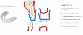
Photo source senivpetro/freepik
A beautiful smile is an indicator not only of beauty, but also of health. Facial surgery can correct the effects of injuries, abnormal jaw structure and birth defects.
A dental technician is the one who deals with such issues. Back in the 7th century BC there were specialists who performed similar operations.
What does a dental technician do?
A dental technician is a medical professional whose work involves the repair and manufacture of dentures and appliances. The technician is not always a doctor; he often has a paramedic education. He works closely with orthodontists, oral surgeons and dentists.
For this reason, it is customary for representatives of the profession to identify narrower areas of activity. For example, there is a direction of orthodontic technician. His responsibility is to create removable jaws and dentures.
Another feature of the dental technician profession is the abundance of tools. In his work he uses:
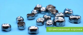
- device for stamping crowns;
- anvils;
- hammers;
- jigsaws.
Dental technicians also actively use 3D printers and programs that help with computer modeling of three-dimensional models of dentures.
What does a specialist do?
Dentists are an ancient profession, but the dental technician as a separate unit has appeared recently.
The main activity of a dental technician is the production of dental crowns, various designs of bridges, maxillofacial and orthodontic structures, clasp and removable plate dentures. The doctor, together with the patient, selects both the materials and the necessary structures. First, he makes an impression of the jaw, from which he designs a model for further production of the prosthesis.
Modern technologies and materials make it possible to make dentures that are close to the appearance of real teeth. The main task of a dental technician is to create a prosthetic structure that is aesthetically beautiful, comfortable and functional.
Content:
- What does a specialist do?
- What is the job
- What problems should you consult a doctor for?
- Treatment methods
Everyone has a unique jaw structure, therefore, it is simply impossible to produce dentures in an in-line manner. The technician works in tandem with the orthopedic dentist, and the result directly depends on the quality and level of each person’s actions.
A dental technician is very concerned about his reputation and tries to fulfill his main duties as efficiently and responsibly as possible. The quality services of an experienced technician are always in demand. In his work, the doctor uses the following professional equipment: a grinding machine, a foundry unit, a vacuum pump, spraying and welding equipment, a sandblaster, a wax furnace, and a melting furnace.
Among the main qualities of a dental technician, it is worth noting mental and physical endurance, good concentration, observation, perseverance, accuracy, attentiveness and sensitivity to people, empathy. It is contraindicated to work as dental technicians for people who suffer from skin diseases, vision diseases, speech and hearing disorders, central nervous system function, mental illnesses, allergies, and infectious diseases.
Responsibilities of dental technicians
Photo source prostooleh/freepik
The list of responsibilities of a representative of this profession can include:
- working with patients;
- creation of prototypes of prostheses or implants and real models;
- repair and restoration of devices.
Also, a dental technician is someone who works with documents: draws up reports on materials, purchasing plans, etc.
To be able to competently and efficiently perform assigned tasks, a specialist must know:
- features of the materials used;
- purpose and characteristics of equipment;
- principles of creating prostheses and devices.
What is the job
A dental technician is a technical and somewhat creative profession. The technician works with the obtained impressions of the patient's mouth. In his work, he talks with the patient about what kind of teeth he would like, calculates the cost and gets to work. After receiving the impression, the doctor goes to his equipped laboratory and begins designing. To produce a model, you need to be able to draw well and have 3D modeling skills.
The next step is the selection of materials, the choice of the required color of the top layer, and the determination of the type of dentures. The selected prosthesis is equipped with certain fasteners, and at the final stage grinding and polishing are performed. Most often, impressions are taken by an orthopedic dentist, and the technician meets with the client only during the period of fitting and fitting of finished dental structures.
What should a dental technician be able to do?
What a dental technician does requires high qualifications and compliance with certain requirements. Here are some of them:
- presence of specialized higher or secondary specialized education in the field of dentistry;
- a valid medical record;
- no medical contraindications.
There are serious health requirements for representatives. In this regard, persons with the following types of diseases cannot work according to their profile:
- impaired finger motor skills;
- chronic infectious diseases;
- epilepsy;
- nervous disorders;
- mental illness;
- visual and hearing impairments;
- dermatological diseases.
Important qualities
A dental technician needs well-developed fine motor skills, accuracy, a penchant for manual labor, observation, high concentration, good working memory, physical and mental endurance. And also sensitivity, empathy, attentiveness to people, the ability to get along with patients. Contraindications: irritability, rudeness, chronic infectious and allergic diseases, cardiovascular diseases, mental illnesses and nervous system disorders, hearing, speech, vision disorders, hand skin diseases.
Where to study to become a dental technician
You can get the necessary education at a medical college. And you can enroll in your chosen specialty only after the 11th grade of school. The duration of training is about three years. A dental technician can also study at a higher educational institution, where more in-depth training in the field will be offered.
Due to the fact that a dental technician is a medical professional, he needs to regularly improve his qualifications, master new working methods and the capabilities of innovative materials.
Salary and prospects
Each dental technician’s career develops depending on the direction he chooses and the development of new methods of work, as well as, of course, on his ambitions and aspirations. The young specialist improves his skills more and more every day, gaining invaluable experience and knowledge.
Once you reach a certain level of professionalism, you can go to advanced training and receive a certificate as an orthodontist. Or, as an option, master management skills and become a leading specialist. If you have an entrepreneurial spirit, you can open your own business. Of course, this will not be easy, but your favorite activity can generate considerable income.
For many young people who want to try themselves as a dental technician, salary plays an important role. Therefore, we note that on average, specialists in this field can count on an income of 30 to 80 thousand rubles per month. As for the demand, due to the ever-increasing interest in specialists in this field, there should be no problems with getting a job.
Career growth of a dental technician
Photo source prostooleh/freepik
The profession of a dental technician is popular and relevant, so a qualified specialist will not have difficulty finding a job.
With the proper skills, a specialist can begin independent practice. This is the only option for his career growth, since the profession involves only internal (horizontal) development and improvement of skills.
Remote learning after college
Distance learning after college implies a stage of advanced training for practicing specialists.
There is no need to visit the educational institution in person and take a workshop, since these courses include only theory:
- various changes in the work of health workers at the legislative level;
- familiarization with new technologies, equipment and materials;
- innovations in the manufacturing process of maxillofacial devices.
Most often, a medical institution sends an employee to undergo advanced training courses if there are special prerequisites for this (mass training of personnel, lack of necessary skills and experience on the part of the employee, the emergence of new equipment).
The employee’s level of professionalism determines his work status and salary level.
The main advantage of distance learning in this case is a flexible educational schedule, allowing a specialist to combine work and taking advanced training courses without any damage or inconvenience.
Dental technician salary
The income of a private practice specialist differs from the salary of a technician who works in an organization. The amounts are individual, as they depend on the chosen pricing policy, client base and region of work of the dental technician.
At the same time, a dental technician at a hospital or clinic with an impressive work experience can earn up to 150,000 rubles.
According to online employee search services, a specialist can count on the following salary:
- Minimum
- 20 thousand rubles. - The maximum
is more than 100 thousand rubles.
These salaries are for salaried employees. When working for yourself, a dental technician's salary can increase significantly.
Features of the profession
There are no identical teeth and jaws. If we draw some analogies, the first thing that comes to mind is fingerprints, which are never identical; There will be at least some difference among similar ones. Therefore, after the dentist makes an impression of the patient’s jaw in his office and sends it to the dental laboratory, the dentist’s painstaking work begins.
So far only models, because to a first approximation it is made from special hard and refractory dental wax. Using this model, the smallest details of the working surfaces of future prostheses or implants are worked out, on which the wearing comfort will depend.
After working out all the parameters of the prosthesis, provided that the matrix in the form of a fingerprint coincides with a copy of the working model, it is converted into ceramics, composite or metal. The tolerances in the manufacture of products are not even millimeter, but micron - this is precisely the class of accuracy required for this work.
Which leads to the following requirements for the chosen profession:
- Perseverance;
- attention to detail;
- sufficient tactile sensitivity;
- stable nervous system;
- good vision.
The advantage will be artistic taste: after all, the jaw prosthesis, especially in its front part, which is demonstrated when smiling, must be not only functional, but also aesthetic.
It is the dental technician, and not the dentist, who determines the material from which the prosthesis will be made.
Interaction between a doctor and a dental technician during aesthetic restoration of teeth with all-ceramic crowns and porcelain multilayer veneers
Kazunobu Yamada, Kanare Dental Center, Nagoya Translation from Japanese and editing by E. Dyakonenko
Introduction I believe that the concepts of “dental aesthetics”, or, more precisely, “aesthetic dental restoration”, have become familiar terms. The generation of porcelain jacket-crowns was followed by metal-ceramics, which made it possible to compensate for the insufficient strength of the porcelain veneer with the high strength of the underlying metal frame and expand the scope of application of ceramics in dentistry, and in the last few years, high-strength dental ceramics, fixed to the supporting teeth with adhesive devices, began to be used to restore teeth. polymers that provide a strong and high-quality connection between the restoration and the hard tissues of the teeth. High artistic skill, dyes for internal coloring, a method of modeling the anatomical shape of a denture using ceramic masses of different colors and shades, the use of opalescent ceramics, etc. – all this was brought to such perfection that the restorations became practically indistinguishable in color and shape from natural teeth. Reports on all-ceramic restorations, initially found only in specialized journals, began to be published in popular publications; advertisements for such restorations often appear in the media. Due to the fact that there is no metal in all-ceramic restorations, their color matches well with the color of natural teeth, and ceramics itself has better biological compatibility with the tissues of a living organism than metal, so I consider the method of restoring teeth with all-ceramic dentures and microprostheses more effective than restoration metal ceramics. In addition, patients themselves believe that the use of metal limits the ability to reproduce the necessary natural color of an aesthetic restoration, and therefore put forward serious objections to the installation of metal-ceramic dentures. And not only this makes the choice of all-ceramic restorations preferable - in many cases, the aesthetic needs of patients can be satisfied solely and exclusively with the help of metal-free ceramics. Such requests include requests to replace unaesthetic restorations that differ in appearance from natural teeth, as well as patients’ wishes to make their teeth lighter, increase their width, shorten their length, improve the anatomical contours of the gums, correct the position of the tooth in the dentition, etc. . So, with the advent of metal-free ceramics, the era of modern aesthetic dentistry has begun, the task of which is to satisfy the aesthetic needs of patients as completely as possible. This article will talk about the art of aesthetic restoration of teeth, in particular with all-ceramic dentures and microprostheses, describe the stages of their manufacture, express the author’s opinion on how the interaction between the doctor and the dental technician should occur at a dental appointment, in addition, it will be shown how several materials for the manufacture of all-ceramic restorations, choose the one that can better satisfy the individual aesthetic needs of a particular patient than others. The article will also provide the author's views and the opinions of experienced dental technicians regarding which of the many dental materials should be preferred in specific clinical situations.
I. Information required by the dental technician Contacts between the office and the dental laboratory are not limited to the production of the working model. It is not permissible if, during the planned production of aesthetic restorations, the information transmitted to the dental technician does not reflect the shape and color of the patient’s teeth, as well as all his wishes. In fact, if restoring the dentition with all-ceramic restorations seems impossible from a biomechanical point of view, taking into account the peculiarities of the patient’s occlusion and the insufficient strength of the material, and a dental examination of the patient will allow a compromise treatment option to be reached, then in this case, with insufficient information, the dental technician will have problems difficulties in completing the task, which will lead to an increase in the number of additional fittings. 1. Necessary conditions for the correct transmission of the selected color
In fact, a dental technician from the laboratory, who came into the office and took part in the selection of ceramic colors, can take upon himself the responsibility of taking photographs and selecting the necessary angles. It is known that if a person continuously looks at any color for 15–20 seconds, then his perception of this color changes, so I believe that having formed his opinion about the color of the patient’s teeth, the dental technician must simultaneously photograph not only the selected color against the background of the teeth a color sample, but also samples similar in color; this may subsequently be useful when choosing porcelain mass for modeling dentures. It is advisable for the dentist to remember the brand and numbers of the porcelain mass with which the dental technician works. In particular, if the location of the dental laboratory is located at a remote distance from the office, and it is difficult or impossible for the dental technician to come to the appointment and make his own judgment about the work to be done, it is advisable that he (the technician) send the orthopedic doctor a task sheet (preferably on an A4 sheet) for photography. A sample of such a task for a doctor is presented in Table No. 1. I would like the dentist to also make slides for the patient. In the photograph (Fig. 1), against the background of the teeth, there are several samples of the color scale, corresponding in color to the patient’s natural teeth, however, due to the reflection of the incident light, the individual characteristics of the color of the teeth are not clearly visible. In the photograph (Fig. 2), the reflection of the incident light does not interfere with obtaining the necessary information about the teeth, however, this picture shows the only example of the color of the ceramics, namely, the sample of color A1, which does not correspond in brightness (lightness) to the color of the patient’s natural teeth, so this picture will also be unacceptable. I believe that to select ceramic colors it is better to use not the VITA color scale, but the brighter Noritake color scale. I believe that the best quality photographs can only be taken by someone who has once been or is involved in photography. Below are specific points to pay particular attention to: 1) When photographing the color of natural teeth, it is important to avoid the appearance of light reflecting off the surface of the teeth in the photograph. 2) After selecting the color, it is placed so that the cutting edge of the selected sample is close to the cutting edge of the natural tooth, and the color number is clearly visible. 3) The photograph must contain not only the selected color, but also color samples with previous and subsequent numbers. It is also recommended to add to these samples the lightest color in the selected group. 4) In cases where the entire anterior area of the dentition is subject to restoration, the color of the ceramics is selected according to the color of the teeth of the opposite row. When creating the surface relief of a future restoration, you should also evaluate the relief of the antagonist tooth of the same name. 5) When choosing a color, the surface of the teeth must be moistened. In order to slow down the evaporation of moisture from the surface of the teeth, it is recommended to use a mixture of glycerin and distilled water, taken in a 1:1 ratio, for moisturizing. I would also like to draw your attention to some important points below:
| Rice. 5. Left tooth after restoration with porcelain multilayer veneer (F.M.V.). Since the surface of the teeth is not moistened, the reflection of the incident light approximately corresponds to the type shown in Figure 3. In general, the coloration appears dull and whitish, so it is difficult to evaluate. However, despite this, the image clearly shows details on the surface of the right tooth. |
| Rice. 6. The surface of both teeth is moistened with water. The reflection of the incident light approximately corresponds to the type shown in Figure 4. From this photograph, the color of the abutment tooth can be easily assessed. In the manufacture of F.M.V. The color of the abutment tooth matches the color of the veneer being made, so obtaining accurate information about the color of the prepared tooth is key to success (both photos were kindly provided by Mr. Masashi Sato of Senmai Dental Clinic). |
| Rice. 3. Schematic representation of the diffuse reflection of incident light. When light hits the flat, rough surface of a ground glass plate, it is reflected in different directions. |
| Rice. 4. Schematic representation of the specular reflection of incident light. When light strikes a flat, smooth surface of an unpolished glass plate, it is reflected in one direction. 1 — angle of incidence 2 — angle of reflection |
1. If the photograph captures the reflection of light from any part of the tooth, then such a photograph will not be able to convey to us information about the coloring characteristics of the tooth in this area. To avoid reflections in photographs, you should use a camera with a round flash, or at least try to take several photographs by placing the camera above or below the teeth being photographed. 2. When taking photographs, in order to be able to compare the main color tone with the color saturation (color density) of the patient’s teeth, it is recommended to shoot with a 2-degree tilt of the camera relative to the upper horizontal plane, identical to Camper’s. 3. The color of the tooth to be restored rarely matches the color of the remaining natural teeth, however, in most cases within the same dentition, the color differences between the teeth will be more likely in the lightness (brightness) of their color than in the saturation of the color tone. Therefore, when photographing selected samples of colors against the background of teeth, it is recommended to add the lightest (brightest) color from the scale used to them so that the relative brightness of the color of all teeth in the photograph can be assessed. 4. In particular, when restoring teeth in the chewing areas, in order for these teeth to not be inferior to the rest in brightness of color, it is recommended to select the tooth color according to the color of the occlusal surface, so this will require photographing the view of the teeth from the side of the occlusal surface. 5. If the film of liquid on the surface of the tooth is homogeneous and uniform, and the scattering of the incident light is insignificant, then assessing the internal structure of the tooth and the depth of color from a photograph will be a fairly simple task. I would also like to draw your attention to some points related to the last (fifth) point, which should be remembered when pointing the camera and taking photographs, otherwise the quality of the photograph may be unsatisfactory. In particular, this applies to taking photographs for the subsequent production of porcelain multi-layer veneers. There are two types of reflection of incident light: diffuse and specular. In Fig. 3 shows a schematic representation of the scattered reflection of incident light, in Fig. 4 – mirror reflection; what is actually observed is shown in Figures 5 and 6. The photograph shown in Fig. 5, was taken unsuccessfully - it does not allow one to recognize and evaluate the color of teeth and choose the appropriate color of ceramics. However, from this photograph it is possible to objectively assess the features of the surface relief of the tooth from which light is reflected, and this information can become very useful at the final stage of correcting the anatomical shape of the restoration. By moistening the surface of the photographed teeth taken in photo 6, we will obtain significantly more accurate information about their color; with the help of this photo we can easily create the required colors for the future F.M.V. by combining the manufactured veneer with the image of the supporting tooth. Therefore, conveying information through photographs is a critical aspect of denture manufacturing, so the photographer must rethink and understand the importance of photographic quality. However, for the successful manufacture of dentures, not only high quality photographs are required, but also an understanding that if the selected material differs in color and structure from the patient’s natural teeth, this will negatively affect the perception of the finished restoration by others, so photography in the office and evaluation of photographs in laboratories should be carried out at approximately the same time of day, under the same weather conditions and under the same room lighting. 2. Necessary means of conveying tooth shape
| Rice. 7. Diagnostic plaster model cast before preparation, used for making F.M.V. This model provides us with the necessary information about the anatomical shape and features of the surface relief of the patient’s teeth, the structure of his gums, etc. |
| Rice. 8. A plaster model, cast from an impression of the teeth after preparation, is intended for the manufacture of F.M.V. From it we can determine the depth of hard tissue removal, which provides us with the necessary information about the thickness and other parameters of the future F.M.V. |
It was previously mentioned in passing that if, when creating a restoration of a front tooth, it is necessary to copy the surface relief of the antagonist tooth of the same name, then this tooth does not need to be moistened in order to better examine the individual details on its surface. In addition, when correcting the position of a tooth in the dentition, when taking photographs, it is recommended to align the vertical baseline of your camera’s viewfinder with the middle axis of the patient’s tooth. Thus, photographs are a means of conveying the color and shape of the patient's teeth, but the image in the photograph is flat (two-dimensional). Unlike photographs, plaster diagnostic pre-treatment models (taken before tooth preparation, Fig. 7) are not flat, but three-dimensional (volumetric), so the information we receive will be extremely important, as will the information that temporary restorations made at models cast after preparation. In cases where the patient does not like the shape of his teeth, and the ultimate goal of the restoration is to correct it, making a pre-treatment model is not necessary, but if making a F.M.V. is aimed at changing the color of teeth or improving their coloring, then in order for the restorations to harmoniously combine in the oral cavity with the remaining natural teeth, it is necessary to accurately reproduce the features of the anatomical shape of the patient’s teeth.
| Rice. 9. Pre-treatment (primary) comparative plaster model. |
| Rice. 10. Presumable restoration of teeth. It represents a possible treatment option that reflects the optimal balance between the dental technician’s ideas and the patient’s wishes. The participation of the dental technician from the very beginning of treatment plays a positive role in ensuring a successful outcome of the aesthetic restoration of the patient’s teeth. |
Using plaster models cast after preparation (Fig. 8), we can determine the depth of hard tissue removal, which will provide us with the necessary information about the thickness and other parameters of the future restoration. 3. Means necessary to correct the position of teeth in the dentition The main objectives of aesthetic dentistry are not only to improve the color and shape of teeth, but to correct their position in the dentition. To accomplish the latter task, when using single crowns and porcelain multilayer veneers in combination, a general plan for the intended treatment of the patient must be outlined. Previously, the so-called “blue film” method was used for these purposes, and now the method of “paired pre-treatment comparative models” was used, which should reflect both the wishes of the patient and the point of view of the doctor and dental technician. Figure 9 shows the pre-treatment diagnostic model, Fig. 10 – view of predicted restorations on a plaster model. These aids (model and restorations) are prepared in a dental laboratory. They are not difficult to produce, but they provide a visual representation of how accurately the predicted restorations meet the patient's expectations. I note that it is important here that the dental laboratory gets involved in the treatment
| Rice. 11. Working model designed to correct the incorrect position in the dentition of two central incisors (orientated in relation to the remaining teeth) using F.M.V. From an aesthetic point of view, wax modeling (in this case on all front incisors) is better done before making a fireproof model. |
| Rice. 12. Type of teeth, the plaster model of which is shown in Fig. 11, before starting treatment. Proximal caries in the middle third of both central incisors and crowding of the anterior teeth. The goal of treatment is to correct the position of the two central incisors using F.M.V. When planning treatment, it was planned not only to correct the position of the central incisors, but also to increase the width of the following lateral incisors, since the insufficient width of the latter could worsen the aesthetic perception of the restoration as a whole. |
patient from the very beginning of the dental appointment. In this case, during the appointment, it will be possible to make temporary restorations using wax-up models formed on the plaster model. The restorations, sent to the office on models, are fixed in the patient’s mouth. In particular, if a three-dimensional assessment (i.e. assessment on a model) is not used when correcting the position of teeth in the dentition, then errors may occur. In addition, if the technician does not have a pre-treatment diagnostic model at his disposal, then when making an aesthetic restoration he will have to reproduce the characteristic differences of the patient's teeth from those teeth that are not intended to be restored. In Fig. Figure 11 shows a working model of the dentition.
| Rice. 13. View of the patient’s teeth after preparation. The lighter color NW0.5 was chosen. The reason for this choice was the planned teeth whitening after restoration. |
| Rice. 14. View of the patient’s teeth after restoration. Porcelain multilayer veneers are fixed on the two central incisors; the shape of the lateral incisors is corrected with a polymer composite, similar to that shown in the model shown in Fig. eleven. |
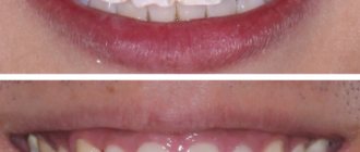
The goal of treatment was to correct the position of two central incisors that were incorrectly positioned in relation to other teeth using porcelain multilayer veneers. In Fig. Figure 12 shows the view of the patient’s teeth before treatment: when correcting the position of the central incisors, a harmonious balance should be created between their shape and the shape of the lateral incisors located behind them. It is clear that insufficient width of the lateral incisors can worsen the aesthetic perception of the restoration as a whole, therefore it is desirable to increase the transverse size of the crowns of the lateral incisors. Here it was decided to adjust the shape of the lateral incisors to the ideal shape with wax before proceeding to the production of a fireproof model. On this model, ready-made F.M.V. were sent to the office. In Fig. Figure 13 shows a view of the patient’s teeth after preparation. Note the smooth preparation, i.e. absence of sharp internal and external linear angles to the preparation border. In Fig. Figure 14 shows a view of porcelain multilayer veneers after fixation in the patient’s mouth. To create aesthetic harmony between the central and lateral incisors, the shape of the lateral incisors was corrected with a resin composite. In Fig. 15 and 16 show photographs of the patient's face before and after aesthetic treatment. I would like to draw your attention to the fact that thanks to the restoration in this case, a harmonious balance was achieved between the width of the patient’s chin and the size of her anterior teeth, in particular her incisors. 4. Other means In order to realize the true wishes of the patient, why not use photographs of the smile and teeth of a famous actor that the patient brought with him, or portraits or photographs (including snapshots) of the teeth of the patient himself at a younger age. In Fig. 17 depicts portraits that the patient brought with her; in Fig. 18 and 19 show photographs of the teeth of the same patient before and after completion of aesthetic treatment.
| Rice. 15. Photo of the patient’s face before treatment. |
| Rice. 16. Photo of the patient’s face after completion of treatment. In this case, a harmonious balance was achieved between the width of the patient's chin and the size of her front teeth. |
II. What should be taken into account when restoring teeth with all-ceramic restorations? The color of a natural tooth is determined by the optical properties of its hard tissues - the reflection and absorption of incident light and its scattering in the internal structures of the tooth. Due to the fact that the optical properties of metal-free ceramics and teeth coincide quite closely, the procedure for manufacturing restorations that are indistinguishable in appearance from natural teeth will not be difficult.
| Rice. 17. Portraits that the patient brought with her. The presence of such aids will be a serious help in the work of a dental technician. |
| Rice. 18. The condition of the teeth of the patient who brought with her the portraits shown in Fig. 17, before the start of aesthetic treatment. Since after starting a new job the patient was constantly in a state of stress due to excessive wear of the upper incisors, she had to constantly keep her mouth closed and clench her teeth. |
Table 2 presents the classification of currently existing metal-free ceramic systems (the classification is given according to the method of manufacturing all-ceramic dentures and microprostheses). One of the most important components used in many metal-free ceramic systems is aluminum oxide, which significantly reduces the transparency of all-ceramic denture and microdenture frameworks. Alumina ceramic materials include: In-Ceram Alumina, GN-1 Alumina, Procera, etc. Compared to them, ceramics based on magnesium spinel - In-Ceram Spinell, GN-1 Spinell and Empress ceramics have greater transparency, therefore, the peculiarity of these materials is the transmission of the color of the underlying tooth. In the experience of the author of this publication, if the structure located under the restoration is light in color, then if there is insufficient space for placing the restoration, preserving living teeth or using
| Rice. 19. To correct the defects shown in Fig. 18, the patient received all-ceramic crowns. It goes without saying that with the treatment, the root cause of anxiety and stress was eliminated. (Photos published with permission from Mr. Nakamura, executive director of the Kanare Technical Center). |
| Rice. 20. Test fitting for water (i.e., before fitting, the tooth surface is moistened with water to hold the restoration - translator's note) of a veneered crown with a frame made of GN-1 Spinell ceramics. The advantage of this crown is that the abutment tooth is visible through the translucent ceramic frame (the color of the abutment tooth can be classified as the lightest color group). |
fiberglass composite posts, the use of translucent, light-permeable ceramic frameworks is indicated, however, if the underlying structure is dark in color and there is sufficient space, then metal-ceramics, or equivalent porcelain-lined opaque all-ceramic alumina-based frameworks, can be used to restore teeth. That is, with adequate preparation (i.e., sufficient removal of hard tissue), the use of all-ceramic systems In-Ceram Alumina, GN-1 Alumina and Procera is indicated. Figures 20 and 21 show the fitting of crowns made of GN-1 Spinell ceramics and Procera ceramics to the same patient. The color of the abutment tooth on which crowns are tried on can be classified as the lightest group of colors. The tooth preparation was done to create enough space to place the porcelain. Despite the fact that it is difficult to judge the brightness and color density of the crowns from the photograph, a visual assessment shows that the crown with the GN-1 Spinell ceramic frame
| Rice. 21. Test fitting for water of a veneered crown with a ceramic frame made of Procera ceramics. The crown looks lighter and more saturated in color than in the first case. |
| Rice. 22. Transparency test of thin samples made from two polymer cements. The transparency of both samples was considered high, but the left sample has greater transparency than the right one. |
is more transparent than with a Procera ceramic framework, however, a crown with a Procera framework is brighter and more saturated in color. I would also like to add that the anatomical shape of both crowns was modeled in the same way using the same ceramic mass for veneering all-ceramic denture frames. This ceramic body was Serabien Noritake . So, if light-colored composite core inlays on fiberglass pins are used to restore a tooth, or if there are no problems associated with the color of the prepared natural tooth, it is advantageous to place restorations on translucent (translucent) ceramic frames. The cement used to cement all-ceramic restorations with translucent frameworks has a major influence on the perceived transparency of the installed crown or veneer. Practice shows that the color of a crown or veneer during a trial fitting with water wetting the surface of the abutment tooth will differ from the color of the same restorations when placed on permanent cement. This practical observation is confirmed by the materials test example shown in Fig. 22. Manufacturers' concepts differ from one another, however, whatever they are, the restoration will look alive if the color of the underlying abutment tooth shines through, so in order to maintain this impression, it is better to use cements with a higher degree of transparency .
| Rice. 23. In order to make a judgment about the “lens effect”, you should first evaluate the shape of the preparation of the abutment tooth. To enhance the “lens effect”, preparation in the area of the cervical edge should be performed with the formation of a slight groove-like shoulder. |
| Rice. 24. Porcelain multi-layer veneer (external and internal views) made for the tooth shown in Fig. 23. To model the veneer in the area close to the neck of the tooth, porcelain colors with a higher degree of transparency were used. |
In the case of discoloration of the abutment tooth or dark color of the metal core, on the contrary, the goal of the restoration is to reduce the unsightly color of the abutment to a minimum, therefore in such cases opaque cements should be used to cement all-ceramic restorations. Recently, reproducing the color of a natural tooth in the cervical area has ceased to be a problem - for this purpose, the restoration is made so that the color of the natural tooth shines through it. This clinical and dental concept is called the “lens effect technique.”4 Typically, the final crown color or F.M.V. represents
| Rice. 25. Fitting on water F.M.V., corresponding to Fig. 24, made to restore the prepared tooth shown in Fig. 23. |
| Rice. 26. Deepening the preparation (increasing removal) in the cervical area of the tooth shown in Fig. 23. This form of preparation will require the production of a new veneer, for which porcelain with a higher degree of transparency will be used. |
a combination of the color of a natural tooth with the color of porcelain, therefore, in cases where the “lens effect technique” is used to model the cervical areas, porcelains with a high degree of transparency are chosen (for example, porcelain masses in the colors Tx, T0, LT0 Noritake ). When making porcelain veneers using this method, only highly transparent cements should be used to cement them. In addition, it will undoubtedly be useful if the doctor discusses with the dental technician the form of tooth preparation for future veneers. In Fig. Figure 23 shows the preparation of teeth under F.M.V., performed in such a way that the high color saturation of hard tissues is preserved in the cervical area. To do this, a light groove-shaped preparation is carried out in the area of the gingival margin. It should be emphasized once again that when making a veneer, this area is modeled using porcelain with a high degree of transparency (Fig. 24) and before fixing with cement, a water test is carried out (Fig. 25). In Fig. Figure 27 shows a veneer re-made for the teeth shown in Fig. 23, after carrying out in-depth preparation (increasing the removal of hard tissue) in the cervical area (see Fig. 26). If we compare Fig. 25 with fig. 28, then we understand that in the first case the restoration looks more natural and aesthetic, since the brightness of its color and color saturation in the cervical area better coincide with similar indicators in natural teeth. With deep preparation, on the contrary, obtaining the intended color will become impossible. In my opinion, this is due to the fact that due to the presence of a thicker layer of transparent porcelain, the reflection of light from the surface of the prepared tooth is weakened (i.e., the transmission of tooth color is deteriorated), and the restoration will appear darker and its aesthetic appearance will be inferior to what was achieved In the first case. There are other concepts that I would like to explore in as much detail as this one, but each is based on specific clinical situations. For example, in cases of discoloration of the cervical area, one should consider the situation and decide whether a preparation suitable for cases similar to the one presented in Fig. 23? Is it necessary to reflect the changed color, and is it not better to form a straight ledge, reminiscent of tooth preparation for all-ceramic crowns? It seems to me that the use of the "lens effect technique" will be useful in situations where the color of the cervical region has not been assessed in daylight, and also where problems may arise in reproducing the color of the cervical region due to the presence of greenish tones in the color of the lower areas of the abutment tooth or when the patient's gums are pale. In the previous sections I and II, we talked about the participation of the dental technician in the aesthetic restoration of the patient’s teeth before making a diagnostic model and that if you have complete and detailed information about the color of the tooth, then it will not be difficult to reproduce the color of the restoration in the area of the cervical edge.
| Rice. 27. Veneer made for the prepared tooth shown in Fig. 26. | Rice. 28. Fitting on water F.M.V., corresponding to Fig. 27, made to restore the prepared tooth shown in Fig. 26. We see that the coloring of the restoration shown in Fig. 25 looks lighter and in terms of color saturation and brightness matches the color of natural teeth more closely than in the second case (Fig. 28). |
Part 2