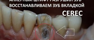
Clients periodically come to the clinic with this question. The answer cannot be unambiguous; it all depends on the situation, the condition of the tooth, the root canals, and the remaining intact walls of the tooth (on the other hand). You can save a tooth in different ways: put a high-quality filling, make an inlay and a crown. The third option is the most deplorable - removing the remains of the tooth, but it is only suitable if the wall of the tooth has broken off much below the level of the gum and there is nowhere to attach the crown.
Why Teeth Chip – Potential Causes of Injury and Crown Failure
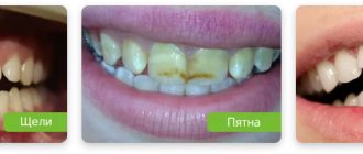
Enamel is considered the hardest tissue in the body. But even chips can appear on it, and there are many possible reasons for this. A chip is an injury in which the integrity of the crown is compromised. The damage may be very minor, even unnoticeable. But in other cases, the chip affects not only the enamel, but also the dentin.
If a tooth breaks off near the gum, such a defect cannot be ignored. Moreover, in case of large-scale damage, the pulp may also be injured, and this will inevitably provoke severe piercing pain - you need to see a doctor as soon as possible.
A chip is an injury in which the integrity of the crown is compromised
As a rule, teeth are injured as a result of mechanical impact. For example, the front incisors and canines are at high risk - they can easily be damaged if you fall or get hit. Molars more often chip off during meals, when chewing solid foods. A cracker, a fish bone, or, for example, a nut shell can injure the chewing crown. However, there are other reasons that can provoke such trouble:
- insufficient hygiene, abundant plaque and hardened deposits - the active activity of pathogenic microorganisms in this case leads to the gradual destruction of hard tissues, the development of caries and pulpitis,
- the habit of biting off thread with teeth, cracking nut shells, gnawing seeds,
- demineralization of enamel - as a result of systemic failures within the body, hormonal changes, pathological and age-related changes,
- incorrect bite – leads to uneven distribution of the chewing load, which causes crowns to wear down and chip, which are subject to increased pressure,
- consequences of dental treatment - after depulpation, that is, removal of the nerve, the tooth becomes “dead”. This means that it is deprived of constant replenishment, which is why it darkens and becomes brittle. Therefore, chips on pulpless incisors and molars are not uncommon.
Improper dental treatment can cause a problem.
If the enamel is healthy and strong, it will be quite resistant to mechanical stress, temperature and chemical factors. But if the top protective layer has weakened due to constant traumatic exposure, sudden temperature changes in food and drinks, hormonal imbalances, deficiency of vitamins and microelements, or systemic pathologies, it will be more susceptible to cracks and chips.
Causes
The question of why teeth chip off worries many, especially when it comes to children’s teeth. Not only does this bring some aesthetic discomfort; but often the surrounding tissues are also injured, and this can be much more dangerous. There are several most common reasons why a person’s tooth may break off:
- As a result of injury;
- Anomaly or pathology;
- Lack of useful elements, such as calcium;
- Hormone imbalance;
- Reduced immunity.
In addition, according to some scientists, teeth can chip due to the fact that the acidity in the mouth is reduced. Often the tips of the incisor also break off when trying to bite through something hard.
Types of chips and characteristic symptoms
Experts in the field of dentistry distinguish several types of chips, which differ from each other in the nature and depth of damage. An enamel chip is a minor violation of the integrity of the crown, for example, when a small piece of the cutting edge breaks off. With such damage, patients usually do not experience pain or even discomfort. But such a defect may well affect the aesthetics, especially if it falls into the smile zone. In addition, if you ignore the problem, the tooth will continue to decay over time.
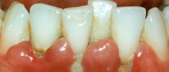
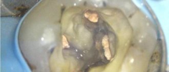
Dentin damage is usually diagnosed when the wall of a tooth near the gum, a piece of an incisor, or even half of a crown breaks off. It is much more serious if a deep chip affects a large part of the crown or, for example, leads to its vertical fracture. In this case, the person inevitably faces severe pain, and emergency dental care is required. If the root is damaged, complete removal will have to be carried out.
The photo shows damage to the dentin of the tooth.
What can you do before visiting the dentist?
Even if the damage is minor, it is better to consult a dentist as soon as possible. The integrity of the tooth must be restored, otherwise the hard tissues will continue to deteriorate. Sooner or later, a tiny chip will lead to the development of caries and significant destruction of the crown.
If we are talking about a serious injury, it is better to see a specialist as soon as possible. Here's what you need to do right away if a tooth breaks off near the gum:
- rinse your mouth with water to remove small food particles and enamel fragments,
- briefly apply ice to the cheek on the side of the sore spot, wrapped in a few napkins - this will help reduce swelling and relieve pain,
- in case of bleeding, apply a sterile gauze swab to the wound,
- Take a painkiller tablet if symptoms are severe.
Before going to a specialist, you can take painkillers.
If there are significant injuries to the baby incisors and molars, the first thing you need to do is try to calm the child down and ask him to rinse his mouth with boiled water at room temperature. Immediately after this you need to urgently go to the doctor.
Restoration methods in dentistry
The depth of damage directly affects the choice of methods of treatment and restoration of the tooth. So, if we are talking about large chips right up to the gums, you can consider the following options for solving aesthetic and functional problems:
- artistic restoration (direct composite veneers) – a specialist restores the shape of the crown by layer-by-layer application of a light-curing composite material, exactly the same as that used for light fillings. This is the best option if the chip is located near the gum, but does not affect most of the tooth,
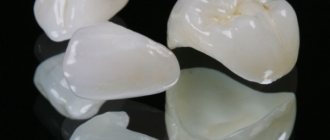
The defect can be corrected by installing a light-composite material - restoration tab – you can restore the shape using the tab, among other things. Essentially, it is a large filling that is made from impressions in a laboratory. It replaces part of the crown, thereby restoring its shape and functionality,
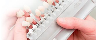
- veneers and lumineers - the possibility of using this method should be considered based on more accurate characteristics of the clinical picture. In some cases, it is impossible to hide large defects with thin ceramic or zirconium plates. Veneers and ultra-thin lumineers are usually used to completely transform your smile. With the help of such plates you can successfully disguise minor external flaws and make your smile perfect. But before installing such onlays, you will have to grind off the enamel layer in order to preserve the anatomical parameters of the teeth. However, lumineers allow you to do without this procedure, since their thickness is only 0.3-0.4 mm1,
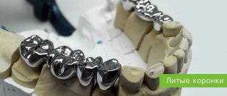
Veneers can be used to treat this type of damage. - artificial crown - if the visible part is almost completely destroyed, it will be necessary to install a crown, possibly with the preliminary installation of a pin or stump tab to strengthen the connection of the prosthesis with the root system. Well, if, on top of everything else, the root is seriously damaged and requires removal, then the next best solution would be to install a crown on the implant. As an alternative, one can also consider bridge-like prosthetics based on row elements adjacent to the defect.
“I had this happen. One tooth on the upper jaw first chipped. It wasn’t noticeable, I didn’t feel any pain either, so I decided it was nothing to worry about. After a couple of months I ate nuts, bit poorly, and in the end the half tooth was gone! I had to get a crown, but the doctor said it wouldn’t be possible to build it up. True, he was initially dead for me, but it’s still a shame.”
Alexey, from correspondence on the 32top.ru forum
In all these cases, appropriate treatment is carried out first. In case of chips and cracks in the enamel, it is worth undergoing an examination to determine the condition of the enamel layer. Perhaps the doctor will consider it necessary to prescribe a course of fluoridation and remineralization to strengthen it.
If the pulp is seriously affected, it is likely that it will have to be removed. Partially or completely - depends on the depth of the damage. This is a neurovascular bundle that provides nutrition and vital activity to the crown. Without the nerve, it darkens, becomes brittle and breaks down faster. After depulpation, the canals are also treated and filled, and a pin or stump inlay can be installed.
Sick teeth: save or remove
First of all, I want to say that every doctor should understand that preserving the tooth itself is, for many reasons, better than removing it. Implantation is not a panacea. You can always get an implant – in any case and at any age, when there is no other option. But trying to save your tooth is an art!
Today, many doctors say that conservative treatment of large cysts is impossible. They are removed and a resection of the root tips is performed - that is, the cyst is removed along with part of the root. Then artificial bone material is placed there. To do this, you need to cut the gum. That is, the treatment of such pathologies in the “standard order” is surgical. The life expectancy of such a tooth is 5 years. After this, a relapse very often occurs, that is, the inflammatory process begins again.
I am a supporter of endodontics, in other words, I try to treat teeth where a cyst or granuloma has formed conservatively through a canal, without cutting the gum. And I, of course, have my own technique in these cases: in fact, it is enough to rinse the canal well and remove the infection with therapeutic drugs. In this case, of course, it is necessary to know anatomy, chemistry, and also microbiology - that is, to understand what microflora and what to kill with, because the success of treatment depends on this. For the doctor, in theory, it is no secret that in the case of cysts and granulomas, the causative agent of infection is gram-positive and gram-negative cocci. And believe me, if the doctor understands these things, then there will be nothing magical in endodontic treatment. True, such treatment takes from 1 month to six months, depending on the size of the cyst. Today there is an opinion that such teeth can be treated in one visit, without prior calcium treatment. Based on my own experience, I believe that it is difficult to achieve positive dynamics in this case, we’ve been through it - we know! After all, it also happens: the tooth seems to be improving, and then suddenly there is a sharp relapse. Therefore, once a month the patient visits the clinic, I rinse the tooth, apply medicine, perform an X-ray control and... put a temporary filling. And so on until a positive result is confirmed by the results of a computed tomography scan. After six months, a crown can be placed on the tooth. In difficult cases, we install a temporary crown for six months, after which we perform another computed tomography scan and after that we cover the tooth with a permanent crown. After this treatment, I have no relapses in 100% of cases.
Or here’s another example: a fracture of the crown of a tooth below the level of the gum (gingival attachment). You can, of course, remove the tooth, as is usually the case. But I prefer to clinically lengthen the neck of the tooth due to additional tissue located in this area. How? I'll try to explain. The gum has a movable layer that covers the neck of the tooth, and there is also a layer partially attached to the bone tissue. Using a laser (that is, surgically), I remove the gum around the tooth, thus lengthening its neck and providing space for the installation of an orthopedic structure designed to protect the tooth. I know that for many this sounds like something unreal. Fortunately, I can afford to save teeth with a crown fracture even below the gum attachment, but only if this fracture does not reach the level of the interradicular septum. If the interroot septum is affected, then infection will constantly get there. As a result, the tooth will still have to be removed.
Yes, such treatment is not for one day. After all, it takes 2-3 weeks just to form a new gum. In general, the entire process can take from 1 to 2 months - in complex cases (more often when the painters break, that is, large molars in the area of the palatal or lingual wall), in simple cases - 2-3 weeks. But during all this time the patient will need to come to me 2 times. First time I lengthen my neck. The second is when I restore the wall of the tooth, so that the gums grow correctly: this will provide enough space where you can then put a stump inlay, and put a crown on top. But this is the work of an orthopedist...
With root fractures, a lot depends on how badly the root is damaged. Root fractures in single-rooted teeth are not dangerous, especially if the root is broken, for example, 4 millimeters vertically. In these cases, we combine surgical and orthodontic treatment approaches. And then we restore the tooth using orthopedics. But if the crack is deep, then there is no point in saving such a tooth, because complications may arise later.
Perforations of teeth can be closed in any part of the roots, provided that the tooth is sterile (not periodontitis): with a competent approach, the prognosis of such a tooth will be 100% successful. But if the perforation is located in the area of the interradicular septum, a large cyst has formed there, then this perforation can, of course, be closed, but the success of treating such a tooth will be 50 to 50. It is important to seal the periodontium hermetically so that infection does not get there. I close the perforations with a special cement that was created for this purpose and is also used to close wide apical foramina. It seals the perforations hermetically, hardens quickly, and behaves well in a humid environment. However, I would like to make a reservation right away: working with perforations is only possible under a microscope - after all, I need to see how I close the hole! This is the job of an endodontist. Perforations are treated in one visit, meaning you don’t have to wait several weeks or even months for results. But if the bottom of the cavity is severely broken by a bur, when the tooth is not only perforated, but also the area of the interradicular septum is open and infection constantly gets there, then closing such perforations may be impractical.
When removing an instrument from a tooth root, it is also necessary to use a microscope. Although often, for example, when an instrument is broken in a bend in a canal, even a microscope is powerless. What can I say here? We use... intuition and think through every step well. Sometimes it is necessary to use instruments that were not originally designed for work in the canals, or even several instruments, creating “in-house” designs that ultimately allow the removal of the recalcitrant fragment. In general, everything “that comes to hand” is used - in the good sense of this expression. Why not if it can help? When doing this work, it is very important not to overexpand the channel. In principle, I work without expanding the tooth canal - I work only with the tool! The channel remains intact on all sides. There is also the so-called by-pass technique - its essence is that we “bypass” the instrument broken in the canal, leaving it in the tooth canal system. If there is full access to the canal and the instrument can be bypassed by properly rinsing the canal with aggressive solutions, cleaning it and passing it with “cement,” then the foreign body no longer causes any harm to the tissues, since it ceases to be a source of additional infection. And this is also considered a positive result of treatment! To be honest, many doctors look and do not understand how I manage to remove instruments from the apical foramina - for them it is a mystery, shrouded in darkness =).
What complications can there be?
Sometimes patients deliberately postpone visiting a doctor if the injury does not cause them the slightest discomfort. After all, a defect may not be too noticeable, but this does not mean that it can be ignored. Hard tissues will continue to deteriorate, and soon, in such conditions suitable for this, active pathogen activity will begin. Then caries, pulpitis, periodontitis - at this rate, after a while you can lose a tooth.
There are other consequences too. For example, sharp carved crowns can injure the inner surface of the cheeks, which can lead to infection through scratches and the development of chronic stomatitis. Therefore, the problem cannot be ignored, even if the damage is minor and does not in any way affect the appearance of the smile. The sooner you see a doctor, the easier and faster the doctor will be able to eliminate the defect.
Example 3. Chronic periodontitis, significant root exposure, tooth mobility
If chronic periodontitis (periodontal disease) is accompanied by significant loss of bone tissue (more than ½ of the root length), tooth mobility, and exposure of root bifurcations, tooth extraction is the only possible solution to the problem.
In the case of periodontal disease, the main problem is the presence of periodontal pockets and loss (atrophy) of the bone tissue around the teeth. The roots are gradually exposed, and tooth mobility appears. If the periodontal pockets are so deep that even periodontal surgery cannot solve the situation, it is necessary to remove such teeth, because Millions of microbes accumulate in the pockets, which further “corrode” the bone. It is better not to wait until the teeth fall out on their own, but to remove the source of infection, thereby preventing further loss of bone tissue and leaving the possibility of implantation.
What can be done for prevention
No one is immune from accidental injuries, but if the enamel is strong and the teeth are healthy, the chances of avoiding serious damage will be much higher. Therefore, for prevention, it is important to maintain oral hygiene and visit the dentist every six months for prevention and professional cleaning of plaque. It is worth reducing the consumption of sugar and simple carbohydrates, which contribute to the growth of bacteria in the oral cavity and the development of caries.
Excessive consumption of sweets can lead to tooth decay
On a note! If the enamel has thinned and become sensitive, be sure to consult a specialist. To restore it, fluoridation and remineralization procedures can be prescribed - hard tissues are coated with fluoride-containing compounds to nourish and strengthen them.
Enrich your diet with vegetables and fruits, seafood high in fluoride and calcium. Do not bite the seeds or try to crack the nut shell - treat your teeth with care and, if possible, eliminate all traumatic factors. Remember that if you have problems with your bite, it is better not to delay orthodontic treatment, and if any suspicious symptoms appear, you should immediately consult a doctor.
1Magne, P. Adhesive ceramic restorations of anterior teeth, 2012.