Features of the masticatory muscles:
- All chewing muscles are attached to the lower jaw (mandibula);
- All masticatory muscles elevate the lower jaw, performing chewing and facilitating the correct pronunciation of words;
- The chewing muscles, unlike the facial muscles, have fascia.
These features are not indicated in atlases and textbooks, and, as a rule, they are not required to be mentioned in tests. I brought them specifically to make it easier for you to navigate - as soon as we talk about any masticatory muscle, you immediately understand that it attaches to the lower jaw and lifts it. That is, one name of a muscle group is enough to remember the function and place of attachment.
Hypertonicity of the masticatory muscles and its correction with BTA for aesthetic problems of the lower half of the face
Soykher M.I., Orlova O.R., Mingazova L.R., Soykher M.G.
The aesthetic appearance of the lower half of the face reflects the morphology of the dental system and the function of the masticatory muscles. In aesthetic medicine, the problems of correction of the lower third of the face remain relevant, despite the accumulated many years of experience. The functional state of the masticatory muscle is associated with the configuration of the lower third of the face and possible asymmetry, which hides a combination of dental and neurological problems that require a detailed examination and adequate treatment of the patient. Some diseases, such as bruxism and oromandibular dystonia, are accompanied by hypertrophy of the masticatory muscles, which occurs as a result of their forced contraction, and the massive lower third of the face becomes an aesthetic manifestation here.
Keywords:
Facial disproportion; hypertrophy of the masticatory muscles; bruxism; oromandibular dystonia; myofascial pain syndrome; botulinum toxin type A
Soykher Marina Ivanovna, candidate of medical sciences, dentist, head. Doctor of the Center for Interdisciplinary Dentistry E-mail
Orlova Olga Ratmirovna, Doctor of Medical Sciences, Professor of the Department of Nervous Diseases of the Faculty of Faculty of Physics, First Moscow State Medical University named after. I. M. Sechenova, President of the MoESBT E-mail
Mingazova Leniza Rifkatovna, candidate of medical sciences, neurologist, employee of the department of nervous diseases of the State Educational Institution of Higher Professional Education MMA named after. I. M. Sechenova E-mail
Soykher Mikhail Grigorievich, candidate of medical sciences, dentist, leading specialist of the Center for Interdisciplinary Dentistry E-mail
INTRODUCTION
“Everything in a person should be beautiful.” Today, more and more people are striving to implement this principle in their own lives <2>. Almost every person pays attention to his appearance, and attaches special importance to how his face looks <3, 4>. Dissatisfaction in this case can become quite a serious problem and affect both the psychosomatic state (causing depression, uncertainty, neuroses...), and professional status, family and personal relationships.
In aesthetic medicine, the problems of correction of the lower third of the face remain relevant, despite the accumulated many years of experience. According to a survey of patients, the main complaint they present is facial disproportion, in particular the “square face” problem. What is meant by the expression “square” (or “trapezoidal”) face?
In domestic and foreign medicine, for a brief answer to this question, they use such characteristics as protruding angles of the lower jaw, hypertrophy of the masticatory muscles themselves, prominent contours of the lower zone of the face, angular contours of the face (prominent mandibular angle, hypertrophy of the masseter, lower facial contour). The contours and shape of the lower half of the face are determined by the relative position of the upper and lower jaws (occlusal relationship), the size and shape of the lower jaw, as well as the condition of the masticatory muscles.
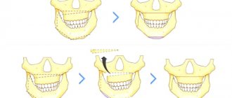
The lower jaw is suspended in space to the fixed bones of the skull with the help of muscles and ligaments. The only support for it is the chewing teeth. It is the teeth that fix the position of the jaw in three mutually perpendicular planes. When the position of the teeth and, accordingly, the dentition changes or their loss, the position of the jaw in space also changes. In most cases, there is a decrease in the lower third of the face, distalization of the bite, with characteristic facial manifestations. There are disturbances in the coordination of the masticatory muscles and temporomandibular joints. The dentition, the geometry of which is normally designed to compensate for the complex biomechanics of the chewing function of the cranial-maxillary system, simultaneously serves as support for the soft tissues of the face, which must be taken into account during the aesthetic rehabilitation of patients.
The four chewing muscles on each side are interconnected genetically (they originate from one branchial arch - the mandibular), morphologically (they are all attached to the lower jaw, which they move during their contractions) and functionally (they perform chewing movements of the lower jaw, which determines their location) .
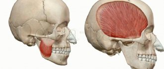
M. masseter is a chewing muscle that starts from the lower edge of the zygomatic bone and zygomatic arch and is attached to the tuberositas masseterica and to the outer side of the ramus of the lower jaw. It has the shape of an irregular rectangle and consists of a superficial part and a deep part. The strongest muscle in the human body in terms of force generated - on molars it develops a force of up to 72 kg.
M. temporalis is the temporal muscle, with its wide origin it occupies the entire space of the temporal fossa of the skull, reaching at the top to the linea temporalis. The muscle bundles converge in a fan-shaped manner and form a strong tendon, which fits under the zygomatic arch and is attached to the processus coronoideus of the lower jaw.
M. pterygoideus lateralis - lateral pterygoid muscle, starts from the lower surface of the greater wing of the sphenoid bone and from the pterygoid process and is attached to the neck of the condylar process of the mandible, as well as to the capsule and to the discus articularis of the temporomandibular joint.
M. pterygoideus medidlis - medial pterygoid muscle, originates in the fossa pterygoidea of the pterygoid process and is attached to the medial surface of the angle of the mandible, symmetrically m. masseter, to the tuberosity of the same name..
M. masseter, m. temporalis and m. pterygoideus medialis, with the mouth open, pull the lower jaw towards the upper, in other words, they close the mouth.
With the simultaneous contraction of both muscles of the pterygoidei laterales, the lower jaw moves forward. The reverse movement is produced by the most posterior fibers of m. temporalis, running almost horizontally from back to front. If m. pterygoideus lateralis contracts only on one side, then the lower jaw moves to the side, in the direction opposite to the contracting muscle. M. temporalis gives a certain position to the lower jaw during speech, thereby ensuring the articulation of the latter.
The masticatory muscle, in addition to chewing movements, takes part, together with the facial muscles, in the articulation of speech sounds, facial expressions, yawning, and swallowing. We can say that this muscle is in a state of “chronic fitness”. Excessive prolonged activity of the masticatory muscles leads to their hypertrophy, which is characterized by an increase in strength and muscle mass (Fig. 5).
The functional state of the masticatory muscle is associated with the configuration of the lower third of the face and possible asymmetry, which hides a combination of dental and neurological problems that require a detailed examination and adequate treatment of the patient. Some diseases, for example, bruxism and oromandibular dystonia, are accompanied by hypertrophy of the masticatory muscles, which occurs as a result of their forced contraction, and their aesthetic manifestation is a massive lower third of the face <6–10>.
The purpose of the study is to study the relationship between the condition of the masticatory muscles and the aesthetic appearance of the lower half of the face; to evaluate the effectiveness of using the botulinum toxin type A drug "Lantox" in order to reduce hypertonicity and correct hypertrophy of the masticatory muscles under the control of surface electromyography.
MATERIALS AND METHODS
40 patients were examined. The average age is 35 years. In order to understand whether problems of an aesthetic nature are caused by pathological processes in the dental system, we conducted a dental and neurological study, which included:
analysis of anamnestic data; clinical study of the masticatory muscles, muscles of the neck and upper shoulder girdle, and the area of the temporomandibular joint (TMJ);
- occlusiogram, analysis of the static and dynamic organization of occlusion;
- orthopantomogram;
- TMJ tomogram;
- teleradiography (TRG) of the lateral surface of the head with markers;
- photoanalysis (portrait and intraoral photographs);
- axiography;
- functional analysis of jaw models in an articulator;
- diagnostics of parafunctions using brookscheckers;
- electromyography (EMG) of the masticatory and neck muscles.
When examining the patient, attention was paid to the following clinical signs: head position, range of active movements in the cervical spine; facial expression, state of the facial muscles when speaking, swallowing, signs of blepharospasm, oromandibular dystonia, facial asymmetry; corneal reflex and reflex from the nasal mucosa, the state of the muscle ridges at rest and when clenching the teeth; volume of active movements of the lower jaw - the distance between the incisors (in cm) when opening the mouth, the trajectory of movement of the lower jaw; mandibular reflex; volume of active movements of facial muscles, brow and orbicular reflexes; Chvostek's sign; sensitivity on the face, oral mucosa and tongue.
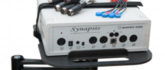
Myograph "Synapsis" dental
Electroneuromyograph for maxillofacial studies and monitoring the effectiveness of therapeutic measures.
More details
The study of the state of the musculoskeletal system included: identification of biomechanical static disorders - scoliosis, asymmetry of the shoulders, shoulder blades and other deformities; identification of the “short leg”.
During palpation examination, we used a 3-point scale for assessing muscle tension and soreness:
0 points - no tension and no pain;
1 point - slight muscle tension, no pain on palpation;
2 points - moderate muscle tension and pain on palpation;
3 points - severe muscle tension and sharp pain on palpation, the presence of painful muscle tightness and/or trigger points.
EMG indicators were recorded using an electromyograph “Synapsis” (NMF “Neurotech”, Taganrog), supplemented with special software. Injections of botulinum toxin into the masticatory muscles were carried out under the control of electromyography; for this purpose we used the Mist device (NMF Neurotech, Taganrog).
During the clinical study, 2 groups of participants were identified:
Group 1 - 30 patients who suffered from bruxism: 25 women, 5 men;
Group 2 - 10 patients with signs of focal muscular dystonia (the leading syndrome of oromandibular dystonia): 8 women, 2 (two) men.
"MIST" professional
Myographic control of injections, biofeedback training, anesthesia regimen.
More details
Clinical features
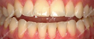
1st group. In dental practice, bruxism is defined as parafunctional activity of the masticatory muscles. The following parafunctions of the masticatory muscles have been described (arranged in descending order of frequency): clenching the teeth, moving the lower jaw forward or to any side, inserting the tongue between the teeth, biting the tongue and lips, grinding the teeth, rhythmic movements of the tongue and submandibular muscles <12–13> . Only 10 patients from group 1 noted grinding and grinding of teeth at night. The rest had the habit of clenching their teeth tightly during the day in response to even minor emotional stress. A hereditary factor (bruxism in close relatives) was determined in all patients in this group. The history also included episodic pain in the face of muscular origin (myofascial pain syndrome of the face), frequent headaches caused by tension in the pericranial muscles. All patients complained of fatigue of the masticatory muscles in the morning. When studying the aesthetic appearance of the face, a massive lower third was determined due to hypertrophy of the masticatory muscles, which caused considerable concern, especially in women.
Dental status in patients of group 1: violation of the integrity of the dentition (absence of one or more teeth), pathological abrasion of teeth, the presence of wedge-shaped defects in the cervical area.
Clinical examination of the masticatory muscles revealed signs of hypertrophy; the muscles are dense, tense, with the presence of painful muscle compactions (myofascial trigger points). When palpating the masticatory muscle itself, in 20 patients the pain radiated to the upper and lower jaws, upper and lower molars, ear, frontal region, TMJ, and neck. In 22 patients, palpation in the area of the tubercle of the upper jaw was sharply painful.
Tenderness or discomfort in the medial pterygoid and digastric muscles was also noted. All patients had muscle tension in the floor of the mouth and limited mobility of the hyoid bone; in 10 patients there was tension and slight hypertrophy of the sternocleidomastoid muscle. Palpation of the muscles on the opposite side was painless or moderately painful.
In 40% of patients, pain occurred on palpation of the lateral pole of the joint head during rotation on both sides, and the temporomandibular ligament on both sides. Discomfort when palpating the lateral pole of the joint head in static conditions on both sides.
In 25% of patients, there was a restriction in mouth opening due to pain to 1.5±2.2 cm between the incisors (normally from 4.6 to 5.6 cm). Further lowering of the lower jaw due to the appearance of sharp pain became almost impossible. There was also a restriction in the movement of the lower jaw forward and to the side.
Only 20% of patients experienced a clicking or crunching sound when opening their mouths. Pain sensitivity of the facial skin and oral mucosa was not changed.
When conducting surface electromyography of the masticatory muscles and neck muscles, the following results were obtained: asymmetry in the work of the temporal, masticatory muscles, high rates of the total biopotential of the muscles under study.
When analyzing a chewing test, a violation of the symmetry of chewing, frequency, amplitude, phase and total biopotential of chewing is noted.
2nd group. Oromandibular dystonia (OMD) is hyperkinesis involving the muscles of the perioral region and masticatory muscles. In patients of group 2, the following clinical forms of OMD were determined: spasm of the muscles that close the mouth and compress the jaw (dystonic trismus) - in 6 people; constant trismus with lateral jerking movements of the lower jaw, bruxism and hypertrophy of the masticatory muscles - in 4 people. Subjectively, all patients of the 2nd group complained of unpleasant sensations, which were described as “periodic movement of the lower jaw”, “the jaw moves to the side”, “it is impossible to find a comfortable position of the jaw”, “forcible clenching of the teeth”, “teeth chattering against each other” friend."
Clinical examination of the masticatory muscles revealed signs of hypertrophy; muscles are dense, tense, with the presence of painful muscle thickening (myofascial trigger points). As a rule, the masticatory muscle itself undergoes pronounced changes, while the temporal and pterygoid muscles undergo smaller changes. Three patients had asymmetric dystonia. Visually, this phenomenon was manifested by asymmetry of the lower half of the face (the volume of the hypertrophied masseter muscle on one side is more pronounced than on the other).
Also, in all patients of group 2, subcompensated signs of dystonic phenomena in other areas were determined: blepharospasm, mild and moderate forms of cervical dystonia, dystonic tremor of the head and upper extremities, writer's cramp.
Dental status: in patients of the 2nd group, there was a violation of the integrity of the dentition (the absence of one or more teeth), pathological abrasion of teeth, and the presence of wedge-shaped defects in the cervical region. When conducting surface electromyography of the masticatory muscles and neck muscles, the following results were obtained: asymmetry in the work of the temporal, masticatory, and neck muscles, torsional twisting of the lower jaw, an increase in the functional activity of the neck muscles and the total biopotential of the muscles under study. When analyzing the chewing sample, a violation of the symmetry of chewing, frequency, amplitude, phase and total biopotential of chewing was noted.
For therapeutic and aesthetic purposes, all patients received injections of botulinum toxin type A (BTA) “Lantox” (Lanzhou Institute, China). It was injected into the masseter proper, temporalis, medial and lateral pterygoid muscles. Large doses were injected into the masticatory muscle itself from the outside (5–10 units at one point). For a more uniform distribution of the drug throughout the muscle, it is injected into several points (from 4 to 8), 1-2 injections are carried out from the oral cavity. In patients with an asymmetric form of dystonia, large doses of the drug were also administered on the side of the larger muscle. The average total dose of BTA (Lantox) was 100 units per procedure.
RESULTS
Analysis of clinical data showed that in patients of group 10 suffering from bruxism, on days 2–10 after injections, the feeling of fatigue in the masticatory muscles in the morning disappeared and headaches stopped. By days 14–21, a decrease in the massiveness of the lower half of the face became noticeable. Against this background, the necessary dental procedures were successfully carried out.
"Hummingbird" dental
Express assessment of the level of bruxism, study of facial muscle tone, wireless technologies.
More details
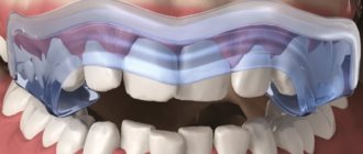
Due to the weakening of the masticatory muscles, the phenomena of bruxism and the phenomenon of clenched jaws ceased for some time. During this time, patients were advised to fix their attention on the facial muscles, consciously relax the lower jaw, open the dentition and try to form a new motor stereotype of the masticatory muscles (based on the principle of biofeedback). For this purpose, disconnecting splints were used on the lower jaw.
In patients of group 2 (OMD), the signs of hyperkinesis were also leveled out already on days 10–12 after injections due to weakening of the activity of the masticatory muscles, which significantly improved the quality of life of patients. Unpleasant subjective sensations disappeared, and the opportunity for dental treatment appeared. In patients with asymmetric dystonia, the symmetry of the lower half of the face was restored.
The study demonstrated the positive effect of BTA (Lantox) on the functional and morphological state of the masticatory muscles, and a good clinical effect alleviated the condition of patients and made it possible not to use any medications.
CONCLUSIONS
The aesthetic appearance of the lower half of the face reflects the morphology of the dental system and the function of the masticatory muscles. In aesthetic practice, it is necessary to conduct a detailed clinical analysis of the condition of the masticatory muscles, especially in patients with a massive lower half of the face. Patients with increased tooth wear and failing dentures require a detailed neurological examination to exclude bruxism and oromandibular dystonia. Injections of botulinum toxin into the masticatory muscles are the method of choice for this group of patients. The drug botulinum toxin type A (Lantox) at a dose of 100 units per procedure is effective and safe for the treatment of hyperactivity of the masticatory muscles in neurological, dental and aesthetic practice.
LITERATURE
1. Budylina S.M., Degtyareva V.P. Physiology of the maxillofacial region. - M: Medicine, 2001. - P. 87–156.
2. Goldstein R. Aesthetic dentistry. Volume 1. - M.: Stbook. - 2005. - P. 10–14.
3. Kalyuzhny D.V. Physiological mechanisms of regulation of pain sensitivity. - M.: Medicine, 1984. - P. 102–114.
4.Karlov V.A. Neurology of the face. - M.: Medicine, 1991. - 284 p.
5. Kupriyanov V., Stovichek G. Human face. - M.: Medicine, 1988. - 272 p.
6. Mingazova L.R. Pathogenesis and treatment of myofascial pain syndrome of the face // Clinical materials. conference of young scientists FPPO MMA named after. I.M. Sechenov “Current issues of clinical medicine.” - M., 2002. — pp. 54–58.
7. Mingazova L.R. Clinical and physiological analysis and treatment of myofascial pain syndrome of the face. Abstract of thesis. dis…. Ph.D. honey. Sci. - M., 2005. - 25 p.
8.Orlova O.R. Focal dystonia: clinical picture, pathogenesis, treatment using botulinum toxin // Diss.... Dr. med. Sci. - M., 2000. - P. 13–29.
9.Orlova O.R., Mingazova L.R., Vein A.M. Facial pain of a muscular nature: clinical and physiological characteristics and treatment with botulinum toxin type A (dysport) // Abstracts of the Russian scientific and practical conference “Clinical and theoretical aspects of acute and chronic pain”. - Nizhny Novgorod, 2003. - pp. 113–115.
10.Orlova O.R. Focal dystonia: clinical picture, pathogenesis, treatment using botulinum toxin. Dissertation for the degree of Doctor of Medical Sciences. - Moscow, 2000. - pp. 13–29.
11.Orlova O.R., Yakhno N.N. The use of Botox (botulism toxin type A) in clinical practice. - M., 2001. - P. 143–147, 161–163.
12. Orlova O.R., Mingazova L.R., Vein A.M. Myofascial pain syndrome of the face: new aspects of the clinic, pathogenesis and treatment // New in dentistry. - 2003. - No. 1. — pp. 25–29.
13.Petrov E.A. Electrophysiological characteristics of pain syndrome of temporomandibular joint dysfunction // Ross. dental magazine. - 2002. - No. 6. - P. 34–35.
14.Petrosov Yu.A., Skorikova L.A. Prevention of TMJ dysfunctions by eliminating parafunctions of the masticatory muscles // Abstracts of the V All-Russian. Congress of Dentistry: Prevention of dental diseases. - Novosibirsk - M., 1988. - P. 156–157.
15.Al-Ahmad HT, Al-Qudah MA. The treatment of masseter hypertrophy with botulinum toxin type. — A. Saudi Med. J. - 2006. - 27. R. 397–400.
16.Gurney CE Chronic bilateral benign hypertrophy of the masseter muscle //Am. Surg. - 1947. - No. 73. - P. 137.
17.Moore AP, Wood GD The medical management of masseteric hypertrophy with botulinum toxin type A // Br. J. Oral Maxillofac Surg. - 1994. - No. 32. - P. 26–28.
18. Sannomiya E., Goncalves M., Masseter muscle hypertrophy // Bras. Dent. J. - 2009. - No. 17 (4). — R. 347–350.
19. Slavicek R. The masticatory organ: Function and Dysfunction.
— Kloster neuburg: Gamma Med. — wiss. Fortbildungs - GmbH, 2006. - R. 59–90. toxin type A // Saudi Med. J. - 2006. - No. 27. - P. 397–400. come back
Anatomy of the masticatory muscles:
I. Temporal muscle (musculus temporalis)
The largest and most visible muscle of mastication. Unlike all other chewing muscles, its abdomen is located on the cranial vault (fornix cranii).
In green I highlighted the outline of the temporal muscle itself, in blue - the outline of the powerful tendon with which the muscle is attached to the coronoid process (processus coronoideus) of the lower jaw. I outlined the coronoid process in yellow, I hope you remember it from craniology. When you show the location of the temporal muscle during the test, you should outline the entire contour, that is, both the muscle and its tendon.
The temporal muscle is located in the temporal fossa (fossa temporalis). If you take a dissected skull, or even a high-quality artificial specimen, you will see impressions on the sides. These impressions are shallow, but occupy a large area. It is these depressions that are called the temporal fossae. In the picture from Wikipedia, the temporal fossa is highlighted with a black frame:
Origin: The temporalis muscle begins on the temporal surface of the frontal bone, as well as on the squamous surface of the temporal bone.
Attachment: The temporalis muscle attaches to the coronoid process of the mandible (the sharpest process of the mandible). Don't forget that the chewing muscles start from the stationary part and are attached to the bone that they move.
Function: the temporalis muscle closes the mouth, raising the lower jaw until the teeth are closed. Also, if you push your lower jaw forward, like a drawer in a table, it is the temporalis muscle that will push it back to its normal position.
II. Chewing muscle (musculus masseter)
It is also a very noticeable and large muscle. If we look at a tablet with an image in profile, it is impossible to confuse the masticatory muscle with anything else. See:
The chewing muscle is extremely important for survival, so our distant ancestors developed this muscle to be very powerful and strong. The masticatory muscle consists of two parts - superficial (pars superficialis) and deep (pars profunda). You can easily distinguish the superficial part - it is attached to the anterior part of the zygomatic arch (arcus zygomaticus). The deep part is also attached to the zygomatic arch, only not in front, but in the back.
I highlighted the superficial part with a green outline, and the deep part with a blue outline:
Origin: Both parts of the masticatory muscle begin from the zygomatic arch.
Attachment: both parts of the masticatory muscle are attached to a special place on the lower jaw - to the masticatory tuberosity (tuberositas masseterica). Let me remind you that the chewing tuberosity is located on the outer surface of the branch of the lower jaw (ramus mandibulae).
Function: raise the lower jaw, chewing and helping to pronounce words. Also, the superficial part of the masseter muscle helps push the jaw forward. We will push it back using the temporal muscle, as you remember.
III. Lateral pterygoid muscle (musculus pterygoideus lateralis).
Now we need a tablet with a skull, which we look at as if from inside the oral cavity (cavitas oris). Something like this:
We turned the skull with the back of the head towards us and sawed it in the frontal plane so that we could look from the inside at the oral cavity, gums and teeth.
Let's look at the lateral pterygoid muscle. Here we can see both lateral pterygoid muscles, so I've highlighted both:
The lateral masseter muscle has two parts - upper and lower.
a) Upper part.
Origin: Infratemporal crest of the greater wing of the sphenoid bone. Let's remember the sphenoid bone (os sphenoidale) - it has a body (corpus), large wings (alae majores) and small wings (alae minores), as well as pterygoid processes (processus pterygoidei).
For some reason, many people like to confuse large wings and pterygoid processes. Let's look at a simplified diagram (front view) so as not to confuse these anatomical formations:
We are interested in the large wing. It is on this that the small infratemporal crest (crista infratemporalis) is located. This is where the origin of the superior portion of the lateral pterygoid muscle is located:
The upper part of the lateral pterygoid muscle is attached to the temporomandibular joint (articulatio temporomandibularis).
b) The lower part of the lateral pterygoid muscle also begins with the sphenoid bone. Now we need to shift our attention from the large wing a little lower - to the pterygoid process (processus pterygoideus). It seems to split into two plates - medial and lateral. Look at the penultimate picture - there I highlighted the pterygoid processes in brown.
Let's find the lateral plate (lamina lateralis) of the pterygoid process. This is, in general, not difficult:
It is from this plate that the lower part of the lateral masseter muscle begins.
So, the beginning: the lateral muscle (its lower part) begins with the lateral plate. Great memory, by the way.
Attachment: pterygoid fossa (fovea pterygoidea) of the lower jaw. Not a particularly well-known anatomical formation, so I found this diagram:
Our pterygoid dimple is located at number 4.
The function of the lateral pterygoid muscle is to raise the jaw (like all masticatory muscles), as well as to abduct the lower jaw in the direction opposite to the muscle. For example, the left lateral pterygoid muscle, when contracted, moves the lower jaw to the right side.
IV. Medial pterygoid muscle (musculus pterygoideus medialis). Let's take a tablet we already know to look at this muscle:
Beginning: Pterygoid fossa (fossa pterygoidea) of the sphenoid bone. By the way, the lower jaw also has, as you remember, a pterygoid fossa, only it is called fovea pterygoidea, so as not to be confused. Ancient anatomists are very prudent guys, definitely.
To be honest, I find it difficult to show it on a tablet. I don’t have a skull at hand at the time of writing this article, but the respected Sinelnikovs indicate in their atlas that “... the medial and lateral plates limit the pterygoid fossa.” Moreover, in the illustration closest to the text, the fossa pterygoidea is indicated on the lateral plate (two pictures ago I highlighted the lateral plates of the pterygoid processes in red). The Sinelnikovs are right in any case - they are the kings of anatomy, after all. I advise you, dear readers, on occasion to ask your teachers about the location of this hole.
In general, speaking about the beginning of this muscle, we focus on the lateral plates of the pterygoid processes of the sphenoid bone and clarify the fossa with the teachers.
Insertion: pterygoid tuberosity of the lower jaw. There won't be any difficulties here anymore, here's where this place is:
Function: Raises the mandible, as well as “pushes” the mandible to the opposite side (i.e., similar to the lateral pterygoid muscle).
Chewing function and the muscles responsible for it
Without these muscles, we would have to be fed intravenously.
Their well-coordinated teamwork allows us to dig into our favorite burger and enjoy a cold milkshake with appetite. The four main paired muscles of mastication - the masseter proper, medial pterygoid, lateral pterygoid and temporal - are attached to the skull and lower jaw. They are responsible for movements of the temporomandibular joint .
The buccal and mylohyoid muscles play an auxiliary role in the process of eating and drinking.
Muscles of Mastication : These powerful muscles lift your lower jaw so you can close your mouth and chew food.
One of the strongest facial muscles, the masseter muscles are thick, flat, and rectangular in shape and are attached to the lower jaw and cheek bones on either side of the face.
Medial pterygoid muscles : these muscles have three functions.
Being also located on both sides of the head, they work as follows:
- With the simultaneous contraction of both muscles, the lower jaw moves forward.
- Contraction of one medial pterygoid muscle leads to movement of the jaw in the opposite direction - this is how we move the jaw left and right.
- Thanks to the simultaneous work of the medial pterygoid, masticatory and temporal muscles, we are able to close our mouth and bite.
These muscles are attached to the pterygoid processes of the sphenoid bones of the skull and the inner surface of the angles of the lower jaw.
Lateral pterygoid muscles: the work of these muscles is no less important - they are responsible for opening the mouth, and after you bite off a piece of, for example, pie, they help chew it.
These short, wing-like muscles are located above the medial pterygoid muscles on either side of the head.
Temporalis muscles: The temporalis muscles, which look like large fans, are located, as you might guess, at the temples and help close the mouth.
Cheek muscles: we don’t bite our cheeks when chewing food This is due to the fact that the cheek muscles keep them at a safe distance from the teeth.
You can understand where the cheek muscle is located by touching with your fingers the depression on the inside of the cheek between the upper and lower jaws.
The cheek muscles have three functions, two of which are not related to mastication:
- Helps you avoid biting your cheeks while eating.
- They control the movement of air flow in the oral cavity when whistling, inhaling and exhaling, which is necessary for normalizing breathing and playing wind musical instruments.
- Together with other muscles, they help us smile.
Mylohyoid muscles : swallowing movements play an important role not only in nutrition, but also in the act of articulate speech. When you swallow, a pair of mylohyoid muscles help raise the floor of your mouth, making this action easier.
Lexical minimum
- Mandibula;
- Musculus temporalis;
- Fornix cranii;
- Processus coronoideus;
- Fossa temporalis;
- Musculus masseter;
- Arcus zygomaticus;
- Tuberositas masseterica;
- Ramus mandibulae;
- Musculus pterygoideus lateralis;
- Cavitas oris;
- Os sphenoidale;
- Alae majores;
- Alae minores;
- Processus pterygoidei;
- Crista infratemporalis;
- Articulatio temporomandibularis;
- Lamina lateralis;
- Fovea pterygoidea;
- Musculus pterygoideus medialis;
- Fossa pterygoidea.
ATLAS OF HUMAN ANATOMY - G.L. Bilic - 2014
Muscular system
MUSCLES AND FASCIA OF THE HEAD
Chewing muscles
The masticatory muscles, developing from the mesenchyme of the first visceral (mandibular) arch, begin on the bones of the skull and attach to the lower jaw, ensuring its movement in the temporomandibular joint. All masticatory muscles are innervated by branches of the trigeminal nerve. The masticatory muscles include the masticatory muscle itself, the temporalis muscle, the medial and lateral pterygoid muscles (Table 38).
Table 38. Muscles of mastication (innervated by branches of the trigeminal nerve)
| Name | Start | Attachment | Function | Blood supply |
| Chewable muscle | Lower edge of the zygomatic bone zygomatic arch | Chewing tuberosity of the lower jaw | Raises the angle of the mandible | Aa.: chewing, transverse facial |
| Temporal muscle | Temporal surface of the frontal bone, parietal bone, greater wing of the sphenoid bone, temporal fascia | Coronoid process of the mandible | Raises the lower jaw, the posterior fascicles pull the jaw back | Aa.: maxillary, superficial temporal, deep temporal |
| Medial pterygoid muscle | Pterygoid fossa of the pterygoid process of the sphenoid bone | Pterygoid tuberosity of the lower jaw | Raises the angle of the mandible | Aa.: maxillary, facial |
| Lateral pterygoid muscle | Infratemporal crest of the greater wing of the sphenoid bone, outer surface of the lateral plate of the pterygoid process | Neck of the mandible, intraarticular disc and capsule of the temporomandibular joint | With unilateral contraction, the lower jaw moves to the opposite side; with bilateral contraction, the lower jaw moves forward | Aa.: maxillary, facial |
The chewing muscle (m. masseter) is a thick quadrangular plate located in the parotid region, covering the outside of the branch of the lower jaw (Fig. 229). The muscle begins as a wide, thick tendon on the lower edge of the anterior two-thirds of the zygomatic arch, as well as muscle bundles on the lower edge and inner surface of the posterior third of the zygomatic arch. According to the location of the fibers, the muscle is divided into two parts: superficial (larger) and deep (smaller).
The bundles of the superficial part follow downwards and backwards, the deep part - almost vertically from top to bottom. The muscle is attached to the masticatory tuberosity and to the lateral surface of the coronoid process of the mandible. Adjacent to the outer surface of the masticatory muscle is its dense fascia, and to it in its posterior part is the parotid salivary gland. The excretory duct of this gland crosses the anterior part of the muscle in the posteroanterior direction at a level between its upper and middle thirds.
Function: the masticatory muscle lifts the lower jaw, mainly the angle of the lower jaw (presses the lower molars against the upper molars), developing great force ("crushing muscle"). The superficial part of the muscle is involved in moving the lower jaw forward.
Rice. 229. Masseteric and temporal muscles, right view:
1 - Lateral ligament; 2 - Styloid process; 3 — Joint capsule; Articular capsule; 4 - Mastoid process; 5 — External acoustic meatus; 6 - Temporalis; Temporal musclc; 7— Parietal bone; 8 - Frontal bone; 9—Zygomatic arch; 10 — Masseter, deep part; 11 - Masseter, superficial part
Blood supply: masticatory artery, transverse facial artery.
The temporal muscle (t. temporalis) is thick, wide at the top and narrow at the bottom, occupies the entire temporal fossa. It begins on the temporal surface of the parietal bone and the squama of the temporal bone, as well as on the inner surface of the temporal fascia. The muscle bundles go from top to bottom, passing into a short, thick and narrow tendon, which is attached to the coronoid process of the lower jaw (see Fig. 229).
Function: the temporalis muscle raises the lower jaw, acting mainly on the front teeth (“biting muscle”). The posterior muscle bundles pull the lower jaw forward forward.
Blood supply: superficial and anterior deep temporal arteries.
The medial pterygoid muscle (m. pterygoideus medialis) is a thick quadrangular plate located in the infratemporal and pterygoid fossae, adjacent to the inner surface of the ramus of the mandible (Fig. 230, 231). The muscle begins in the pterygoid fossa of the pterygoid process of the sphenoid bone. The muscle bundles pass from top to bottom, laterally and posteriorly in the same direction as the fibers of the masticatory muscle. The muscle is attached to the pterygoid tuberosity on the inner surface of the angle of the lower jaw.
Function: the medial pterygoid muscle lifts the lower jaw and pushes it forward.
Blood supply: maxillary, facial arteries.
The lateral pterygoid muscle (m. pterygoideus lateralis) is a thick short plate located in the infratemporal and pterygoid fossae. The muscle fibers are oriented in the anteroposterior direction. The muscle has two heads - upper and lower. The upper head begins on the maxillary surface and the infratemporal crest of the greater wing of the sphenoid bone, the lower head begins on the outer surface of the lateral plate of the pterygoid process of the sphenoid bone. Both heads of the muscle, converging, form a muscle that is directed posteriorly and laterally and is attached to the anterior surface of the neck of the lower jaw, the articular capsule of the temporomandibular joint and to the intra-articular disc. The buccal nerve and buccal artery pass between the two heads of the muscle (see Fig. 230, 231).
Function: the lateral pterygoid muscle, with unilateral contraction, moves the lower jaw to the opposite side; with bilateral contraction, the lower jaw moves forward. The muscle pulls forward the articular capsule and articular disc of the temporomandibular joint, protects the capsule from pinching, and displaces the disc along with the articular process of the lower jaw.
Blood supply: maxillary, facial arteries.
The nature of the movements of the lower jaw under the influence of the masticatory muscles is presented in Fig. 232.
Rice. 230. Muscles of mastication, right view (the zygomatic arch and the coronoid process of the lower jaw are sawed off and removed, the tendon of the temporal muscle is cut off and raised upward):
1 - Medial pterygoid; 2 - Lateral pterygoid, lower head; inferior head; 3 - Lateral pterygoid, upper head; superior head; 4 - Temporalis; Temporal muscle; 5 - Temporal muscle; Tendon; 6 — Articular disc
Rice. 231. Chewing muscles, rear view from the inside:
1 - Head of mandible; Articular surface; 2 — Articular disc; 3 — Lateral ptcrygoid, upper head; superior head: 4 - Temporalis; Temporal muscle; 5 - Lateral pterygoid, lowcr head; inferior head; 6 - Masseter, deep part; 7— Masseter, superficial part; 8—Medial ptcrygoid
Rice. 232. Directions of movements of the lower jaw in the temporomandibular joint, right view (A - diagram of movements of the lower jaw as a whole, B - movements of the head of the mandible within the joint are shown):
1— Lateral pterygoid; 2 - Articular tubercle; 3 — Articular disc; 4 - Mandibular fossa
content .. 91 92 ..Chewing muscles (human anatomy)
The group of masticatory muscles consists of 4 muscles that produce movements of the lower jaw in the temporomandibular joint (Fig. 70).
Rice. 70. Muscles of the head. 1 — tendon helmet; 2 - frontal belly of the supracranial muscle; 3 - temporal fascia; 4 - orbicularis oculi muscle; 5.6 - muscle that lifts the upper lip; 7 - canine muscle; 8, 10 - orbicularis oris muscle; 9 - muscle that lowers the angle of the mouth; 11 - muscle that lowers the lower lip; 12 - mental muscle 13 - buccal muscle; 14 - sternocleidomastoid muscle; 15 - parotid salivary gland; 16 - duct of the parotid salivary gland; 17 - chewing muscle
1. Masseter muscle
, m. masseter, quadrangular in shape, lies on the outer surface of the lower jaw branch, consists of three layers: superficial, intermediate and deep. The superficial layer begins with a tendon from the lower edge and inner surface of the zygomatic arch, goes down and attaches to the masticatory tuberosity of the lower jaw. The size of the surface layer and the direction of the muscle fibers depend on the shape of the head: with a brachycephalic head, the surface layer is wide and short, the muscle fibers go down in a divergent direction, with a dolichocephalic head, it is narrow and long, the muscle fibers run parallel. The intermediate layer begins from the inner surface of the zygomatic arch and from the anterior slope of the articular tubercle of the temporal bone. The anterior bundles go down and backward, the posterior bundles go down and forward, parallel to the posterior edge of the ramus of the lower jaw, protruding somewhat (11-23 mm) from under the posterior edge of the surface layer. The bundles of this layer are attached to the outer surface of the branch of the lower jaw, below its notch. The intermediate layer of the muscle protrudes maximally with a dolichocephalic head shape. The deep layer starts from the inner surface of the zygomatic arch and the temporal fascia, goes down and forward, and attaches to the tendon of the temporal muscle (Fig. 71).
Rice. 71. Individual differences in the structure of the masticatory (a, c) and temporal (b, d) muscles (according to N. N. Mosolov). a, b - in brachycephalics; c, d - in dolichocephals. 1 - zygomatic bone and zygomatic arch; 2 - chewing muscle; 3 - temporal muscle; 4 - tendon of the temporal muscle; 5 - coronoid process of the mandible
Between the superficial, intermediate and deep layers of the masticatory muscle there is fiber that connects with the fiber of the subgaleal space of the temporal region and the fatty lump of the cheek. The neurovascular bundle passes through the fiber located between the superficial and intermediate layers.
Function: raises the lower jaw, the surface layer moves it forward.
Innervation: chewing nerve (from n. trigeminus).
2. Temporalis muscle
, m. temporalis, fan-shaped, performs the temporal fossa, consists of three layers: superficial, middle and deep. The superficial layer begins tendinously from the inner layer of the temporal fascia in the area of the temporal line, and is attached tendinously to the apex and outer surface of the coronoid process of the mandible. The middle layer begins muscularly from the site of the temporal bone, goes down and is attached by a powerful tendon to the coronoid process and the branch of the mandible in the region of the mandibular notch and oblique line. The deep layer begins muscularly from the temporal surface of the greater wing and the infratemporal crest of the sphenoid bone, parietal, squama of the frontal, temporal and temporal surface of the zygomatic bones. The front beams go down and back, the middle ones - almost horizontally. Converging, they form a common powerful tendon that passes behind the zygomatic arch and is attached to the coronoid process and the inner surface of the ramus of the mandible. With a dolichocephalic head shape, the temporal muscle is wide, and with a brachycephalic head, it is narrow.
Function: the anterior and middle fascicles lift the lowered lower jaw, the posterior fascicles pull the jaw pushed forward; participate in the act of speech, giving a certain position to the lower jaw (see Fig. 71).
Innervation: deep temporal nerves (from n. trigeminus).
3. Lateral pterygoid muscle
, m. pterygoideus lateralis, triangular in shape, lies in the infratemporal fossa, medially from the branch of the lower jaw. It begins muscularly with two heads: superior and inferior. The superior head originates from the infratemporal surface and the infratemporal crest of the greater wing of the sphenoid bone, from the tendon of the deep layer of the temporal muscle. It goes back and laterally, located parallel to the infratemporal surface of the large wing of the sphenoid bone, separated from it by a layer of connective tissue containing the masticatory and posterior deep temporal nerves and part of the pterygoid venous plexus. Connecting to the lower head, it is attached by short tendons to the articular capsule and articular disc of the temporomandibular joint. The lower head extends from the outer surface of the pterygoid process of the sphenoid bone and attaches to the pterygoid fossa of the condylar process of the mandible. The upper and lower heads of the brachycephalic form of the head are short and wide, while the dolichocephalic form of the head is long and narrow. The buccal nerve passes through the gap between the upper and lower heads.
Function: moves the lower jaw in the direction opposite to the contracted muscle, and with bilateral contraction pushes it forward.
Innervation: lateral pterygoid nerve (from n. trigeminus).
4. Medial pterygoid muscle
, m. pterygoideus medialis, quadrangular in shape, narrow and long with a dolichocephalic head shape, short and wide with a brachycephalic head. Located on the inner surface of the lower jaw in the same direction as the masticatory muscle. It begins with tendon and muscle bundles from the walls of the pterygoid fossa of the pterygoid processes of the sphenoid bone, the outer surface of the pyramidal process of the palatine bone, goes back, down and laterally, attaching to the pterygoid tuberosity on the inner surface of the angle of the lower jaw, symmetrically with the masticatory muscle. The size of the medial pterygoid muscle is variable. The direction of the muscle bundles is to form an angle of 37-48° with the sagittal plane; a larger angle occurs with a brachycephalic head shape (Fig. 72).
Rice. 72. Individual differences in the structure of the lateral (a, c) and medial (b, d) pterygoid muscles, a, b - in dolichocephals; c, d - in brachycephalics), (according to N. N. Mosolov). 1 - lateral plate of the pterygoid process; 2 - upper jaw; 3 - lower jaw; 4 - medial pterygoid muscle; 5 - lateral pterygoid muscle; 6 - condylar process
Function: with unilateral contraction, it shifts the lower jaw to the opposite side, with bilateral contraction, it lifts it and pushes it forward.
Innervation: medial pterygoid nerve (from n. trigeminus).
The masticatory muscles, depending on the location of the punctum mobile in relation to the punctum fixum, produce various movements of the lower jaw: forward, backward, lifting and lateral movements - moving the jaw to the side. Thus, when the moving point is located anterior to the fixed one, the jaw moves backward due to contraction of the posterior bundles of m. temporalis, posteriorly - advancement of the jaw forward, carried out by bilateral contraction mm. pterygoidei medialis et lateralis and superficial layer, m. masseter, below - raising the jaw as a result of bilateral contraction mm. masseter, temporalis, pterygoideus medialis, laterally the jaw moves to the side; at the same time m are reduced. pterygoideus lateralis of the opposite side and posterior bundles of m. temporalis of the same side. With a general contraction of all chewing muscles, the traction forces of the muscles that push the jaw forward and backward are mutually balanced, as a result of which the lower jaw rises vertically,
content .. 91 92 ..
Facial muscles.
The facial muscles are closely intertwined and are located around the eyes, nose, ears, and mouth. The facial muscles include the muscles of facial expression and mastication, which are attached to the skin and soft tissues of the face. The overall structure includes about 20 flat skeletal muscles lying under the skin of the face and scalp. All of them, with the exception of the buccal, are not surrounded by fascia.
The structural features allow the facial muscles to move the skin and soft tissues relative to the bony surface of the skull. This contributes to the formation of furrows, folds, wrinkles and dimples, which change facial expression and play a big role in matters of appearance. The facial muscles are connected to the nervus facialis—the facial nerve. When the facial nerve is damaged and inflamed, the corresponding muscles partially or completely lose their functions and do not work properly, which leads to visible changes in appearance.
Chewing group of facial muscles.
It provides the chewing process. Consists of the temporal - temporalis, chewing - masseter, lateral and medial pterygoid muscles.
The temporal lobe, accordingly, is located in the temporal fossa. The lateral and medial pterygoids are in the infratemporal fossa. The chewing muscle is located in the cheek area. The muscles of this group are attached to the lower jaw and are responsible for its movement in the temporomandibular joint during functions such as chewing and grinding.
Mimic group of facial muscles.
Most facial muscles contribute to the expression of emotions. The consequence of contraction of the muscles of this group is a change in facial expression.
Accordingly, the more often the facial muscles are involved, the higher the likelihood of premature wrinkles appearing on the face and neck, since the cervical and facial muscles are closely connected. A distinctive feature is that the muscles in this group are relatively thin and are attached directly to the skin, connecting to each other in separate bundles.
Muscles of the eye circumference.
- The orbicularis oculi muscle consists of several parts: orbital, secular, lacrimal.
- The orbital muscle is responsible for closing the palpebral fissure, forming folds in the orbital area and transverse wrinkles in the frontal part.
- The century muscle is responsible for closing the eyelids.
- Lacrimal muscle - expansion of the lacrimal sac, filling with tear fluid.
- The corrugator muscle is responsible for bringing the eyebrows closer together and takes part in the formation of interbrow folds.
- The proud muscle is responsible for the middle part of the eyebrows and wrinkles the skin between the eyebrows.
- The depressor brow muscle lowers the eyebrow and promotes the formation of transverse folds in the area of the root of the nose.
Muscles of the mouth circumference.
The muscles of the mouth belong to the buccolabial group and are a complex structural unit. The main functions are to control the movements of the lips and mouth. In total, there are 2 main categories of muscles in this group, which have different functional purposes: squeezing, raising and extending the lips, lowering the corners of the mouth, and others.
The functions performed by the muscles around the mouth correspond to the name.
- Orbicularis oris muscle
- Greater zygomatic
- Zygomatic minor
- Laughter muscle
- Depressor muscle - depressor anguli oris
- Lowering lower lip
- Elevator of the upper lip
- Raising the corner of the mouth
- Mentalis muscle
- Transverse chin muscle
Muscles of the nasal circumference
The nasal muscle begins in the upper jaw, attaches to the nasal bone and is a group of muscles that consists of two parts. The outer one goes around the wing of the nose, then widens slightly at the midline and passes into the tendon. The inner part is attached to the posterior end of the cartilage of the nasal wing.
- The nasalis muscle is responsible for narrowing the nostrils.
- The depressor septum muscle is responsible for widening the nostril at the entrance.
Source: https://bbtape.ru/
The structure of the neck muscles.
The neck muscles are a large array of superficial, middle and deep muscles. The neck muscles are primarily responsible for tilting and other head movements in all directions. In addition, they perform several other functions: keeping the head in balance, helping with swallowing and pronouncing sounds. The neck muscles are closely connected to a number of important structures and, unlike the facial muscles, are covered with dense fascia.
Superficial muscles of the neck.
The superficial muscle group consists of two parts: the subcutaneous muscle and the sternocleidomastoid muscle.
The sternocleidomastoid muscle is a long splenius muscle with two heads. The muscle originates from the sternal head (the anterior surface of the manubrium of the sternum) and the clavicular head (the upper surface of the middle third of the clavicle). The place of its attachment is the mastoid process of the temple bone, or rather the outer surface of this process.
If both halves contract, the muscle pulls the head forward and bends the neck. When you inhale deeply, it lifts the ribs and sternum up. If one half contracts, the muscle tilts the head forward on the side of contraction. Responsible for rotating the head up and in the opposite direction.
The saphenous muscle is located just under the skin and is flat and thin. It begins in the chest area below the collarbone, passes medially and upward, occupying almost the entire anterolateral area of the neck. Only a small triangle-shaped area located above the jugular notch remains unclosed.
Bundles of the subcutaneous muscle rise into the facial area and are woven into the masticatory fascia. Some of them attach to the laughter muscle and the muscle that depresses the lower lip. This muscle pulls back the skin and protects the veins from compression. It can also pull the corners of the mouth down, which is important for human facial expressions.
Middle or median muscles of the neck.
This muscle group includes: the lateral and medial pterygoid muscles, the buccal muscle, the median, anterior and posterior scalene muscles, the digastric, the thyrohyoid and sternohyoid, the sternothyroid and trapezius muscles.
The mylohyoid muscle has the shape of an irregular triangle and is flat. It begins in the area of the lower jaw, where the mylohyoid line is located. The muscle bundles go from top to bottom, as well as from back to front. Reaching the midline, they connect with the bundles of the same muscle on the opposite side and form a suture of the mylohyoid muscle. The posterior bundles join the anterior portion of the hyoid bone. The left and right mylohyoid muscles form the floor of the mouth and are called the diaphragm of the mouth.
The main task of the mylohyoid muscle is to lift the hyoid bone upward. If the muscle is fixed, it helps lower the movable (lower) jaw and is an antagonist of the masticatory muscles. When the muscle contracts while eating, it lifts and presses the tongue against the roof of the mouth, allowing the bolus of food to pass into the throat.
The digastric muscle is the tendon that connects the posterior and anterior belly and is attached to the greater horn and body of the hyoid bone using a fascial loop.
The digastric muscle helps with active opening of the mouth (with resistance, for example), lowering the lower jaw while the hyoid bone is fixed.
When swallowing, it lifts the hyoid bone to the mastoid process and the lower jaw (if the latter is fixed by the masticatory muscles).
The stylohyoid muscle has a thin, flattened belly, starting in the area of the styloid process of the temporal bone, running forward and downward, located along the digastric muscle (the anterior surface of its posterior abdomen). The distal end of the muscle splits, covers the tendon of the digastric muscle with legs, attaches to the greater horn, the body of the hyoid bone and plays an important role in the process of articulate speech.
The sternohyoid muscle is located deep. The function of the muscle is to lower the hyoid bone. When the suprahyoid muscles (located between the mobile jaw and the hyoid bone) contract, the sternohyoid muscle, together with the maxillary and sternothyroid muscles, moves the lower jaw.
The geniohyoid muscle begins in the area of the mental axis of the lower jaw, then goes down and back. Located above the mylohyoid muscle, it is attached to the body of the hyoid bone (its anterior surface).
Raises the hyoid bone upward. To a fixed state, it helps to lower the movable jaw, which makes it an antagonist of the masticatory muscles.
The scapulohyoid muscle is part of the subhyoid muscle group and is a paired muscle on the anterior surface of the neck. It has a long, flattened shape and a tendon that divides it into two abdomens. It pulls down the hyoid bone and provides tension on the pretracheal plate of the cervical fascia.
The sternothyroid muscle has a flat shape. It originates from the posterior surface of the first cartilage and the manubrium of the sternum, goes upward and attaches to the thyroid cartilage of the larynx (an oblique line of its lateral surface). The main task of this muscle is to lower the larynx.
The thyrohyoid muscle starts from the oblique line of the thyroid cartilage. Attached to the greater horn, the body of the hyoid bone. Raises the larynx with the hyoid bone fixed.
Deep muscles of the neck.
The deep neck muscles are a complex of lateral and medial (prevertebral) muscles. The list of deep tissues includes the anterior, posterior, middle scalene, longus colli, lateral rectus, anterior rectus and longus capitis muscles.
The anterior scalene muscle originates from the anterior tubercles of the third and fourth cervical vertebrae, goes down and forward, and is attached to the anterior scalene muscle of the first rib in front of the groove of the subclavian artery.
This muscle plays an important role in the functioning of the body. It ensures the elevation of the upper rib during breathing, rotation of the neck in different directions, and forward bending of the cervical portion of the spinal column.
The middle scalene muscle begins in the region of the posterior tubercles of the six lower vertebrae of the neck, goes down behind the anterior scalene muscle and is attached to the upper surface of the 1st rib, behind the groove of the subclavian artery.
The middle scalene muscle acts as an inspiratory muscle (raises the first upper rib). With fixed ribs, it contracts on both sides and bends the cervical part of the spinal column forward. With a unilateral contraction, it bends the same part of the spine and turns it to the left or right.
The posterior scalene muscle originates from the transverse processes of the 6th, 5th, 4th and 3rd cervical vertebrae, moves down behind the middle scalene muscle, and is attached to the outer surface of the second rib.
The posterior scalene muscle acts as an inspiratory muscle. With fixed ribs, it bends the cervical spine anteriorly (since it contracts on both sides). With unilateral contraction, it bends and turns this section in a certain direction.
The longus colli muscle occupies the entire anterolateral surface of the vertebral bodies, from the atlas to the 3rd and 4th thoracic vertebrae. The middle sections of the muscle are slightly expanded. The length of the muscle bundles varies, so the muscle is usually divided into three parts: superior oblique, medial vertical, inferior oblique.
The longus capitis muscle is located in front of the longus colli muscle. The origin is the transverse processes of the 3rd to 6th cervical vertebrae. The place of attachment is the occipital bone (the muscle is located in front of the foramen magnum of this bone). The function of the longus muscle is to tilt the head and flex the upper half of the cervical spine.
The anterior rectus capitis muscle is short. It begins where the lateral mass of the atlas and the anterior surface of the transverse process are located. From here the muscle goes up and is attached to the bottom of the basilar part of the occipital bone, in front of the foramen magnum. The task of the muscle is to tilt the head to one side or another (unilateral contraction) or tilt the head forward (bilateral contraction).
The origin of the lateral rectus capitis muscle is the anterior part of the transverse process of the atlas. From here the beams are directed outward and upward. The muscle ends in the area of the paramastoid process of the jugular process of the occipital bone. The function of the lateral rectus muscle depends on the type of contraction. With unilateral contraction, it tilts the head to the side, and with bilateral contraction, it tilts forward.