Reasons for the development of atheroma
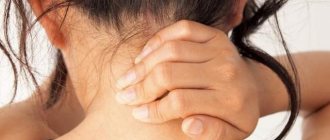
Atheroma develops in those places where there is an accumulation of a large number of sebaceous glands - in the area around the nose, on the eyelids, neck, scalp, chest, back and earlobe. Atheroma behind the ear is the most common of all types of atheroma, and, as a rule, affects both men and women equally.
The occurrence of atheroma of the earlobe is characterized by an unpredictable course and frequency of occurrence due to constant rubbing of the tumor with foreign objects - various hats, scarves, headphones, collars of shirts and blouses. In medical practice, cases of degeneration of a benign tumor into a malignant one are described, which occur in cases where proper surgical treatment is not carried out.
Diagnostics
Consultation with an otolaryngologist is necessary first of all to exclude ear, nose and throat diseases. If necessary, the doctor will refer the patient to specialists.
At your appointment, the ENT doctor will conduct a survey and examine the lump. To collect anamnesis, the doctor will check whether there is pain when pressing, whether there have been infectious diseases. In addition, he will take into account whether the bump behind the ear is soft or hard, whether it is pedunculated or tightly adjacent.
Next, the doctor will order a blood test to diagnose inflammation. To determine the cause of the tumor, an ultrasound or x-ray is performed.
What does atheroma behind the ear look like?
The reasons for the development of atheroma include the following factors:
- Disturbance of metabolic processes in the body.
- Harmful factors of external and internal ecology.
- Excessive sweating (hyperhidrosis).
- Diseases of the endocrine system and hormonal imbalance.
- Increased production of sebum by the glands and oily skin.
- Infection of the sebaceous gland duct (during ear piercing, for example).
- Presence of acne and seborrhea of the scalp.
Factors that can trigger the appearance of atheroma include improper and insufficient hygienic care and hypothermia of the body.
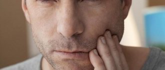
Tumor under the ear
Tumor under the ear: treatment, causes of occurrence
The appearance of a tumor under the ear may not cause serious harm to health, but in order to avoid possible complications, it is necessary to diagnose the type of this tumor by a doctor.
Tumors often develop due to malfunctions in the immune system, due to a lack of microelements and vitamins in the human body, and due to violations of hygiene rules. Tumors above the ear can also develop due to viral infections and injuries.
A tumor under the ear can occur due to various diseases: lymph nodes, lipomas, neoplasms, boils, nevi, papillomas, cysts, lyfangioma and hemangioma, cancer. Therefore, it is necessary to go to a specialist who will make a diagnosis and select treatment.
Typically treatment in these cases is with medications or surgery.
Pain remaining after treatment and hearing loss should be of concern.
Lymphadenitis
Due to the enlargement of the lymph nodes, a tumor may appear under the ear. The symptoms will be as follows: swelling, severe pain of a pulsating nature, localized on the left or right under the lower jaw when pressed.
Furuncle
This tumor is a purulent, severe inflammation, it differs in color and is easily palpated, the pain on palpation is very strong.
The boil is in the shape of a cone, with a purulent area in the center. The pus comes out on its own; there is no need to press on the boil and cause an infection.
Wen
The tumor occurs due to blockage of the sebaceous glands. As it swells and grows, the formation becomes painful on palpation. It can grow up to several centimeters. Requires timely treatment.
Swelling of the ear
Edema or perichondritis is a tumor localized in the auricle and in the center of the ear, in which the cartilage becomes inflamed. The disease is infectious. It usually causes swelling of the entire ear, severe pain and pus.
Lipoma
This type of tumor under the ear is harmless and benign. Its growth is slow, the lipoma does not cause pain, and its composition is soft. Often does not require treatment.
Cancer
Most often, a cancerous tumor under the ear is a sarcoma. It is formed from connective tissue. It looks like a dark colored knob-shaped lump. Usually painful. May contribute to the formation of pus and fistulas.
Symptoms to see a doctor may include:
- Strong pain;
- Throbbing pain;
- Enlarged lymph nodes;
- Increase in tumor size;
- Inflammatory processes;
- Presence of pus;
- Change in tumor color.
First, you need to come for an examination to a therapist or dentist. A knowledgeable specialist is needed to diagnose the cause of a tumor under the ear.
In order to make a diagnosis, you need:
- Undergo an ultrasound examination;
- To make an X-ray.
If lymphadenitis is present, it is necessary to eliminate the infection or overcome the virus that caused the inflammation. The patient is prescribed antiviral and immunomodulatory drugs.
If the lymph nodes are inflamed, then treatment will be with medication.
If there is a purulent process, the doctor prescribes antifungal medications and antibiotics. For hygiene of the sore spot, you need to use antiseptic drugs.
If a patient has a wen and needs to remove it, then first a cortisol injection is given. The operation will be performed with local anesthetics.
Small lipomas can be eliminated without surgery.
If cancer is suspected, treatment will be carried out by an oncologist.
Tumor formations can be removed either by surgery or by laser. When a tumor becomes malignant, surgical removal is usually done.
To avoid complications, timely treatment is necessary. If the compaction is small in size, then, as a rule, after its elimination, complications do not arise.
But even with an ordinary boil, severe inflammation can occur, and the boil must be removed by a surgeon.
Complications may be as follows:
- Inflammatory tissue processes;
- Tissue suppuration;
- Infectious diseases;
- Diseases of the mouth and ears;
- Inflammatory processes in nerve tissues;
- The appearance of malignant neoplasia.
To avoid complications, you need not to overcool or heat the diseased area, not to press on pus and fat formation, and not to physically influence them.
Pelvic tumor in women
To avoid the appearance of a tumor above the ear, you must adhere to the following rules:
- Dress according to weather conditions;
- Do not overcool;
- Avoid drafts;
- Treat viral infections in a timely manner;
- Try to stress less;
- Strengthen the body;
- Massage your face;
- Eat right.
If a small tumor appears above the ear, you should not let the disease progress, do not treat yourself, but contact a specialist at the Onco.Rehab clinic to make the correct diagnosis and select the necessary treatment program.
Symptoms of atheroma
Typically, atheroma reaches from 5 to 40 mm in size. In the initial stages, until infection sets in, atheroma may not bother patients. If suppuration occurs or the atheroma increases in size, the following symptoms may occur:
- Pain and redness in the area of atheroma.
- Swelling and increased body temperature.
- Burning and itching in the area where the atheroma is located.
- Feeling of free fluid when pressed.
In some cases, these symptoms of inflammation disappear within 10-15 days, after which the nature of the tumor changes. It becomes more dense, homogeneous and immobile. This occurs due to the replacement of the secretion of the sebaceous glands with connective tissue cells. At this stage, atheroma can give active growth, but can also remain in an unchanged position for several years. Metastasis to other organs is not observed in atheroma.
If a person’s immunity has enough strength to cope with the infection, then the atheroma spontaneously opens, as a result of which its contents drain from the cavity. This may be secretion from the sebaceous glands mixed with blood and pus. There is nothing wrong with this, except for the fact that after the autopsy, unsightly scars and scars remain on the body.
Of course, the presence of tumors such as atheroma of the earlobe or other area of the body should be an indication for surgical intervention and removal, since the tumor can grow into adjacent tissues and organs, as well as become inflamed as a result of a secondary infection.
The appearance of noticeable painful lumps
I was advised to contact Farid Firudinovich, as an ENT surgeon, by an ENT doctor from a private clinic in Vidnoye, recommending him as a doctor with God’s hands. I was looking for a doctor for surgery to correct the nasal septum (septoplasty). I suffered for 20 years. At the local clinic and hospital they told me that they didn’t see anything serious on the x-ray of the paranasal sinuses, and that fixing it would only spoil it. But I can’t breathe normally, either on my own or on hormonal sprays, my head hurts constantly. Dryness, runny nose, snoring. Antihistamines didn't help either. Coincidentally, my husband and I did a CT scan of the jaws for the dentist, and there the septum was clearly visible. And she looks terrible. I specifically compared it with my husband’s CT scan (photos in two projections before the operation are attached to the review). I went to a private clinic that I trust. Naturally, with such a curvature and lower shells - just straighten it. As a result, I came to the Sinai clinic for a consultation with F. F. Kurbanov, taking with me a CT scan of the sinuses. And it also shows that due to the curvature of the septum, polyps began to sprout in the right sinus, and there was some kind of swelling in both sinuses (I remember the names). In general, I arrived on time. The doctor told me all this, showed me, explained. A professional who is confident in his knowledge is very important. I felt that I trusted him. It was scary, of course, because I read everything on the Internet over the years. But the doctor is sure, and I am sure. I was afraid that there might be complications, perforation, for example. Everything there is so small and fragile! But I no longer wanted to go through compulsory medical insurance, since they didn’t see the obvious what they could do during the operation - only God knows. They didn’t send me for a CT scan to clarify the diagnosis; they didn’t see the problem visually either. But Farid Firudinovich saw enough even without a CT scan, the program with a CT scan took a long time to load, and he already told me what he saw: the left eardrum, for example, began to deform (retract). Well, the rest/main picture. In general, we discussed the date, passed all the tests and arrived for the operation in two weeks. General anesthesia, a good anesthesiologist (special thanks to him, unfortunately, I forgot his last name) and an assistant, and an hour later I was already in the ward, waking up from sleep with a bandage on my nose. A couple of hours later the doctor came, cheered me up, told me how things were going and what would happen next. We were in touch all the time, which is also important. It was a hard night, it’s hard to sleep when your nose is stuffed with tampons, but it’s all over, after a week the stitches were removed. And I'm finally breathing! On her own, without drops, pills or headaches. My nose still hurts, but these are minor things; every day I feel more and more like a human being. I am writing a month after the operation. And it will only get better! I express my deep gratitude to Farid Firudinovich Kurbanov for his professionalism, experience and human attitude. Thank you! I recommend that anyone who is hesitant to take such a step should not delay. Quality of life is very important. Also, I want to thank the clinic staff for their care!
Liked: Professionalism, experience, human attitude.
I didn’t like it: In the Sinai clinic itself, the general administrative organization is a bit difficult, but the professionalism of the doctors makes up for this drawback.
Treatment and prevention of atheroma
Treatment of atheroma is predominantly surgical, with removal of the cavity along with its contents. To do this, a small skin incision is made on the body, through which access to the atheroma is provided. After removing the contents, the cavity is washed, and if necessary, a turunda is inserted for several days to facilitate the release of blood, pus and fat.
The operation is performed not in a hospital setting, but on an outpatient basis. Local anesthesia is used for pain relief. For small atheromas, sutures are not applied, since the skin incision heals on its own after 5-6 days. For large atheromas, cosmetic sutures are applied, which are regularly processed after 1-2 days. The operation itself is painless for the patient, but the danger is that without eliminating the causes that caused the appearance of atheroma, there is a high probability of relapses (almost 50% of all cases). That is why it is so important not only to eliminate the cause of atheroma, but also to take measures to prevent the disease. These include personal hygiene measures and early consultation with a doctor. It is not recommended to treat atheroma on your own.
Does your child need specialist help?
You can make an appointment with a doctor by phone
8-(4822)-33-00-33
or using the online registration system on the website
To make an appointment with a doctor
Read also: Dermatitis in children
Parotid fistula
Occurs when there is an intrauterine disorder in the formation of the hearing organ. A fistula is a canal that opens behind the ear in the area of the cartilaginous part of the auricle, and the other end goes into the oral cavity, middle ear or neck area. A lump at the site of the fistula outlet is formed only when the tissues of the canal become inflamed.