Mechanical damage to a tooth due to injury without violating its integrity is called a bruise. This type of injury is common in children and adults, especially athletes. According to statistics, among all injuries to primary teeth, bruises account for 2.5%; bruises of permanent teeth account for 1.5% of the total number of traumatic injuries in the permanent dentition.
Even if the tooth looks unchanged after a bruise, it is necessary to visit a dentist to prevent the development of complications associated with the injury.
Symptoms
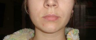
A strong blow to a tooth is accompanied by damage to periodontal tissue, and some fibers and small blood vessels rupture. There are no visible structural damage to the tooth; upon visual inspection, it appears intact. After a bruise, the tooth remains motionless, and minor mobility is rarely observed. The gums in the area of the injured tooth swell.
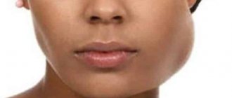
In the first hours after the impact, patients experience pain in the tooth, which intensifies when biting; the pain is aching in nature. The tooth feels high, and slight bleeding may occur from under the gum near it. When a bruise occurs, the neurovascular bundle of the tooth can be damaged, that is, the pulp is injured, and hemorrhage occurs in the pulp chamber, and the enamel becomes pink in color. A severe bruise can lead to the death of the pulp.
Often, when a bruise occurs, cracks appear on the tooth enamel, which can only be detected with a special examination.
Causes
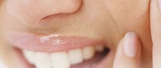
There are many reasons for a tooth bruise. The most common is a strong direct blow to the jaw. Athletes often receive such injuries; they are almost always the consequences of accidents. It is impossible not to notice it, since after a blow the front tooth hurts, and the pain is usually sharp.
There is another reason - the so-called chronic jaw injury. The peculiarity of such injuries is that they are insignificant in their impact, but a person is constantly exposed to them. Examples include some human habits: biting nails, holding a pen or pencil in the mouth, cracking nuts, etc. Under the influence of such seemingly insignificant factors, the enamel becomes thinner and microchips form on it. The result of these innocent habits is tooth loss.
Injury can also occur during dental procedures performed by an inexperienced dentist or using old equipment. Any violation of the treatment or removal technique can lead to damage to healthy tissue. That is why our clinic employs experienced, highly qualified specialists. We are responsible for the health of our patients.
Diagnostics
- Patients with a tooth injury are referred for x-ray diagnostics to exclude a root fracture. To obtain information using targeted radiography, it is sometimes necessary to take several images from different angles, which is undesirable for the patient.
The most accurate information about the condition of the roots is provided by computed tomography - an x-ray examination method that allows you to obtain a three-dimensional image of the tooth. The examination results are displayed on the computer screen and must be transferred to a CD or USB flash drive.
An X-ray examination of a tooth bruise reveals a slight widening of the periodontal fissure.
- The condition of the pulp after injury is monitored using EDI - electroodontodiagnosis. The method consists in determining the reaction of the nerve endings of the pulp to the influence of electric current. The level of electrical excitability of the pulp depends not only on its condition, but also on the degree of formation of the tooth root.
The examination is carried out 2 or 3 days after the injury, since on the first day the pulpal response may be reduced due to traumatic neuritis. Be sure to perform an electrical test on adjacent healthy teeth to compare sensitivity levels. 3-4 weeks after the injury, EDI is repeated.
- Another method of examination for a tooth bruise is transillumination, the essence of which is to pass a beam of cold light through the tooth and evaluate shadow formation. If there are cracks in the enamel after an impact, they will be clearly visible in the stream of light; The technique also helps to detect pulpitis. In modern clinics, all dental units are equipped with light guides for transillumination examination.
Providing first aid for broken, knocked out and dislocated teeth
One of the most common injuries is a fracture. The types of fracture are as follows:
- Chipping of the enamel of a tooth crown.
- Partial tooth destruction without exposing the pulp.
- Chipped crown exposing dental pulp.
- Tooth root injury.
Additionally, the mouth and gums, the jaw itself and the bone nearby can be damaged. If this happens, the first thing you need to do is calm down - the accompanying injuries create additional stress, which significantly interferes with the doctors and the victim himself. In addition, you should try to find broken fragments and leave them in the water until you see a doctor. The situation does not require an urgent visit to the dentist, so you can make an appointment a little later, for example the next day, but try not to involve the damaged tooth until it is restored.
As for diagnosis, everything is simple - the doctor will first conduct an examination. To detect the risk of root injury, the victim is sent for x-rays and electroodontometry. In this way, the condition of the pulp (tooth pulp) is checked. The goal of treatment is to restore the crown of the tooth. If the fracture led to pulpitis, the patient will be treated with subsequent restoration of the pulp. If an x-ray shows that there has been root trauma, complete extraction of the tooth and subsequent placement of a post is often recommended.
The second most common injury is dislocation. This is a partial or complete loss of connection between the tooth and the socket. Dislocation occurs:
- Partial. The tooth moves greatly or slightly, but does not fall out completely.
- Full. The tooth falls out.
- Impacted, in which the dental crown is driven into the gum.
If a temporary tooth has been completely dislocated (falls out of its socket), do not under any circumstances try to return it to its place - you may damage the new permanent one or its rudiment. And in order to look for and insert a molar into place, you will have only 20 minutes. If you manage to do this, the chance of re-engraftment becomes much greater. What is the procedure? As soon as you have discovered the lost tooth, take it with clean hands (it is also advisable to wash it) by the white part, without touching the root, and return it to the hole, orienting parallel to the neighboring teeth. When you return the lost item to its place, bite on a gauze pad or a piece of cloth, such as a scarf.
There are situations when the tooth cannot be inserted back, for example, if the victim is unconscious or the socket is severely damaged. In this case, place the found tooth in milk or saline solution. You cannot put it in water or any other liquid; you can hold it in the victim’s mouth, but there is a risk of swallowing.
Unlike a fracture, in a situation with complete dislocation, it is necessary to urgently visit a specialist for examination and subsequent treatment.
For partial dislocation, the procedure is slightly shorter. It is important to try not to spit (or do it as little as possible) and under no circumstances take out the tooth. If it prevents your mouth from closing, carefully insert it back in with clean hands, focusing on the adjacent teeth. To hold it in place, bite down on gauze or any other piece of tissue.
In the case of an impacted dislocation, first aid is practically useless; the only option is an emergency visit to the dentist.
Panoramic radiography
How are dislocations identified and treated? The doctor first examines and records the complete absence or displacement of a tooth. Next, the victim is sent for an X-ray and radiovisiographic examination, where the location and condition of the alveoli are clarified. In case of incomplete dislocation, the patient is recommended treatment aimed at preserving the tooth and fixing it with the help of special mouthguards and other structures. In addition, medications are recommended to prevent inflammation and infection in the socket, for example antibacterial ones.
If, after complete dislocation, the tooth does not take root, the specialist is forced to resort to prosthetics and implantation. In case of impacted displacement, you can either remove the affected tooth and replace it with an implant, or perform immobilization using local anesthesia.
A knocked out tooth is a dislocated or broken crown due to a mechanical injury. For example, a blow to the jaw caused the victim to completely dislocate or chip the dentin, exposing the pulp. Depending on the injury detected, you can provide first aid as described above. That is, if a tooth falls out, find it, try to put it back in, or place it in saline before visiting a doctor.
Complications after injury
With bruises, the prognosis is usually favorable, but in some cases the injury can lead to the following complications:
- Darkening of the enamel. After a bruise, the cause of darkening is hemorrhage into the pulp chamber: the pink color of the tooth darkens over time, the enamel acquires a brownish or gray tint. Depulped teeth tend to darken due to the fact that metabolic processes in them stop, the teeth become “dead”, and the enamel becomes dull.
- The death of the pulp leads to the development of pulpitis: the pulp decomposes, inflammation develops in the tooth, which without treatment turns into periodontitis.
- Periodontitis is inflammation of periodontal tissue. It can be: post-traumatic, occurring a short time after the injury; chronic, develop as a consequence of pulpitis. When a purulent infection occurs, there is a high risk of tooth loss and purulent blood poisoning.
- The appearance of a post-traumatic odontogenic cyst at the root apex during the development of post-traumatic periodontitis.
- Stopping the development of roots of permanent teeth in children.
- In case of injury to milk teeth, the following are possible: disruption of the formation of the rudiments of permanent teeth, their death.
What causes bruises?
Jaw injury may result from:
- falls;
- blow;
- fights;
- children's games;
- accidents, etc.
The severity of the injury is determined taking into account the following factors:
- features of the surface or object that caused the injury;
- impact intensity;
- affected area of the face;
- condition of bone tissue before the incident.
The strength of the bruise and possible complications depend on the listed indicators. Regardless of the severity, it is important to see a doctor to evaluate the condition and prescribe appropriate treatment. This will avoid unforeseen consequences and quickly restore the damaged area.
Treatment
- For mild bruises, treatment consists of resting the tooth for 3-4 weeks by reducing the load during chewing: the menu includes soft and semi-liquid foods, and a blender is used to grind hard foods.
- To ensure rest for baby teeth, temporary bite separation with the help of mouth guards is used; if a permanent tooth is bruised, splinting is performed. The splint allows you to immobilize an injured tooth and redistribute the load during chewing onto healthy teeth.
- If the pulp dies due to an impact, the tooth cavity is opened and the pulp is removed, after which the root canals are filled and a permanent filling is installed. If the crown of a tooth darkens, it can be whitened.
- When a baby tooth is bruised, grinding of the cutting edge of the crown of the antagonist tooth is used to prevent tooth contact and reduce pain. This method is not used for permanent teeth.
- To relieve pain, it is recommended to take an anesthetic tablet (ibuprofen, ketorolac, nimesulide) and apply an anesthetic gel (Dentol, Kamistad) to the gums around the tooth.
- When swelling of the soft tissues of the face accompanies a bruise, cold compresses are applied: a plastic bottle with cold water (not lower than +4°C) is wrapped in a cloth and held on the area of swelling for 15-20 minutes.
- A course of magnetic laser therapy is carried out - a combined effect on injured tissue of a magnetic field and low-intensity laser radiation. The method helps improve healing processes, relieves swelling and inflammation. The course consists of 10 daily procedures lasting 5 minutes.
- UHF therapy is indicated to accelerate tissue regeneration.
Prevention
To prevent dental injuries, you must:
- Observe safety precautions at work, arrange the workplace and carry out potentially hazardous work in accordance with established rules.
- Properly equip playrooms for children and supervise children while playing.
- Follow traffic rules and use seat belts when traveling, as even minor emergency braking of the car carries a high risk of injury.
- Avoid conflict situations that lead to fights.
- Ensure the safety of sports games using special equipment (helmets, masks, dental guards).
- Carefully select sports grounds: they must comply with safety requirements.