Hemostatic (antihemorrhagic, hemostatic) drugs are a group of drugs that are used to stop bleeding (hemorrhages) and prevent excessive blood loss.
Normally, blood is a fluid liquid - a suspension of formed elements (erythrocytes, leukocytes, platelets) in plasma. However, during bleeding, blood is able to change its rheological (fluid) properties - it becomes excessively thick and viscous, and then solidifies at the site of damage to the vessel wall (coagulates), forming a clot (thrombus). The thrombus closes the lumen of the wound and thereby prevents further bleeding and loss of blood from the body.
In case of extensive bleeding or in diseases that are accompanied by a violation of the formation of blood clots, the blood does not have time or is not able to clot in time. This leads to excessive blood loss, and then to shock, oxygen starvation of organs (especially the brain and heart) and the development of various kinds of negative consequences, including death. In this case, hemostatic drugs are used.
Indications for use
Hemostatic drugs are used for conditions associated with blood clotting disorders and bleeding.
Fibrinolysis inhibitors are used to stop severe bleeding during operations (especially on the heart, lungs, large vessels), extensive injuries, birth hemorrhages, reduced blood clotting in liver diseases, as well as in case of overdose of fibrinolytic drugs (fibrinolysin, streptokinase, alteplase, urokinase, tenecteplase and etc.).
Aprotinin is additionally used for diseases of the pancreas: acute pancreatitis (including alcoholic), severe injuries and pancreatic cancer.
Vitamin K preparations are used for bleeding associated with vitamin K deficiency in the body: hemorrhagic syndrome of newborns, liver diseases (cholelithiasis and obstructive jaundice) and intestinal diseases (enterocolitis, Crohn's disease). In addition, vitamin K preparations are used for bleeding caused by an overdose of indirect anticoagulants - warfarin, acenocoumarol, phenindione.
Herbal hemostatics are indicated for uterine bleeding (menorrhagia), minor bleeding from small vessels of the stomach and intestines.
Hemostatic drugs for local use are used to stop capillary and parenchymal bleeding (from internal organs) during open abdominal operations.
Etamsylate is used for the prevention and treatment of surgical bleeding, as well as for uterine, nosebleeds, bleeding gums due to periodontal disease, and gingivitis.
Eltrombopag and recombinant thrombopoietin are indicated for the treatment of idiopathic thrombocytopenic purpura - a chronically low level of platelets in the blood, which is dangerous due to frequent bleeding - when the spleen is removed.
Emicizumab is used to treat hemophilia, an inherited disease associated with blood clotting disorders and life-threatening bleeding.
Clotting factor drugs are used for severe, extensive bleeding, as well as for the treatment of hemophilia.
Types of hemostatics
Hemostatic agents are divided into resorptive and local agents. Hemostatic effect of hemostatic agents
- resorptive action occurs when drugs of this group are introduced into the blood, and
- local action - in direct contact with bleeding tissues.
Based on their effect on the hemostasis mechanism, hemostatic agents of both groups are divided into specific and nonspecific agents.
Specific products contain blood clotting components or substances that directly affect coagulation. Specific resorptive drugs include platelet-rich plasma, erythropoietin, which stimulates the formation of red blood cells, etc.
Nonspecific agents have an indirect effect on the blood coagulation system - adrenaline (or epinephrine) locally, which constricts blood vessels, hydrogen peroxide.
Depending on the nature of the bleeding, the characteristics of the patient and the location of the wound on the body, the following are used as local hemostatic agents in modern medicine:
- synthetic or fibrin glue (sealant);
- gelatin-based products;
- oxidized reduced cellulose.
Classification of hemostatic drugs
Hemostatic drugs are classified into:
- fibrinolysis inhibitors: amino acids (tranexamic acid, aminocaproic acid), protease inhibitors (aprotinin);
- vitamin K preparations: menadione, phytomenadione;
- herbal preparations: liquid extract of water pepper, nettle leaves, yarrow herb;
- hemostatic agents for local use: thrombin, fibrinogen, Ambien, calcium chloride;
- other hemostatic drugs for systemic use: etamsylate, eltrombopag, emicizumab, recombinant thrombopoietin.
In addition, preparations of proteins that regulate blood coagulation - coagulation factors II, VII, VIIa, VIII, IX, X and their combinations, von Willebrand factor, nonacog alpha - have a hemostatic effect.
Vital amputation using ferrous sulfate-based drugs in pediatric dentistry
At the present stage of development of dentistry, treatment methods that involve preserving the vitality of the pulp and restoring its functions are becoming increasingly widespread. This is especially true when treating pulpitis of primary teeth with unformed roots, when it is important to preserve the pulp to complete root growth.
Treatment of pulpitis of primary teeth in children should be timely and adequate. Temporary teeth play an important role in the formation of dentition and jaws, timely eruption and correct placement of permanent teeth, normal development of the functions of the dental system, while early removal leads to a failure of normal formation processes.
The choice of treatment method for pulpitis depends on the diagnosis of the disease, the age of the child and the possibility of establishing psychological contact with him. With the development of new technologies in dentistry, there has been a tendency to increase the frequency of use of vital treatment methods, which have a number of advantages: the number of patient visits to the dental office is reduced, and the use of drugs with a resorptive effect is eliminated. Among the vital methods, the most widely used method is vital amputation (pulpotomy), based on morphological differences in the structure of the root and coronal pulp.
When treating pulpitis of primary teeth using the vital amputation method, preparations based on 35% formocresol and 2% glutaraldehyde are used. However, according to studies, in many cases the use of these drugs was unsuccessful. There is also an opinion about the possible cytotoxic and mutagenic effects of formocresol. In this regard, there was a need to search for new, more effective drugs for the treatment of pulpitis of primary teeth. Today these are preparations based on ferrous sulfate (ViscoStat, Astringedent).
According to the literature (R.E. McDonald, D. Avery, M.S. Duggal), treatment of primary teeth using the vital amputation method using 35% formocresol was unsuccessful in approximately half of the cases: during the study period (12 months) Children in this group complained of pain in the area of the treated tooth, changes were found on the radiograph in 5-6 teeth out of 50 (destruction of bone tissue in the periapical region, in the root furcation zone). Accordingly, the percentage of successful treatment was 89%. When using ferrous sulfate preparations during the entire observation period (1 year), only in one tooth out of 50 changes were detected on the radiograph: destruction of bone tissue in the periapical region and in the root furcation zone. Accordingly, the success of endodontic treatment was 98%. Thus, the percentage of successful treatment when using ferrous sulfate preparations is much higher than when using formocresol-based preparations.
Materials and research methods
Vital pulp amputation is a method of removing inflamed and infected coronal pulp in order to preserve the vital root pulp.
The method of vital amputation consists in removing the coronal pulp, rich in cellular elements, and preserving the root pulp, which ensures the normal physiological course of the growth and development of the temporary tooth and its surrounding structures. The use of pulpotomy is based on differences in the structure of the coronal and root pulp of teeth: the coronal pulp has a looser structure due to the large number of vascular anastomoses and the presence of cellular elements. Consequently, during inflammation, more significant changes in microcirculation occur in the coronal pulp. In the root pulp there are practically no cellular elements, connective tissue fibers predominate, therefore, in the root pulp tissue swelling is less pronounced, there is no compression of blood vessels and the phenomena of congestive hyperemia. This structural feature allows amputation of the coronal pulp with subsequent preservation of the function of the viable root pulp.
Ferrous sulfate preparations used for vital amputation in primary teeth are ViscoStat and Astringedent.
ViscoStat is a viscous hemostatic gel containing 20% ferric sulfate, which has a gentle coagulating effect on soft and hard dental tissues. Hemostasis is achieved mainly due to the formation of coagulation plugs (thrombi) in the lumens of the capillaries. This product is used to stop capillary bleeding and ensures high-quality pulpotomy of primary teeth in one visit.
The effectiveness of the ViscoStat solution (Fig. 1) increases significantly when using the special Dento-Infusor device, since the effect of hemostatic agents depends on the method of their application.
Rice. 1. ViscoStat - viscous, containing 20% ferric sulfate, hemostatic gel, has a gentle coagulating effect.
Using a brush at the end of the Dento-Infusor nozzle, the hemostatic is “rubbed” into the capillaries, which leads to the formation of blood clots. This also removes blood clots outside the lumens of the capillaries. This procedure protects the formed vascular blood clots from being removed when washed off.
As a result, we have a clean, dry surface.
Description of clinical observations
Clinical case: patient A. A. Frolov, 4.5 years old. He complained of pain in the area of the 74th tooth that occurred during meals. When examining 74 teeth, a carious cavity filled with softened dentin was discovered. The tooth cavity was opened at one point, touching which caused pain. The color of the tooth was not changed. Percussion is painless, the mucous membrane and the transitional fold in the projection of the roots of the causative tooth are without pathology. As a result, a diagnosis of chronic fibrous pulpitis of 74 teeth was made and treatment was carried out using the vital amputation method using ViscoStat gel according to the following scheme:
Local anesthesia: Ultracaine D-S 1.7 ml, 1:200,000.
Preparation of a carious cavity and opening of the cavity of 74 teeth. Necrotic dentin is removed from the walls and bottom of the carious cavity. It is important to carefully prepare the carious cavity before opening the pulp chamber. The cavity is then opened wide to create a direct transition into the tooth cavity. Resection of the arch of the dental cavity is carried out with a sterile bur. In temporary molars, after opening the hole with a ball-shaped bur, the overhanging edges are cut off with a cylindrical bur. This manipulation requires the doctor to know the topography of the pulp chamber in order to prevent perforation and provide direct access to the orifices of the root canals. Antiseptic treatment (chlorhexidine bigluconate 0.05%).
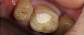
Removal of coronal pulp (pulpotomy) using a ball bur at low speed. Next, the mouths of the root canals are processed, forming additional platforms to relieve excess pressure from the root pulp. Then a deep pulpotomy is performed with a sterile carbide bur on an extended stem (Fig. 2).
Rice. 2. Pulpotomy was performed.
Using the Dento-Infusor tip, ViscoStat solution is applied to the orifice pulp. Hemostasis is achieved within 10-30 seconds.
During the process of rubbing the solution, additional water is sprayed so that coagulation clots do not stick to the treated tissue. The work area is thoroughly rinsed and cleaned with a saliva ejector. The amount of hemostatic required for one tooth is 1/3-1/2 of the volume of the tooth cavity. After the bleeding has stopped, the ostial pulp is covered with a brown scab, there is no bleeding (Fig. 3, 4).
Rice. 3. Application of ViscoStat gel to the canal orifices.
Rice. 4. Coagulated ostial pulp.
Applying a thin layer of zinc oxide eugenol cement to the treated tissue and the bottom of the pulp chamber. Next, an insulating gasket made of glass ionomer cement is made (Fig. 5).
Rice. 5. Placement of zinc oxide eugenol cement and a gasket made of glass ionomer cement on the bottom of the tooth cavity.
Carrying out tooth restoration (Fig. 6, 7).
Rice. 6. Tooth restoration.
Rice. 7. The final appearance of the restoration using photopolymer material.
Conclusion
Ferrous sulfate preparations appeared on the dental materials market relatively recently. In the course of research and clinical experiments, the physical and mechanical properties of ferrous sulfate preparations were studied, and their greater effectiveness was proven over formocresol preparations (98% and 89%) in the treatment of pulpitis of primary teeth in children using the vital amputation method.
The percentage of successful treatment with ferrous sulfate preparations is much higher than with formocresol-based preparations. The use of ferrous sulfate preparations (ViscoStat) makes it possible to quickly stop bleeding and ensure high-quality pulpotomy of primary teeth in one visit, avoiding complications and the need for repeat visits for patients.
Literature
- V. K. Leontiev, prof. L.P. Kiselnikov. Pediatric therapeutic dentistry.
National leadership. - M.: GEOTAR-Media, 2010. - Dentistry of children and adolescents /
edited by Ralph E. MacDonald, Daveig R. Avery (translation by T. V. Vinogradova). - M.: MIA, 2003. - M. S. Duggal, M. E. J. Curzon, S. A. Fail, K. J. Toumba, A. J. Robertson. Treatment and restoration of baby teeth
(translation by T. V. Vinogradova). - M.: MEDpress-inform, 2006. - L. S. Persin, V. M. Elizarova, S. V. Dyakova. Pediatric dentistry.
- Medicine, 2005. - R. Beer, M. A. Bauman, Andrei A. Kielbasa. Illustrated guide to endodontology.
- M.: MEDpress-inform, 2008. - T. V. Vinogradova. Guide to pediatric dentistry.
- Medicine, 1987. - M. V. Kuryakina .
Therapeutic dentistry for children . - M., Nizhny Novgorod: NGMA, 2001. - A. A. Kolesov. Pediatric dentistry.
- M., 1991.
A complete list of references is in the editorial office.
Causes
Reasons for the development of alveolar bleeding:
- systemic disorders;
- damage to the alveoli, interradicular septum;
- tissue ruptures, crushing, other damage to the soft tissue of the gums;
- premature cessation of the action of an anesthetic drug containing adrenaline, which causes vasoconstriction;
- inflammatory processes in the area of tooth extraction;
- alveolitis, purulent melting of tissues;
- vascular damage%
- disruption of the integrity of the blood clot due to mechanical intervention due to the fault of the patient;
- hemangioma (vascular erosive tumor).
Systemic disorders that can lead to the development of alveolar bleeding include:
- hypertension;
- infectious diseases;
- scurvy;
- vascular pathologies;
- hemorrhagic diathesis;
- blood diseases that negatively affect clotting;
- hepatitis.
