Author of the article:
Soldatova Lyudmila Nikolaevna
Candidate of Medical Sciences, Professor of the Department of Clinical Dentistry of the St. Petersburg Medical and Social Institute, Chief Physician of the Alfa-Dent Dental Clinic, St. Petersburg
The human oral cavity has its own microflora and its own specific diseases arise. Let's talk about one of them - fibrous epulis of the gums: what kind of disease it is, what are the causes of its occurrence, how is the treatment carried out.
What it is
Epulis is a tumor-like neoplasm that develops on the alveolar process of the jaw. It also has other names - epulid, supragingival, giant cell granuloma. More often it is localized in the area of incisors, canines and small molars of the upper jaw, less often the tumor is detected in the lower jaw. The growth has a round or irregular shape and a wide stalk; the size of the formation varies from 3 mm to 5 cm.
Despite the fact that such a tumor looks frightening, it practically does not cause concern to the patient, unlike, for example, a burn, with the exception of a violation of aesthetics and the sensation of a foreign body in the oral cavity. Large tumors may cause difficulty chewing and swallowing food.
If epulis is accompanied by bleeding, infection may occur, which will lead to complications.
The disease mainly affects adults, but the neoplasm can also appear in children; in the latter, it occurs during teething. Studies have shown that women are more susceptible to developing epulis than men.
Prevention
There are a number of rules, the observance of which can prevent the appearance and further development of epulis:
- maintaining oral hygiene,
- avoiding, if possible, situations leading to damage to teeth, as well as soft periodontal tissues.
If there is any suspicion of the presence of epulis, you should urgently seek help from a professional of the appropriate profile. This is the only way to prevent more serious, including irreversible, consequences with a high degree of probability.
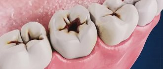
Varieties of epulis
Conventionally, there are 3 types of epulis:
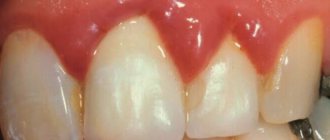
- Fibrous. The neoplasm is dense, the basis is coarse fibrous tissue. Characterized by slow growth. There is no pain or bleeding.
- Angiomatous. The tumor contains a large number of blood vessels and is localized mainly in the lateral parts of the jaw. With this form of the disease, bleeding is observed even with slight pressure, for example, with a toothbrush or food. When pressed, the tumor is painless and has a soft consistency.
- Giant cell. The tumor can reach large sizes, displacing teeth that are located in the epulis growth zone. The mucous membrane of the epulis is bluish-red in color. On palpation, the neoplasm is painless, but may bleed moderately if injured.
Depending on the nature of the process, epulis can be benign or malignant. In the first case, the neoplasm is characterized by slow development and, as mentioned above, in most cases does not cause pain. The tumor size in this case rarely exceeds 10–20 mm. When the process becomes malignant, rapid tissue growth is observed. In this case, the process is often accompanied by pain, bleeding gums and affects the root canals of neighboring teeth.
Why does epulid develop?
Of one hundred percent of all cases of epulis development, eighty are provoked by trauma to the mucous membrane. The alveolar ridge can be damaged by a splinter from a cracked or chipped tooth, an improperly installed filling, a removable denture, or braces. An incorrect bite can also cause damage.
Among other causes of epulide on the gums, the following are often found:
- injuries of the maxillofacial apparatus as a result of falls, fights, accidents,
- changes in hormonal levels: for example, with diabetes, pregnancy, menopause, adolescence,
- the presence of plaque, hard deposits and stone on the teeth,
- thermal and chemical burns,
- decreased local immunity,
- in childhood – teething.
Constant irritation of the mucous membrane leads to the active proliferation of its cells and the appearance of a tumor.
Diagnosis and treatment of epulis
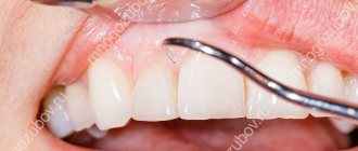
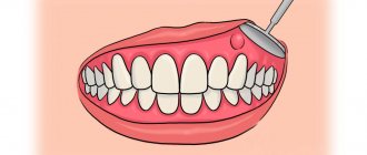
Diagnosis of epulis comes down to collecting complaints, clinical examination, radiography and histological examination of the material.
The primary task in making such a diagnosis is to eliminate the provoking factor: professional cleaning of dental plaque, treatment of caries and its complications, replacement or correction of orthopedic structures according to indications. In fibromatous and angiomatous forms, dynamic observation is indicated, since after sanitation of the oral cavity and elimination of the causes of the disease, the tumor may decrease in size until it disappears completely.
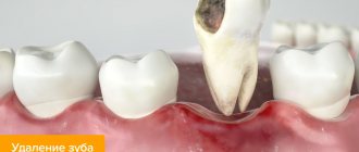
Treatment of epulis is carried out surgically. The growth on the gum under local anesthesia or general anesthesia is excised with a scalpel or laser within the healthy tissue along with the periosteum. The second method is preferable, since the laser simultaneously coagulates the vessels and stops bleeding. With giant cell epulis, the area of bone tissue involved in the process is also removed. To do this, use a bur or cutter. The wound is closed with gauze with an iodoform mixture or a formed mucoperiosteal flap. If necessary, the material is sent for histological examination.
Intact teeth in the area of tumor localization must be removed with a high degree of mobility and severe exposure of the roots. If the bone lesion is extensive or epulis recurs, partial resection of the alveolar part along with the teeth is performed.
To speed up the healing of a fresh wound, the doctor prescribes compresses with medicinal ointments and rinses to the patient.
Forecast
If the causative factor that provoked the appearance of granuloma is not eliminated in a timely manner, the consequences can be very disastrous. For example, the probability of recurrence is very high even if surgical excision was performed.
Provided a comprehensive diagnosis is carried out, as well as qualified treatment, the prognosis in the vast majority of cases turns out to be quite favorable.
In addition, preventing the situation from worsening in the future is possible through proper prevention. This means preventing injury to the gums, competently performed dental prosthetics, thorough fitting of orthodontic appliances, as well as a systematic qualified examination in a professional dental office.
Preventive actions
Monitoring the condition of the teeth and oral cavity will reduce the likelihood of tumors.
- Timely sanitation of the oral cavity. Visiting the dentist for a preventive examination and professional hygiene twice a year will prevent the growth of caries, the appearance of defects in fillings and the formation of tartar, leading to gum injury and the appearance of epulis.
- Prevention of gum injury. If systematic injury to soft tissue occurs as a result of poorly fitted orthopedic or orthodontic structures, consult your doctor about this problem. He will adjust the crowns or dentures.
If these preventive measures are followed, the prognosis is favorable and epulis does not recur.
We hope that our article about epulis will be for informational purposes only. And if you want to soothe your gums, make them strong and strong, try the unique two-component mouth rinse ASEPTA ACTIVE.
This is the only rinse with a combination of chlorhexidine + benzydamine for the treatment of inflammatory periodontal diseases. The product has a combined effect: antimicrobial, anti-inflammatory and analgesic. Instant anesthetic effect allows you to quickly reduce pain.
Dentists' recommendations
Epulis is a gum tumor that poses a great danger to the health of teeth and the entire body. Treatment of the pathology is complex and lengthy, but even with high-quality treatment, the risk of relapse cannot be ruled out.
It is not always possible to prevent the occurrence of epulis on the gums of a child or an adult, but with timely consultation with a specialist, complications, complex treatment, long recovery and aesthetic defects that remain after surgery can be avoided.
The best prevention is to visit the dentist every 6 months. If you are concerned about discomfort and a feeling of rubbing of the mucous membranes or gums, you should immediately consult a dentist, without waiting for a tumor or other pathologies to occur.
Clinical researches
Repeated clinical studies have proven that the two-component mouth rinse ASEPTA ACTIVE more effectively combats the causes of inflammation and bleeding compared to single-component rinses - it reduces inflammation by 41% and reduces bleeding gums by 43%.
Sources:
- Clinical and laboratory assessment of the influence of domestic therapeutic and prophylactic toothpaste based on plant extracts on the condition of the oral cavity in patients with simple marginal gingivitis. Doctor of Medical Sciences, Professor Elovikova T.M.1, Candidate of Chemical Sciences, Associate Professor Ermishina E.Yu. 2, Doctor of Technical Sciences Associate Professor Belokonova N.A. 2 Department of Therapeutic Dentistry USMU1, Department of General Chemistry USMU2
- The effectiveness of the use of Asept “adhesive balm” and Asept “gel with propolis” in the treatment of chronic generalized periodontitis and gingivitis in the acute stage (Municipal Dental Clinic No. 4, Bryansk, Kaminskaya T. M. Head of the therapeutic department Kaminskaya Tatyana Mikhailovna MUZ City Dental Clinic No. 4, Bryansk
- Study of the clinical effectiveness of treatment and prophylactic agents of the Asepta line in the treatment of inflammatory periodontal diseases (A.I. Grudyanov, I.Yu. Aleksandrovskaya, V.Yu. Korzunina) A.I. GRUDYANOV, Doctor of Medical Sciences, Prof., Head of Department I.Yu. ALEXANDROVSKAYA, Ph.D. V.Yu. KORZUNINA, asp. Department of Periodontology, Central Research Institute of Dentistry and Maxillofacial Surgery, Rosmedtekhnologii, Moscow
- The role of anti-inflammatory rinse in the treatment of periodontal diseases (L.Yu. Orekhova, A.A. Leontyev, S.B. Ulitovsky) L.Yu. OREKHOVA, Doctor of Medical Sciences, Prof., Head of Department; A.A. LEONTIEV, dentist; S.B. ULITOVSKY, Doctor of Medical Sciences, Prof. Department of Therapeutic Dentistry of St. Petersburg State Medical University named after. acad. I. P. Pavlova
Causes of occurrence (Etiology)
In general, the etiology of this disease is quite natural and therefore easily explainable. Such a tumor is formed as a result of the influence exerted by local irritating factors. Injuries to the gum margin by destroyed walls of dental units, unpolished prosthetic bases, metal clasps, protruding components or ribbed edges of orthodontic structures are fraught with the progression of chronic inflammation of the productive type. Predisposing factors leading to tumor formation include pathologies of occlusion, hormonal imbalances, and narrowing of the dentition.
Giant cell
The name is scary, isn't it? And rightly so, because giant cell granuloma is considered the most dangerous type of disease, which threatens to develop into a malignant tumor. Epulis of this type is most often found in middle-aged and elderly women. What worries patients: the rapid increase and growth of a hard and very dense neoplasm to the touch, its bluish or dark brown tint, pain when pressing and chewing, bleeding.
Giant cell form of epulis
Is it possible to use traditional medicine?
Treatment with folk remedies is allowed during the rehabilitation period after removal of the epulis. Basically, rinsing the gums with solutions that have an antiseptic, healing, and anti-inflammatory effect is used. For this purpose, decoctions and water infusions of medicinal plants are used: chamomile, calendula, sage. Procedures are carried out up to 5-7 times a day, especially after meals. In addition to decoctions of beneficial herbs, solutions of baking soda and salt are used (one teaspoon per 200 ml of warm water).
Reviews
Epulis in medicine is classified as a tumor neoplasm, but there is no need to be afraid of this disease. Unattractive appearance and aesthetic changes are perhaps the biggest problem during the course of the disease.
However, for many, the tumor causes a lot of inconvenience and is difficult to treat with medication. In addition to the information in this article, readers will be interested to know how the operation is performed and how long the recovery period lasts.
What medications helped reduce the tumor or get rid of it completely? Share your experience and impressions in the comments.
If you find an error, please select a piece of text and press Ctrl+Enter.
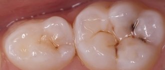
Fibrous
Fibrous epulis is the safest of all existing ones. When it appears, the damaged gum is practically indistinguishable from the rest of the mucous membrane. The tumor itself is quite dense, most often localized externally, that is, vestibular, noticeable in the smile area. It looks like a small ball or bump. This neoplasm grows extremely slowly, and the capillaries on it do not bleed.
Fibrous epulis
Angiomatous
This is a “soft” tumor. It is completely permeated with blood vessels, so it often has a pronounced color. When subjected to mechanical action, blood may ooze from it. Most often, such a neoplasm occurs in children during the period of the appearance of the first permanent teeth, between the ages of 5 and 10 years. What is characteristic of it is the likelihood of relapses after treatment.
Angiomatous form of epulis
If the tumor appeared during pregnancy
The development of epulis in women during pregnancy has a high probability of relapse. This is due to changes in hormonal levels. Any damage to the gum mucosa can not only lead to pathology, but also accelerate its growth due to endocrine disruptions and metabolic disorders in the body. After childbirth, due to the stabilization of hormones, the neoplasm may slow down its development, but will not disappear.
“When I was pregnant with my second child, I was diagnosed with epulis. It’s good that there was only a little time left before giving birth, I was so worried... I was afraid that it would develop into cancer. As soon as I gave birth, two weeks later I went to remove it with a laser. Everything worked out well, so far there are no relapses, and I hope there won’t be…”
Elena P., 32 years old