Why are things made of metals removed before an MRI?
Features of titanium implants Crowns and pins Conclusions If you have ever had a magnetic resonance imaging scan, you know that before the examinations you are asked to remove all metal objects. With rings and earrings everything is simple. Is it possible to do an MRI with dental implants? After all, they cannot be removed during the procedure.
Why are things made of metals removed before an MRI?
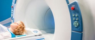
The principle of operation of a magnetic resonance imaging scanner is to create its own magnetic field in which the patient is located. Hydrogen atoms in the patient's body resonate with a magnetic field and the result is recorded as a series of images. These are virtual sections of organs and tissues that help make an accurate diagnosis.
Doctors ask that metal objects be left outside the room with the tomograph so that any metal or alloy that can be magnetized does not accidentally enter the magnetic field. All substances are divided into:
- Diamagnets
. Their magnetic susceptibility is negative. Silicon, gold, silver are diamagnetic. - Paramagnets
. They are capable of magnetization, but their magnetic susceptibility is very weak and difficult to detect, so only scientists understand the difference between dia- and paramagnets. Platinum and aluminum are paramagnetic. - Ferromagnets
. The magnetic susceptibility of such metals is high; they are capable of magnetization and change location under the influence of a magnetic field. Iron, cobalt, nickel are ferromagnetic.
Based on these properties, doctors ask to remove metal objects during magnetic tomography. Ferromagnets not only distort images and heat up in a magnetic field, but can also shift under its influence.
With which prostheses is it unacceptable to perform magnetic resonance diagnostics?
Although it is permissible to do an MRI with dental implants. During this examination, “internal” images of the body are obtained. A magnetic field is created in the tomograph, and the patient is placed in it.
The patient does not feel pain, and MRI does not damage tissues. The magnetic field created by the tomograph is 10 thousand times stronger than the Earth's magnetic field. It has the ability to attract objects that contain metal. If there are elements made of ferromagnetic materials in the magnetic resonance examination field, a potential threat arises. For this reason, people who have metal clamps on the aneurysm are not allowed to be diagnosed. The magnet can move the clamp - this will damage the vessel.
MRI diagnostics cannot be performed in the presence of the following types of implants:
- Cardio;
- Neuro;
- Ear;
- Containers that provide dosed dispensing of medications.
All electronic elements in the body, whose functioning can be disrupted under the influence of a magnetic field, the movement of which can create a health hazard - a limitation for MRI. But there are always exceptions, these include implants whose passport indicates their compatibility with the procedure.
Is it possible to do MRI with titanium implants?
Titanium is paramagnetic
.
Theoretically, it is magnetized. In practice, its magnetic susceptibility is 10 -5
. Such values are so small that in everyday life we say that titanium is not magnetized. It interacts with the magnetic field within limits that do not in any way affect the examination results. It is possible to do MRI with dental implants of all organs, including MRI of the brain. Fears that implants will move are unfounded, for the simple reason that titanium is not ferromagnetic. It does not heat up or change location under the influence of a magnet. Implantation is not a contraindication for tomography.
What about crowns and pins?
The question arises: what about the crowns that are placed on the implant? Some of them contain metals.
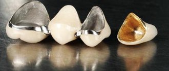
Modern metal dental crowns are created from alloys with weak or negative magnetic susceptibility. However, crowns that are many years old can distort the image and negate the diagnosis. Moreover, the more crowns, the stronger the distortion.
In principle, it is possible to do an MRI with metal crowns, provided that the patient warns the doctor about their presence. The doctor will take into account all factors: the examination organ, the fusion of the prosthesis, and will decide whether to carry out or refuse the procedure.
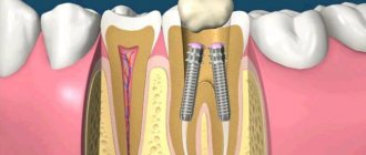
MRI with a pin in a tooth (stump inlay) has no contraindications if the pin is made of fiberglass. An MRI with a metal pin in the tooth is performed, because the amount of metal there is minimal and has virtually no effect on the results.
The doctor should be warned about metal crowns and pins. Ideally, he should be told what type of alloy was used. This can be found out in the dentistry where the prosthesis was installed. New tomographs take such information into account and correct the data.
How does a patient with metal ceramics feel during the procedure?
During a diagnostic examination in the presence of metal-ceramic structures, the patient will not experience any discomfort, much less acute pain or other painful sensations. The patient's life is also in perfect order and nothing threatens her.
If for one reason or another you are denied an examination, this will be due to the fact that your metal-ceramic crowns will greatly distort the final result. This will happen due to the resonance of the metal elements of the dental structure and the magnetic field of the tomograph. As a result, the doctor will either not be able to see the tissues he needs, or marks will appear on them that will give a false diagnosis.
Attention! Patients with metal-ceramic crowns are often denied examination due to the high cost of MRI. Since it is difficult to predict the final result, some laboratories prefer to refuse treatment to the patient, especially when it is carried out on a budgetary basis, when it is not the patient who pays, but the clinic.
conclusions
- There is no reason not to get an MRI if you have implants. Dental titanium is practically not magnetized. Titanium implants do not affect the examination results.
- Metal pins are also not a contraindication to tomography. The amount of metal there is minimal, and in fiberglass products it is completely absent.
- Metal dental crowns can distort results. This depends on the metal from which they are made, the number of prostheses and the organ being examined. Modern materials for prosthetics have low magnetic susceptibility. They are safer. In general, it is better to choose metal-free designs when installing crowns. This will prevent problems with MRI in the future.
Expert of the article Alexey Pavlovich Nesterenko Implant surgeon
Experience more than 10 years
What can replace MRI?
If a patient is contraindicated to undergo magnetic resonance imaging, and he does not want to remove metal-ceramic crowns, you can try to find an alternative. To do this, the following methods can be used, which can be found in the table.
| Procedure | Information content |
| Second place in terms of information content | |
| Relatively effective, sometimes requires confirmation with MRI | |
| Effective in some cases | |
| Not very informative, used only in combination | |
| It is advisable to use only in combination. |
Attention! Despite the possibility of using an alternative option, only magnetic resonance therapy can give the most accurate result, identifying even the slightest abnormalities that are not yet visible on ultrasound and any other tests.
Regardless of how many metal-ceramic crowns you have and what part of the body you are undergoing an MRI procedure for, you should always warn your doctor about the presence of a foreign body in your teeth.
If it is impossible to undergo an examination, the specialist will prescribe other procedures that can give an accurate picture of the state of health. If magnetic resonance imaging cannot be done, a computer diagnostic method is usually used.
How long does a crown stay on a tooth?
The service life of entire crowns primarily depends on the material. So:
- metal ones last an average of 5 years;
- metal-ceramic – 8-10 years;
- ceramic and zirconium dioxide - 10-15 years and above.
The duration of wearing dentures is also affected by: how well the supporting tooth was treated and ground, the skill of the dental technician and the quality of oral care. The dentist is visited every six months to remove plaque from products and adjust them. And after 7-10 years, most crowns become unusable and are replaced with new ones.
But loose and broken dentures will not last long: from several days to a couple of weeks. Therefore, people contact the clinic immediately: the faster the doctor adjusts or replaces the bridge or crown, the greater the chance of saving the abutment tooth.
This is how a crown is sawed
Other problems with crowns
In addition to affecting MRI results, metal crowns cause other problems: they break, chip, and the root canals and gums underneath may become inflamed, or the tooth stump may collapse. But most often, fixed dentures become loose or fly off.
What to do if the crown of a tooth is loose or has fallen off?
Crowns or bridges made of diamagnetic or ferromagnetic metals may begin to wobble after MRI. This is due to the displacement of the prostheses during the examination and subsequent decementation. In the future, the structures will fall out.
The presence of metal structures does not affect the results of a CT examination
However, the causes of poor fixation are not always related to magnetic resonance imaging. Sometimes dentures become loose and fly off due to:
- washing out the cement from under the crown - if its edge does not fit tightly to the neck of the tooth, the bond (adhesive composition) is gradually destroyed under the influence of saliva and drinks;
- poorly made design - it will not fit tightly if it does not follow the shape of the tooth in the smallest detail;
- small volume of the stump or its destruction - the remains do not hold the prosthesis, and it flies off.
Regardless of the reason, contact an orthopedic dentist. He will correct a loose or fallen crown and re-fix it. If the prosthesis cannot be repaired, a new one will be made and installed.
Swallowed a crown...
When a crown falls out, it is often swallowed along with saliva, food or drinks. There is no reason to worry - it will come out on its own along with the feces in a few hours or 1-2 days.
The only dangerous situation is when the prosthesis splits. In this case, an x-ray is taken, the location of the bridge or crown is determined, and then the stomach is washed out or an enema is given.
If you swallow a crown, it will be passed out in your stool within a maximum of 2 days.
But, in any case, the swallowed crown is reported to a gastroenterologist, surgeon or traumatologist. Magnetic resonance imaging is postponed until the structure is removed from the body.
How can MRI affect iron crowns?
Prostheses made of iron, steel, cobalt alloys and other ferromagnetic materials are still most often installed. Yes, there is titanium, porcelain and zirconium dioxide - but they are expensive, so not everyone can afford them.
Iron orthopedic structures are durable and cheap. But they become a problem if you need to do an MRI for 4 reasons:
- They distort the picture. The results will not be accurate. This is especially critical when diagnosing oncological pathologies: MRI is the best method for identifying tumors at the initial stage, when recovery success is high.
- They get very hot. This leads to a burn of the mucous membrane.
- They move from their place. After displacement, the crowns will have to be adjusted, re-attached, or new ones made.
- Possible loss. The magnetic field attracts metal crowns - it literally rips them out of your mouth.
Iron crowns get very hot and distort MRI results
Patients with metal prostheses are recommended to remove the structures before MRI or undergo another study - CT, PET. The above applies to closed-type tomographs or when examining the head, cervical or thoracic region. There is no risk when diagnosing other parts of the body.
Should you tell your doctor that you have dental implants?
A patient with installed dental implants must notify the doctor about this. Then the specialist makes the necessary adjustments, taking into account the location and composition of the prostheses. These settings eliminate the appearance of blurred distortions (also called field artifacts). And then the results of the examination will be clear and precise.
But if a person decides to keep silent about “non-native” teeth, then the picture will turn out blurry, because implants, although slightly, do affect the quality of the pictures. And such a careless patient will have to pay for the procedure and undergo diagnostics again. Why does the blame in this case fall on the patient? Because before the MRI, he will definitely be asked about the presence of implants and what kind of metal they are made of.
“Mom 2 years ago had an MRI of the head and blood vessels, using a high-field tomograph. And she’s had implants installed in her upper jaw for about 5 years now. And nothing, everything went fine. There are no special sensations during the procedure. Just be sure to tell the girls at the reception that there are implants before starting the procedure. They warn the doctor themselves, and then the device is reconfigured.”
Vitalina, from correspondence on the woman.ru forum.
Is it possible to do an MRI if you have dental implants?
Many patients: “Can I do MRI with dental implants?” After all, today every third person has pins installed.
Indications for use
The reason for prescribing this procedure may be a suspicion of a tumor, disruption of vascular activity or blood supply to tissues. Most often, MRI is prescribed as the main examination of the brain, spine or spinal cord to diagnose diseases of the osteoarticular system and abdominal organs. The doctor can prescribe diagnostics for ligament injuries, gynecological diseases, kidney diseases, suspected tumors and metastases.
Features of the procedure
This procedure can be carried out both for the patient’s entire body and for a separate part. The patient lies down on the couch with the head fixed. The couch is then moved inside the MRI machine. The procedure takes from 30 to 90 minutes. The patient spends this time in a relaxed and motionless state. A special feature of the procedure is the loud noise of the device, so the patient is provided with either headphones or earplugs. Throughout the procedure, the patient has access to an emergency interrupt button. Before the procedure, all metal objects must be removed. In some cases, the procedure is performed under anesthesia. The images are usually ready immediately after the examination or the next day.
