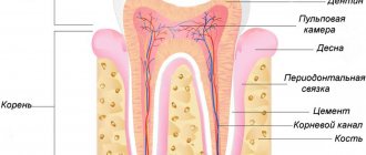
The structure of the periodontium.
Fixation of the tooth, as noted, is carried out using periodontal tissue, the fibers of which are stretched between the cement and the bone alveolus. The combination of three elements (osseous dental alveolus, periodontium and cementum) is designated as the supporting apparatus of the tooth. The width of the periodontal fissure ranges from 0.1 to 0.55 mm. The direction of the bundles of collagen fibers of the periodontium is not the same in its various parts. At the mouth of the dental alveolus (marginal periodontium) in the retaining apparatus, dentogingival, interdental and dentoalveolar groups of fiber bundles can be distinguished (Fig. 1-48).
Rice. 1-48. Periodontal fibers : 1 - interpapillary; 2 - circular; 3 - interdental; 4 - dentogingival
Dental fibers begin from the cementum of the root at the bottom of the gingival pocket and spread fan-shaped outward into the connective tissue of the gums. The thickness of the beams does not exceed 0.1 mm.
Interdental fibers form powerful bundles 1.0-1.5 mm wide. They extend from the cementum of the contact surface of one tooth through the interdental septum to the cementum of the adjacent tooth. This group of bundles maintains the continuity of the dentition and participates in the distribution of chewing pressure within the dental arch. Dentoalveolar fibers start from the cementum of the root along the entire length and go to the wall of the dental alveolus. Bundles of fibers begin at the apex of the root, spread almost vertically, in the apical part - horizontally, in the middle and upper third of the root they go obliquely from bottom to top (see Fig. 1-48). The orientation of bundles of periodontal collagen fibers, as well as the structure of the spongy substance of the jaws, is formed under the influence of functional load. In teeth devoid of antagonists, over time the direction of the periodontal bundles from oblique becomes horizontal and even oblique in the opposite direction. The periodontium of non-functioning teeth is more loose.
content .. 104 105 106 107 108 109 ..Concept of dentofacial segments (human anatomy)
As noted above (see section General description of teeth and dental organs, this edition), the dentofacial segment combines the jaw area and the tooth with the periodontium. The segments of the 1st and 2nd incisors, canine, 1st and 2nd premolars, 1st, 2nd and 3rd molars are distinguished. The boundary between the segments is a line drawn through the middle of the interdental space.
The dentofacial segments of the upper and lower jaws include various components (Fig. 89). Thus, the incisive segments of the upper jaw include the alveolar and palatine processes. The dentofacial segments of the premolars and molars contain the processes of the jaw with the lower wall of the maxillary sinus located in them.
The basis of each segment is formed by the alveolar process. The cross section of the alveolar process in the area of the incisal segments is close to a triangle, the base of which faces upward. In the area of the premolar and molar maxillary segments it approaches a rectangle. The outer and inner walls of the alveolar process consist of a thin layer of compact substance, between which there is a spongy substance, a tooth root with periodontium. The outer compact plate is thinner than the inner one, especially in the area of the incisal and canine segments. The palatine process in the area of the incisor-canine segments consists of the upper and lower plates of the compact substance and a layer of spongy substance between them, and at the level of the molar-maxillary segments - only of the compact substance or a compact and insignificant amount of spongy substance. The bone beams of the spongy substance of the segments are located mainly along the height of the jaw and pass from the alveolar process to other processes and walls of the maxillary sinus.
The cross-sectional shape of the incisal segments of the lower jaw is close to a triangle, the base of which faces downwards. In the area of the molars, they have the shape of a triangle with the base facing upward. The shape of the premolar-maxillary segments approaches oval. At the periphery of each segment of the lower jaw there is a compact substance that limits the segment cavity filled with spongy substance, tooth root and periodontium. The thickness of the compact substance is individually different both in different segments and within one of them. The outer plate of the compact substance has the greatest thickness in the region of the molar-maxillary segments, the smallest in the region of the mental foramen. The thickness of the inner wall of the segments is greatest in the area of the canine segments, the smallest in the area of the molar-maxillary. The spongy substance of the lower jaw in the area of the alveolar process is built from straight beams located along the length of the process; in the lower part of the segments they have an oblique direction - from the holes to the lower wall of the segment.
Dentofacial segments of the upper jaw
. Incisor-maxillary segments. With a narrow and high upper jaw, the incisal segments are elongated in height. The 2nd incisal segment includes part of the frontal process. The thickness of the outer compact plate of the alveolar process at the neck of the tooth is 1 mm, at the root level - 1 mm, the inner plate - 1-1.5 mm. The spongy substance consists of long bone beams that are directed into the palatine process, and in the 2nd incisal segment also into the frontal. Oval-shaped cells up to 2.5 mm in size are oriented along the beams. On preparations with a short and wide jaw shape, the incisal segments resemble an equilateral triangle and consist of the alveolar and palatine processes. The thickness of the outer compact plate above the root is no more than 1 mm; in the root area it is almost transparent, and in the area of the inner wall it is no more than 1.5 mm. The beams of spongy substance are short and thin. Starting from the apex and posterior wall of the socket, they are directed to the upper wall of the segment and into the palatine process. The cells are round in shape, their diameter is not more than 1 mm.
Rice. 89. Dentofacial segments (according to L.V. Kuznetsova). a - upper dentofacial segments; b — lower dentofacial segments. I - segments of a narrow and long jaw; II - wide and short jaw segments
Canine-maxillary segments. The shape of the canine segments with a narrow and high upper jaw resembles a truncated cone with the base facing upward, and with a wide and short jaw it approaches rectangular. The extradental part of the segment is formed by the body, frontal and alveolar processes. The nature of the structure of the spongy substance is similar to that in the incisive segments. However, part of the bone beams in both forms of the segment is directed to the frontal process. The thickness of the outer compact plate with a narrow form above the root is at least 1.5 mm, at the level of the root - at least 1 mm. With a wide jaw, the maxillary sinus can be determined at the level of this segment.
Premolar-maxillary segments. The shape of the alveolar process is close to a rectangle, more elongated in preparations of a high and narrow upper jaw. On specimens with a short and wide upper jaw, this segment may contain the corresponding part of the maxillary sinus. The thickness of the outer and inner plates of the compact substance of the alveolar process is about 1 mm. The beams of spongy substance in this form are directed from the top of the buccal root socket (at the level of the 4th tooth) to the area of the anterior, medial wall of the maxillary sinus and to its bottom. From the hole of the palatine root, the beams rush to the base and into the thickness of the palatine process. With a narrow upper jaw at the level of these segments, the maxillary sinus may be absent. The thickness of the compact substance of the alveolar process is about 2 mm. The bone plates of the spongy substance from the corresponding sockets of the tooth go into the palatine process and into the anterior wall of the jaw body.
Molar-maxillary segments. The 1st, 2nd and 3rd molar-maxillary segments usually include the lower wall of the maxillary sinus. The alveolar process of these segments and the maxillary sinus with a high and narrow jaw are elongated in height, the walls of the sinus are located almost vertically. The bone beams are long, directed into the palatine and zygomatic processes. The thickness of the compact plates of the alveolar process and the body are short and wide. The bone plates are short, evenly distributed and directed not only to the processes, but also to the bottom of the medial wall of the maxillary sinus. The thickness of the compact substance of the alveolar process is no more than 1.5 mm.
Dentofacial segments of the lower jaw
. Incisor-maxillary segments. With a narrow and long lower jaw, the incisal segments are elongated along the height of its body. The thickness of the outer compact plate at the middle of the segment height is at least 2 mm, the inner one is at least 2.5 mm. The bone beams are directed along the height of the segment from the walls of the socket, limiting oval-shaped cells measuring 1-2 mm. On specimens with a short and wide lower jaw, the segments are short, with a widened base. The thickness of the outer wall is no more than 1.5 mm, the inner wall is no more than 2 mm. The spongy substance is characterized by thin short bone beams that limit round-shaped cells measuring 1-1.5 mm.
Canine-maxillary segments. The shape of the canine-maxillary segments with a long and narrow lower jaw is close to rectangular. The thickness of the outer wall of the segment hole is 1.5 mm, the inner wall is 3 mm. With a wide and short lower jaw, the segments are shorter and have thinner walls. In the spongy substance, a group of beams can be distinguished, which, starting from the lower wall of the segment, goes to the top of the socket.
Premolar-maxillary segments. On preparations with a narrow and long jaw, the shape of the segments is rectangular. The thickness of the outer and inner walls of the holes is 2 mm. In short and wide jaws, the shape of the segments is close to oval, the thickness of the compact substance along all the walls of the segment socket is somewhat less than on a narrow and long jaw.
Molar-maxillary segments. On preparations with a narrow and long jaw, the 2nd and 3rd molar-maxillary segments have an irregular round shape, the 3rd molar-maxillary segment has the shape of a triangle. The thickness of the compact substance of the outer wall of the hole is at least 3.5 mm, the inner one is 1.5-2 mm. The spongy substance of the molar-maxillary segments is characterized by a coarse cellular structure.
content .. 104 105 106 107 108 109 ..
Tooth surfaces.
For the convenience of describing the relief or localization of pathological processes, a conventional designation of the surfaces of the tooth crown has been adopted. There are five such surfaces (Fig. 1-49).
Rice. 1-49. Surfaces (a), edge (b) and axis (c) of the tooth
1. The closure surface faces the teeth of the opposite jaw. They are found in molars and premolars. These surfaces are also called chewing surfaces. The incisors and canines at the ends facing the antagonists have a cutting edge.
2. The vestibular (facial) surface is oriented towards the vestibule of the oral cavity. In the front teeth, in contact with the lips, this surface can be called labial, and in the back teeth, adjacent to the cheek, - buccal. The continuation of the tooth surface to the root is designated as the vestibular surface of the root, and the wall of the dental alveolus, covering the root from the vestibule of the mouth, is designated as the vestibular wall of the alveolus.
3. The lingual surface faces the oral cavity towards the tongue. For the upper teeth, the name palatal surface is applicable. The surfaces of the root and walls of the alveoli directed into the oral cavity are also called. 4. The contact surface is adjacent to the adjacent tooth. There are two such surfaces: the medial surface, facing the middle of the dental arch, and the distal one. Similar terms are used to refer to the roots of teeth and the corresponding parts of the alveoli. denoting directions in relation to the tooth are also common When examining and describing teeth, the following terms are used: vestibular norm, chewing norm, lingual norm, etc. The norm is the position established during the study. For example, the vestibular norm is the position of the tooth in which its vestibular surface faces the researcher. The crown and root of the tooth are usually divided into thirds. Thus, when dividing a tooth by horizontal planes, the occlusal, middle and cervical thirds are distinguished in the crown, and the cervical, middle and apical thirds in the root. The sagittal planes divide the crown into the medial, middle and distal thirds, and the frontal planes into the vestibular, middle and lingual thirds. The dental system as a whole . The protruding parts of the teeth (crowns) are located in the jaws, forming dental arches (or rows) - upper and lower. Both dental arches contain 16 teeth in adults: 4 incisors, 2 canines, 4 small molars, or premolars, and 6 large molars, or molars. When the jaws are closed, the teeth of the upper and lower dental arches are in certain relationships with each other. Thus, the cusps of the molars and premolars of one jaw correspond to the depressions on the teeth of the same name in the other jaw. In a certain order, opposite incisors and canines come into contact with one another. This ratio of the closed teeth of both dentitions is referred to as occlusion. The contacting teeth of the upper and lower jaws are called antagonist teeth. As a rule, each tooth has two antagonists - the main and additional ones. The exceptions are the medial lower incisor and the 3rd upper molar, which usually have one antagonist each.
Types of teeth:
Typically, a person has 32 permanent teeth: 16 on the upper arch and 16 on the lower arch. Sometimes, due to genetic abnormalities, birth defects, or teething problems, more or fewer teeth appear.
The name of the teeth on each jaw is self-explanatory - the top sixteen are called "maxillary teeth" and the lower half are called "mandibular teeth".
Humans have different types of teeth that perform different functions such as cutting, tearing, grinding and crushing. The teeth are driven by the muscles of the jaw and lubricated by saliva, which is produced by the salivary glands.
Babies are born toothless because their main source of food is mother's milk. As children begin to wean, baby teeth gradually begin to emerge. Among the four types of teeth, the lower incisors are the first primary teeth to develop, and most children will have all twenty primary teeth by age 3. By the age of six, baby teeth are replaced by permanent teeth.
Adults have a total of 32 teeth, called permanent teeth:
- Eight incisors - four maxillary incisors and four mandibular incisors.
- Four canines - two canines on the upper jaw and two canines on the lower jaw.
- Eight premolars - four maxillary premolars and four mandibular premolars.
- Twelve molars - six molars in the upper jaw and six in the lower jaw. It also includes four wisdom teeth.
Teeth have different shapes because each tooth has a specific role during chewing and ultimately digestion. They are also placed at a specific location to better serve their purpose.
Dental formula.
The order of the teeth is recorded in the form of a dental formula, in which individual teeth or groups of teeth are written in numbers or letters and numbers. The complete dental formula is constructed in such a way that the teeth of each half of the jaws are written in Arabic numerals. This formula for an adult is as follows:
Individual primary teeth are indicated in the same way. The order of recording teeth in this formula is as if the recorder is examining the teeth of the person sitting in front of him, which is why this formula is called clinical. When examining patients, clinicians note missing teeth and circle the numbers of the teeth that require treatment. If all the teeth in a row are preserved, such a row is called complete.
The World Health Organization (WHO) has adopted a complete clinical dental formula for permanent dentition in a different form:
According to the WHO classification, the complete clinical dental formula for primary dentition is written as follows:
There are group dental formulas that reflect the number of teeth in each group in the halves of the jaws. This formula is called anatomical. In an adult, the group dental formula looks like this:
Functions of the dental system:
- Chewing is the main function of the teeth and dentofacial system. Chewing is the first stage of the digestion process.
- Aesthetics. The dental system affects the appearance of the face. Teeth also play an important aesthetic role. The position, shape and shade of teeth have a significant impact on the formation of a person’s personality.
- Pronunciation. Teeth help us pronounce words correctly as they play an important role in the pronunciation of consonants. Missing teeth (especially the upper front teeth) or an incorrect bite can somewhat impair normal speech.
Signs of teeth.
The teeth of the same name in the right and left dental arches differ in their structure. There are three signs by which you can determine whether a tooth belongs to the right or left dental arch: 1) sign of the crown angle; 2) a sign of curvature of the crown enamel; 3) root sign. The sign of the crown angle is that in the vestibular norm the angle formed by the closure surface and the medial surface is sharper than the angle between the closure surface and the lateral surface of the cutting edge. The last corner is slightly rounded.
The sign of curvature of the crown enamel is determined by examining the tooth from the side of the closure surface (in the chewing norm), while the medial part of the crown enamel on the vestibular side is more convex than the distal one. The root sign is determined in the position of the tooth in the vestibular norm. If you draw the longitudinal axis of the crown (lower the perpendicular from the middle of the cutting edge) and the longitudinal axis of the tooth (from the apex of the root to the middle of the cutting edge), it turns out that the axis of the tooth is deviated laterally. Consequently, the direction of deviation of the longitudinal axis of the tooth indicates the side of the tooth (Fig. 1-50).
Rice. 1-50. Signs of crown angle and root position
Forms and functions of human tooth types:
Incisors.
The incisors are located at the front of the mouth. These teeth have sharp edges and are adapted for cutting food into small pieces. Humans have eight incisors, four in the upper jaw and four in the lower jaw.
Fangs.
Fangs. They are located in the “corners” of the dental arches. They are characterized by a sharp, elongated and pointed surface. Their main function is to grab and tear food (tough foods such as meat). Humans have four fangs: two on the upper jaw and two on the lower jaw.
Bone tissue
As mentioned above, the jaw bones consist of spongy and cortical bone tissue. Cortical tissue is bone plates, 95% consisting of various mineral salts. These plates are arranged tightly, which is why the cortical bone is hard and durable.
Spongy tissue, on the contrary, is softer, since, for the most part, it consists of soft tissue, has a cellular structure, and the bone plates in it form partitions.
Bone tissue is made up of different types of cells, and each cell performs its own functions. Thus, osteoblasts are responsible for the formation of new tissue, and osteoclasts, on the contrary, for the destruction of old tissue, stimulating the renewal of bone tissue. Osteocytes are the main cells of bone. Interstitial cells are a kind of “lining” of bone.